M
ACIEJK
UŹMIŃSKIThe Influence of Apical Lesions on Electronic
Tooth−Length Measurements – an
in vitro
Study
Wpływ obecności zmian okołowierzchołkowych
na elektroniczny pomiar długości zęba – badania
in vitro
Department of Conservative Dentistry and Endodontics, Medical University of Łódź, Łódź, Poland
Adv Clin Exp Med 2006, 15, 4, 607–611 ISSN 1230−025X
ORIGINAL PAPERS
© Copyright by Silesian Piasts University of Medicine in Wrocław
Abstract
Objectives.Investigating the influence of apical lesions on tooth−length measurements with the Root ZX apex locator (Morita, Japan) and checking a modified method of uncovering the apical part of the canal.
Material and Methods. Eighteen freshly extracted human teeth with attached apical lesions were investigated. Tooth lengths were measured with the Root ZX and a No. 10 K−file in an alginate mould according to the method of Katz et al. (1996) (group A). After removal of lesions, the teeth were embedded in freshly mixed alginate and measured again (group B). Then the file was secured in place with a composite material. The apical 4 mm of the canal of each tooth was exposed by grinding with a water−cooled high−speed diamond bur. Distances from the file tip to the dentino−cemental junction and to the apical foramen were measured with an endodontic microscope (Karr, Switzerland) under ×17magnification.
Results.The tooth lengths measured in group A were longer than in group B in 9 cases (by 0.25 to 1.25 mm). In the presence of the apical lesions, the measurements were too long in 7 canals by 0.15 to 1.3 mm. The mean dif− ference between group A and B was 0.36 mm, with a standard deviation of 0.3 mm. There was a statistically sig− nificant difference between both groups at the probability level of p< 0.05. The modified method of uncovering the apical part of the canal proved to be useful.
Conclusions. The presence of apical lesions negatively affects electronic tooth−length measurements (Adv Clin Exp Med 2006, 15, 4, 607–611).
Key words: apical lesions, electronic tooth measurement, Root ZX.
Streszczenie
Cel pracy. Ocena wpływu obecności zmian okołowierzchołkowych na elektroniczny pomiar długości zębów z użyciem endometru Root ZX (Morita, Japonia). Zbadano ponadto własną metodę szlifu odkrywającego przebieg kanału w okolicy wierzchołka korzenia zęba.
Materiał i metody. Badania przeprowadzono na 18 świeżo usuniętych zębach ze zmianami okołowierzchołkowy− mi, które pozostały na wierzchołkach korzenia zębów po ekstrakcji. Długość zębów zmierzono z użyciem pilnicz− ka o rozmiarze 10 wg ISO w modelu alginatowym zgodnie z metodą Katza (grupa A). Po usunięciu zmian z wierz− chołków korzeni zębów zęby ponownie umieszczono w świeżo zarobionym alginacie i zmierzono długości (grupa B). Pilniczki używane do pomiarów unieruchomiono przez wypełnienie komory płynnym kompozytem. Wykonano szlif odsłaniający wierzchołkową część kanału z użyciem diamentowego wiertła na wiertarkę szybkoobrotową. Zmierzono odległości między końcem narzędzia a otworem anatomicznym pod 17−krotnym powiększeniem, używając mikroskopu endodontycznego (Karr, Szwajcaria).
Wyniki. Długości uzyskane podczas pomiarów w grupie A były dłuższe od tych z grupy B w 9 przypadkach (różnica wynosiła 0,25–1,25 mm). W zębach ze zmianami zmierzone długości były za długie w 7 kanałach (0,15–1,3 mm). Różnica średnich obu grup wyniosła 0,36, a odchylenie standardowe 0,3. Wystąpiła istotna statystycznie różnica między obiema grupami przy poziomie istotności 0,05. Zmodyfikowana metoda szlifu okol− icy wierzchołkowej okazała się użyteczna.
Wnioski. Obecność zmian okołowierzchołkowych wpływa negatywnie na dokładność elektronicznego pomiaru długości zęba (Adv Clin Exp Med 2006, 15, 4, 607–611).
The apex locator has proved to be a useful tool, as it has been demonstrated many times to be more accurate than radiological methods of mea− suring tooth length [1, 2]. The Root ZX, a dividing frequency tool, is not only accurate, but also little affected by difficult clinical situations. There are few data on factors that impact electronic root canal measurements. Until now, authors have shown the influence of foramen diameter, ion con− centrations, and preflaring on tooth length [3], but even these factors have been analyzed in very few studies. In the presence of a minor foramen, apex locator readings are correct. In case of resorption or periapical pathosis, which quite often coexist, the readings can be less accurate [4]. Thus the pre− sent study aims firstly at investigating the influ− ence of apical lesions on tooth−length measure− ments using the Root ZX apex locator (Morita, Japan) and, secondly, at checking a modified method of grinding the apical part of the canal.
Material and Methods
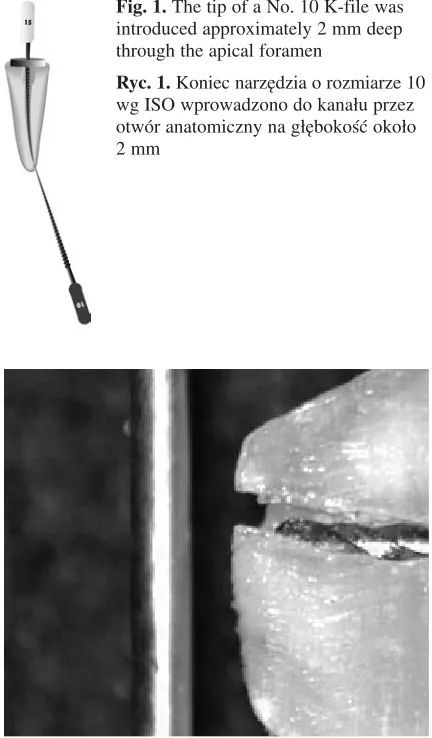
Eighteen human single−rooted teeth which had been previously extracted with attached apical lesions were investigated. The diameter of apical pathosis was from 3 to 6 mm. Following extrac− tion, the teeth were stored in isotonic saline solu− tion for up to 12 hours. Tooth lengths were mea− sured with the Root ZX and a No. 10 K−file in an alginate mould and the data was recorded (group A). This model was developed by Katz et al. [5] and used in other studies [4, 6]. After removal of lesions, the teeth were embedded in freshly mixed alginate and afterwards measured a second time (group B). Then the file was secured in place with some composite material. From this point, each procedure was carried out under two−fold magnifi− cation. To localize the apical foramen, the tip of a No. 10 K−file was introduced without use of force through the apical foramen (Fig. 1). With a strong source of light and the file in place, it was possible to define the location of the canal and the cut surface. The apical 4 mm of the canal of each tooth was exposed by grinding with a water− cooled high−speed diamond drill until the apical part of the file was visible under a thin layer of dentine. Then the rest of the tissue was removed by the means of a scalpel and a No. 10 H−file. The distances from the file tip to the dentino−cemental junction and to the apical foramen were measured under an endodontic microscope (Karr, Switzerland) at ×17 magnification and AverMedia software (Fig. 2). All measurements that were beyond the apical constriction were designated as positive and all which were coronal to the apical
constriction (CDJ) were considered negative [7]. The statistical analysis was carried out using the Student t-test for dependent variables.
Results
The tooth lengths measured in group A were longer than in group B in 9 cases (by 0.25 to 1.25 mm) (Table 1). In the presence of apical lesions the measurements were too long in 7 canals by 0.15 to 1.3 mm. The difference between group A and B on average was 0.36 mm, with a mean deviation of 0.3 mm. There was a statistically sig− nificant difference between both groups at the probability level of p< 0.05.
Fig. 1. The tip of a No. 10 K−file was introduced approximately 2 mm deep through the apical foramen
Ryc. 1. Koniec narzędzia o rozmiarze 10 wg ISO wprowadzono do kanału przez otwór anatomiczny na głębokość około 2 mm
Fig. 2. The microscopic image of the apical portion of a tooth from the study. The needle is an object with known diameter used to calibrate the Aver Media pro− gram before measurements
Ryc. 2. Obraz mikroskopowy odsłoniętej części kanału. Igła do znieczuleń o znanej średnicy
Discussion
Although electronic tooth length measure− ments are precise in 90–95% of cases [8], there are some factors which can make it difficult to take precise readings. Apart from Myers’s phenome− non [9], the lack of a minor foramen (CDJ), perfo− rations, ramifications, and bifurcations have their crucial importance [6, 10]. An important factor is to prevent electric current from escaping through metal or temporary restorations, blood, or pus. Another factor disturbing measurements is pulpi− tis, which is connected with changes in ion con− centrations [11]. The influence of periapical patho− sis is only mentioned in the literature on the basis of clinicians’ experience, but has not yet been sci−
entifically proven [12]. In the present study a No. 10 K−file was used for measurements. Katz et al. [5] have suggested that the use of a larger file can increase accuracy. Another point of view is that a small file is usually used during the first pene− tration of the canal to gain maximal tactile sense. Another study shows that the file size is not so important if an apical foramen is present [13].
Although this was only an in vitrostudy, the outcomes are similar to the author’s clinical expe− rience. The results show that length measurements of teeth without periapical lesions should be decreased by 0.5 mm in order not to enlarge the apical constriction. This has been already proved by Shabahang et al. [14].
No Tooth length Tooth length Difference Distance from Distance from Distance from Distance from (Lp.) in group A in group B, of lengths, file tip to CDJ file tip to file tip to CDJ file tip to
(Długość (Długość mm in group B, apical foramen in group A, apical foramen
zęba zęba (Różnica mm in group B, mm in group A,
w grupie A) w grupie B) długości) (Odległość mm (Odległość mm
mm mm mm między (Odległość między (Odległość
końcem pil− między koń− końcem pil− między koń− niczka cem pilniczka niczka cem pilniczka a otworem a otworem a otworem a otworem fizjologicznym anatomicznym fizjologicznym anatomicznym w grupie B) w grupie B) w grupie A) w grupie A)
1 10 10.5 –0.50 0.4 1.5 0.65 1.75
2 15 14.75 025 0.4 0.8 0.15 0.55
3 14 13.5 0.5 0.1 0.3 –0.4 –0.2
4 16.5 15.5 1 0.5 1 –0.5 0
5 17.5 17.25 0.25 –0.3 0 –0.55 –0.25
6 12 12 0 –0.3 –0.2 –0.3 –0.2
7 14.25 14 0.25 0.2 0.5 0 0.25
8 17.25 17.5 –0.25 0.1 0.7 –0.15 0.45
9 9 9 0 –0.6 0.4 –0.6 0.4
10 23.5 23 0.5 0 0.4 –0.5 –0.1
11 8 8 0 0.3 0 0.3 0
12 10 9 1 –0.3 0 –0.3 –1
13 11.75 11 0.75 0 0.1 –0.75 –0.65
14 11 11 0 0.3 0.5 0.3 0.5
15 8 8 0 –0.2 –0.1 –0.2 –0.1
16 15.25 14 1.25 0.2 0.6 –1 –0.66
17 10.5 10.5 0 0 0.2 0 0.2
18 11.25 11.25 0 0 0.2 0 0.2
Mean 10.8 11.9 2.7 0 0.25 –0.25 –0.33
(Średnia)
Standard 2.5 4.5
deviation (Odchyl. standard.)
Table 1. Tooth lengths. Distances from the file tip to apical foramen and CDJ
Alginate was used as a conducting medium for the electronic apex locator, which was proposed by Katz et al. [5]. Thus, in order to simplify the test procedure in in vitromodels, medium having similarities in resistance to periodontal tissues has been frequently used [15].
In order to reveal the file, grinding of the api− cal part of the root was performed according to the method described by Nguyen [4]. This method was criticized by Stein and Corcora [16] because in this way the apical part of the canal can be acci− dentally removed. In the present study the proce− dure, modified by the author, concerning the local− ization of the apical part of the canal found its application. There was no loss of the apical part of the canal during the grinding. Moreover, the grind− ing method is easier than the method of multilayer sections described by Stein and Corcora [16]. Other authors used SEM to measure distances in the apical part of the canal, which was difficult and caused some damaged samples [17]. The current method is similar in its simplicity to one described by Weiger et al. [18]. In this method, roots that had been previously notched were split into two parts that were later examined separately under the microscope. This, however, does not allow for fix− ing the file in place after the measurements, which is crucial in the opinion of the author of this study. For the precise localization of the apical foramen it proved helpful to mark it with a No. 10 K−file and observe the tooth under a strong light source.
The difference obtained in the present study of 0.36 mm between teeth with and without periapi− cal lesions may be significant in endodontic treat− ment. It is even more important in devices such as the Tri Auto ZX, where a hypothetical minor fora−
men is automatically found and the canal would be enlarged up to this point. On the other hand, the outcomes in group B were clinically acceptable in 94.5% of cases, similar to but slightly higher than those obtained by other authors [19, 20]. This sug− gests that the measurements were not influenced by the apical resorption. The reason for obtaining longer measurements for teeth with apical lesions is unclear. It could be caused by the difference in ion concentrations between the inside of the canal and the apical lesions in group A or alginate (peri− odontal tissue) in group B. According to Kovacević and Tomislav [11], the accuracy of measurements deteriorated with decreasing ratio between the internal and external ion concentra− tions. Another reason could be some differences in conductivity of apical lesions and alginate. Other authors [21, 22] stated in their studies that the length of the canal featuring with larger conduc− tivity is oversized. Thus further studies of lesion conductivity are indispensable.
The current study presents evidence for the conclusion that the presence of apical lesions in the tooth impacts predictably to a minor extent on the accuracy of measurement. However, this state− ment should be proved by further clinical studies as well as experiments on teeth with larger apical lesions and using other apex locators. It should also be considered whether measurements in teeth with apical lesions are influenced by apical resorp− tion. The results of this research suggest that the presence of apical lesions negatively affects tooth length measurements and that the modified method of discovering the apical part of the canal proved to be useful.
References
[1] Pratten DH: McDonald Comparison of radiographic and electronic working lengths. J Endod 1996, 22, 4, 173–176.
[2] Kuźmiński M: Porównanie dokładności pomiaru długości zębów z użyciem endometru i technik radiologicznych. Czas Stomat 2001, 54, 7, 427–431.
[3] Ibarrola JL, Chapman BL, Howard JH, Knowles KI, Ludlow MO: Effect of preflaring on Root ZX apex loca− tors. J Endod 1999, 25, 9, 625–626.
[4] Kuźmiński M, Piątowska D: Ocena wpływu różnych czynników na elektroniczny pomiar długości kanałów zębowych. Czas Stomat 2005, 58, 11, 772–778.
[5] Katz A, Mass E, Kaufman AY: Electronic apex locator: a useful tool for root canal treatment in the primary den− tition. ASDC J Dent Child 1996, 63, 6, 414–417.
[6] Thomas AS, Hartwell GR, Peter CM: The accuracy of the Root ZX electronic apex locator using stainless−steel and nickel−titanium files. J Endod 2003, 29, 10, 662–663.
[7] Dunlap CA, Remeikis NA, BeGole EA, Rauschenberger CR: An in vivoevaluation of an electronic apex loca− tor that uses the ratio method in vital and necrotic canals. J Endod 1998, 24, 1, 48–50.
[8] Lauper R, Lutz F, Barbakow F: An in vivocomparison of gradient and absolute impedance electronic apex loca− tors. J Endod 1996, 22, 5, 260–263.
[9] Myers JW: Demonstration of a possible source of errors with an electric pulp tester. J Endod 1998, 24, 199–200.
[10] Kaufman AY, Fuss Z, Keila S, Waxenberg S: Reliability of different electronic apex locators to detect root per− foration in vitro. Int Endod J 1997, 30, 6, 403–407.
[12] Walton RE, Torabinejad M: Principles and practice of endodontics. W.B. Saunders Company, Philadelphia 1996, 194–198.
[13] Meredith M, Gulabivala K: Electrical impedance measurements of root canal length. Endod Dent Traumat 1997, 13, 126–131.
[14] Shabahang S, Goon WW, Gluskin AH: An in vivoevaluation of Root ZX electronic apex locator. J Endod 1996, 22, 11, 616–618.
[15] Nekoofar MH, Sadeghi K, Akha ES, Namazikhah MS: The accuracy of the Neosono Ultima EZ apex locator using files of different alloys: an in vitrostudy. J Calif Dent Assoc 2002, 30, 9, 681–684.
[16] Stein TJ, Corcora JF: Nonionizing method of locating the apical constriction in root canals. Oral Surg Oral Med Oral Pathol 1991, 71, 1, 96–99.
[17] Pagavino G, Diamante D, Marri M, Pace R: Localization of the apical foramen using the newest electronic instruments: stereomicroscopy and SEM. Minerva Stomatol 1995, 44, 11, 499–506.
[18] Weiger R, John CH, Geible H, Lost Z, Lost C: An in vitrocomparison of two modern apex locators. J Endod 1999, 5, 11, 765–768.
[19] Ounsi HF, Naaman F: In vitroevaluation of the reliability of the Root ZX electronic apex locator. Int Endod J 1999, 32, 120–123.
[20] Ounsi HF, Haddan G: In vitroevaluation of the Endex electronic apex locator. Int Endod J 1998, 24, 2, 120–121.
[21] Vob A, Siebenkees J: Experimentelle und klinische Bewertung der endometregerate Apit und Root ZX. Deutsch Zahnartaltung 1994, 49, 3, 281–284.
[22] Ponce EH, Fernandez JAV: The cemento−dentino−canal junction, the apical foramen, and the apical constriction: evaluation by optical microscopy. J Endod 2003, 29, 3, 214–219.
Address for corresponcence:
Maciej Kuźmiński Zakład Endodoncji
Katedra Stomatologii Zachowawczej, Endodoncji i Periodontologii UM ul. Pomorska 251
92−213 Łódź
e−mail: [email protected] tel.: +48 42 678 31 99
Conflict of interest: None declared Received: 03.06.2005
Revised: 15.03.2006 Accepted: 17.05.2006
Praca wpłynęła do Redakcji: 03.06.2005 r. Po recenzji: 15.03.2006 r.