R E S E A R C H
Open Access
Stable isotope informed genome-resolved
metagenomics reveals that Saccharibacteria
utilize microbially-processed plant-derived
carbon
Evan P. Starr
1, Shengjing Shi
2, Steven J. Blazewicz
3, Alexander J. Probst
4, Donald J. Herman
5,6,
Mary K. Firestone
5,6*and Jillian F. Banfield
5,6,7*Abstract
Background:The transformation of plant photosynthate into soil organic carbon and its recycling to CO2by soil microorganisms is one of the central components of the terrestrial carbon cycle. There are currently large knowledge gaps related to which soil-associated microorganisms take up plant carbon in the rhizosphere and the fate of that carbon.
Results:We conducted an experiment in which common wild oats (Avena fatua)were grown in a13CO2 atmosphere and the rhizosphere and non-rhizosphere soil was sampled for genomic analyses. Density gradient centrifugation of DNA extracted from soil samples enabled distinction of microbes that did and did not incorporate
the13C into their DNA. A 1.45-Mbp genome of a Saccharibacteria (TM7) was identified and, despite the microbial
complexity of rhizosphere soil, curated to completion. The genome lacks many biosynthetic pathways, including genes required to synthesize DNA de novo. Rather, it requires externally derived nucleotides for DNA and RNA synthesis. Given this, we conclude that rhizosphere-associated Saccharibacteria recycle DNA from bacteria that live
off plant exudates and/or phage that acquired13C because they preyed upon these bacteria and/or directly from
the labeled plant DNA. Isotopic labeling indicates that the population was replicating during the 6-week period of plant growth. Interestingly, the genome is ~ 30% larger than other complete Saccharibacteria genomes from non-soil environments, largely due to more genes for complex carbon utilization and amino acid metabolism. Given the
ability to degrade cellulose, hemicellulose, pectin, starch, and 1,3-β-glucan, we predict that this Saccharibacteria
generates energy by fermentation of soil necromass and plant root exudates to acetate and lactate. The genome also encodes a linear electron transport chain featuring a terminal oxidase, suggesting that this Saccharibacteria may respire aerobically. The genome encodes a hydrolase that could breakdown salicylic acid, a plant defense signaling molecule, and genes to interconvert a variety of isoprenoids, including the plant hormone zeatin. Conclusions:Rhizosphere Saccharibacteria likely depend on other bacteria for basic cellular building blocks. We
propose that isotopically labeled CO2is incorporated into plant-derived carbon and then into the DNA of
rhizosphere organisms capable of nucleotide synthesis, and the nucleotides are recycled into Saccharibacterial genomes.
Keywords:Stable isotope probing, Complete genome, Saccharibacteria, Metagenomics, Rhizosphere
* Correspondence:[email protected];[email protected] 5Department of Environmental Science, Policy, and Management, University
of California, Berkeley, CA 94720, USA
Full list of author information is available at the end of the article
Background
The candidate phyla radiation (CPR) comprises a large fraction of the bacterial domain [1,2]. Within the CPR, Saccharibacteria, formerly TM7, is one of the most cap-tivating phyla because of the wide diversity of habitats in which it is found, including activated sludge, human and dolphin oral cavities, seawater, aquifer sediment, soil,
and cockroach guts [3–10]. Much has been learned
about the metabolism of Saccharibacteria and their influence on their environment, for instance, the possible immunosuppressive capabilities of an
episym-biont of Actinomyces odontolyticus in human mouths
and bulking issues caused in wastewater treatment plants [7, 11]. There have been previously proposed subdivisions within the Saccharibacteria phylum, but currently the majority of our genomic data comes from organisms which fall within Subdivision 3 and there ex-ists very little genomic representation of Subdivision 1 [4, 12]. Organisms in Subdivision 1 are predominantly found in environmental samples [13]. One of the reasons why there may be less genomic data available for Saccharibacteria in Subdivision 1 is because of the high microbial diversity of environmental samples, especially soil, which makes sequencing efforts more challenging. Based on 16S ribosomal RNA gene surveys, we know that Saccharibacteria occur in the rhizosphere, but little is known about their metabolism and how they differ from related organisms growing in other environments [14,15]. There are six previously reported, closed, and circular-ized genomes for Saccharibacteria (one from a wastewa-ter treatment plant [16], human mouth [7], two from sediment [1,8], and two from a thiocyanate remediation
wastewater reactor [17]) and some partial genomes.
Based on genomic analyses, it was previously predicted that Saccharibacteria are anaerobic fermenters, despite
the documentation of one genome encoding a
single-subunit of NADH: ubiquinone dehydrogenase and a complete ubiquinol oxidase, which was explained as an
oxygen-scavenging mechanism [8]. It has also been
documented that Saccharibacteria are able to grow in the presence of oxygen [3,7,18]. Previous soil SIP work has shown that Saccharibacteria DNA became labeled in
the presence of 13
C cellulose and toluene [19, 20]. Labeling experiments have indicated that 13CO2can be
traced from plant fixation into the DNA of
rhizosphere-growing microbes [21, 22]. Here, we found that a Saccharibacteria population in rhizosphere soil
in-corporated 13C into its genomic DNA, indicating that
this population was active and dividing. We report the complete genome for this organism and show how analysis of its metabolism sheds light on the pathway by which the label was incorporated. We compare the genome to those of other complete Saccharibacteria to address the question of how this bacterium is
adapted to live in soil and specifically in the
rhizosphere.
Results
After 6 weeks in labeling chambers, Avena fatuashoots
were highly labeled (~ 94 atom% 13C). DNA was
ex-tracted from the rhizosphere and bulk soil samples. By comparing the density separation of the rhizosphere community DNA to the bulk community DNA, we were able to define un-enriched (light), partially13C-enriched
(middle), and highly 13C-enriched (heavy) fractions
(Fig. 1). Based on the cutoff values for these fractions, 32 density-separated fractions for the rhizosphere sample were then combined, generating light, middle, and heavy fractions (the bulk sample only contains light and middle fractions due to the absence of13C-enriched DNA) (Fig.1).
The light, middle, and heavy density separated fractions from the rhizosphere and bulk samples were sequenced and subjected to genome-resolved metage-nomic analyses (Additional file 1: Table S1). From the rhizosphere middle fraction, we assembled 210 Mbp of scaffolds larger than 1 kbp. One especially large scaffold
Fig. 1Stable isotope fraction determination. This plot shows the
was assembled de novo and could be circularized. Local assembly errors were identified and corrected and three scaffolding gaps were filled by manual curation. Manual curation made use of unplaced paired reads that were mapped back to the gap boundaries to fill gaps. The complete, closed genome is 1.45 Mb in length with a GC content of 49.95%. We were able to recover a single chromosome and we detected no integrated phage or plasmids. The genome was most abundant in the rhizosphere middle fraction at 15× coverage, but was also present in the rhizosphere heavy and light frac-tions at ~ 3× normalized coverage. The genome had less than 1× coverage in the non-rhizosphere soil (Additional file 1: Table S1).
DNA buoyant density in a cesium chloride solution is a function of both the extent of isotopic enrichment and the GC content. Low GC has a lower buoyant density as
compared to higher GC DNA [23]. The completed,
closed genome has a lower GC content (49.95%) than the rest of the rhizosphere middle fraction assembly (average GC content of scaffolds larger than 1000 bp is
66%), which indicates 13C may have been incorporated
into the DNA. The rhizosphere middle fraction where the genome was mainly detected had a density of 1.737– 1.747 g/ml. Given that natural abundance DNA with 49.95% GC content would have a density of ~ 1.71 g/ml [24], we estimate that the DNA from which the genome was assembled was at least 50% enriched in13C.
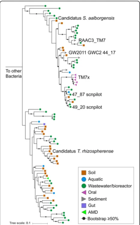
The genome has 1531 protein coding sequences (Add-itional file 2: Table S2) and a full complement of tRNAs (46 in total). The 5S rRNA, 23S rRNA, and 16S rRNA genes are in a single locus that also includes Ala and Ile tRNA genes. Based on the sequence of the 16S rRNA gene, the genome was assigned to be a member of the Saccharibacteria phylum. The closest 16S rRNA gene
sequences in NCBI are from the rhizosphere of Pinus
massoniana(Fig.2) [25]. The most closely related
geno-mically described organism is Candidatus
Saccharimo-nas aalborgensisfrom activated sludge with 84% identity across the full-length 16S rRNA gene [16]. We propose the nameCandidatus“Teamsevenus rhizospherense”for the organism described here, given the derivation of the genome from the rhizosphere. In accordance with the phylogenetic analysis, we renamed Subdivision 1 to Candidatus Soliteamseven because this genome is the first described Candidatus species of this clade. The representatives of this clade are mostly found in soil,
therefore, we propose the complete taxonomic
descriptor: Phylum: Candidatus Saccharibacteria, Class: Candidatus Soliteamseven, Order: Candidatus Teamseve-nales, Family:CandidatusTeamsevenaceae, Genus: Candi-datusTeamsevenus, Species:Candidatusrhizospherense.
We calculated the GC skew and cumulative GC skew
across the closed T. rhizospherense genome and found
the symmetrical pattern typical for bacteria, with a single peak and trough indicative of the terminus and origin of replication (Additional file 3: Figure S1). This re-sult both validates the accuracy of the circularized genome and confirms that Saccharibacteria use the typical bacterial pattern of bi-directional replication from a single origin to the terminus (as do some Per-egrinibacteria, another group of CPR bacteria [26]). The start of the genome was adjusted to correspond to the predicted origin, which lies between the DNA polymerase III subunit beta and the chromosomal replication initiator protein. It has a full set of riboso-mal proteins, except for L30, which is uniformly ab-sent in CPR bacteria [1].
Fig. 2Phylogeny of Saccharibacteria based on 16S rRNA gene
Biosynthetic pathways
TheT. rhizospherensegenome encodes a number of en-zymes for the conversion of nucleotides to NMP, NDP, and NTP and formation of RNA. In addition, we identi-fied genes to phosphorylate G, C, and U. However, the
organism lacks the genes required to synthesize
5-phospho-alpha-D-ribose-1-diphosphate (PRPP).
Fur-ther, it lacks essentially all of the steps for synthesis of nucleotide bases and the pathways that would convert PRPP to inosine monophosphate or uridine monopho-sphate. T. rhizospherense may have a novel nucleotide biosynthesis pathway, but this is unlikely as nucleotide biosynthesis remains highly conserved across domains
[27] and the pathway can be recognized in some CPR
bacteria [26]. Thus, we infer that T. rhizospherense did not de novo synthesize its nucleotides but rather ac-quired them from an external source. The genome
en-codes several nucleases, an external micrococcal
nuclease, and an oligoribonuclease for the breakdown of
externally derived DNA and RNA (Fig.3). The
mechan-ism for DNA and RNA import is unknown, as we did not identify nucleotide transporters. However, there are a number of transporters with unidentified specificity
that could be involved in DNA or nucleotide uptake or the type IV pili could be responsible for this function. A large portion of the genome is dedicated to DNA and RNA repair mechanisms. There are 20 8-oxo-dGTP diphosphatase genes that prevent the incorporation of oxidized nucleotides. These enzymes may be required given that the nucleotides may be scavenged from dead cells and could have accumulated extensive DNA dam-age. Access to damaged DNA may be a consequence of life in a mostly aerobic environment, a seemingly un-usual condition for members of the Saccharibacteria phylum. All other complete genomes were found in mostly anaerobic environments and encode fewer genes with this function.
The T. rhizospherense genome does not encode the ability to synthesize any amino acids de novo from central metabolites. However, it encodes genes to generate amino acids from precursors (e.g., valine and leucine from 2-oxoisovalerate, isoleucine from 2-methyl-2-oxopentano-ate, and histidine from L-histidinol phosphate) and to interconvert some amino acids (serine and glycine). We identified genes for proteases that could breakdown exter-nally derived proteins. No amino acid-specific transporters
Fig. 3Cell diagram ofT.rhizospherense. (A) fusaric acid resistance machinery, (B) unidentified importer, (C) glucan 1,3-beta-glucosidase, (D) NADH
dehydrogenase II, (E) blue-copper protein, (F) cytochrome bo3ubiquinol terminal oxidase, (G) F-type H+-transporting ATPase, (H) peptidase, (I)
were annotated, but several transporters of unknown function could import the amino acids. There is little evi-dence to suggest that externally derived amino acids are broken down for use in the TCA (the only TCA cycle gene identified is a fumarate reductase subunit) or other cycles.
T. rhizospherense appears unable to synthesize fatty acids, yet it encodes three copies of the 3-oxoacyl-(acyl-carrier protein) reductase, five copies of acyl-CoA thioesterase I, and two copies of SGNH hydrolase indi-cating the capacity for fatty acid hydrolysis and con-version. We found that the genome contains a number of genes for sequential steps in the
glycero-phospholipid metabolism pathway. T. rhizospherense
may incorporate phosphatidylcholine (possibly derived from eukaryotes), 1,2 diacyl sn-glycerol-3P (from bac-teria), or phosphatidylethanolamine (the main bacter-ial phospholipid) and may be able to interconvert the compounds using a gene annotated as phospholipase D. We identified a putative phosphatidate cytidylyl-transferase that could add a head group to 1,2 diacyl sn-glycerol-3P forming CDP-diacylglycerol. This may be able to be converted into three products: phosphatidylgly-cerophosphate (via CDP-diacylglycerol-glycerol-3-phosphate 3-phosphatidyltransferase), or to cardiolipin (via cardiolipin synthase), or to phosphatidyl-1D-myo-inositol (via CDP-diacylglycerol-inositol 3-phosphatidyltransferase).
Interestingly, phosphatidyl-1D-myo-inositol is the pre-cursor for generation of phosphatidylinositol manno-sides, glycolipids that are decorated by a chain of mannose molecules and that are found in the cell walls of Mycobacterium [28]. There are several genes for the first step in phosphatidylinositol mannoside biosynthesis, which involves modification of phosphatidyl-1D-myo--inositol by addition of mannose. These include phos-phatidylinositol alpha-mannosyltransferase (three copies) and a single copy of alpha-1,6-mannosyltransferase.
Sub-sequently, a polyprenol-P-mannose
α-1,2-mannosyl-transferase (CAZy glycosylα-1,2-mannosyl-transferase family 87) adds another mannose group. Other mannose additions may involve the three copies of dolichol-phosphate
man-nosyltransferase, which transfer mannose from
GDP-mannose to dolichol phosphate a mannose car-rier involved in glycosylation [29]. Thus, although T. rhizospherense appears to be unable to synthesize fatty acids, it appears to encode a number of genes that may be involved in the interconversion of mem-brane lipids, including phosphatidylinositol manno-sides, if provided 1,2 diacyl sn-glycerol-3P.
Central metabolism and energy generation
Interestingly, the T. rhizospherense genome encodes a simple, two-subunit cellulosome that may be used to attach to and degrade plant or microbially derived cellu-lose to cellobiose. Cellobiose is likely converted to
D-glucose via one of 14 different glycosyl hydrolases. The genome also encodes genes for the production of
D-glucose via breakdown of starch/glycogen and trehal-ose. The genome contains several genes for hydrolysis of 1,3-β-glucan, one of the most common fungal cell wall polysaccharides [30], to D-glucose. A significant portion of plant root exudation are sugars that could be fed directly into the T. rhizospherense metabolism [31].
D-glucose can be converted to D-glucose-6P andD -fruc-tose-6P and fed into the glycolysis pathway. The genome lacks a key step in the glycolysis pathway, the 6-phosphofructokinase enzyme, which takes fructose-6P to fructose 1,6-bisphosphate. However, the missing step is compensated for by the genes in the pentose
phos-phate pathway that convert fructose-6P to
glyceraldehyde-3P which can then continue in the gly-colysis pathway. This is a common workaround strategy in many members of the CPR, which frequently lack 6-phosphofructokinase [32]. The product of the glycoly-sis pathway with the pentose phosphate pathway work-around is pyruvate. The pyruvate is then likely converted to D-lactate by the predicted D-lactate dehydrogenase
protein, which would replenish the NAD+ pool under
fermentative conditions. Pyruvate appears not to be con-verted to acetyl-CoA, since the genome lacks pyruvate dehydrogenase and pyruvate ferredoxin oxidoreductase. Acetyl-CoA metabolism also appears to be lacking in the Saccharibacteria represented by the six other publicly available complete genomes.
The T. rhizospherense genome encodes a xylulose-5-phosphate/fructose-6-phosphate phosphoketolase that may convertD-xylose-5P derived from the pentose phos-phate pathway to acetyl-P. The neighboring gene is annotated as an acetate kinase, which may convert acetyl-P to acetate with the production of ATP. Thus, we predict growth via fermentation under anaerobic
conditions. ATP also can be produced by a F-type H+
transporting ATPase.
In glycolysis, NAD+ is consumed to form NADH that
can be regenerated via what appears to be a linear elec-tron transport chain that includes a single subunit
NADH dehydrogenase (ndh). We identified several
predicted active site residues expected for function (Additional file4: Figure S2A). Modeling revealed a close secondary structure match between theT.rhizospherense protein and the Ndh characterized from Caldalkalibacil-lus thermarum [33]. The large evolutionary distance between T. rhizospherense and C. thermarum likely ac-counts for differences in some active site residues.
The genome encodes a few genes in an incomplete pathway for production of quinone-based molecules, two of which are in multicopy (five copies of genes
an-notated as ubiG and two copies as ubiE). We suspect
identified genes for a cytochrome bo3ubiquinol terminal
oxidase (cyo). Subunit I contains some functional resi-dues as well as the resiresi-dues that distinguish it from cytochrome-c oxidases (Additional file 4: Figure S2B), again this discrepancy in the functional residues may be
due to the evolutionary distance between Escherichia
coliand Saccharibacteria [34]. These genes were also re-ported in a previous study [8]. The terminal oxidase re-quires heme to function; however, only the final step of heme biosynthesis is predicted in the genome with a protein annotated as protoheme IX farnesyltransferase.
We believe T. rhizospherense may scavenge heme from
the environment as we propose it does for nucleotides, amino acids, and quinones. Five FNR family transcrip-tional regulators may serve to detect O2. If O2 is
avail-able, it may be possible for electrons to be passed linearly from the Ndh to the cytochrome bo3ubiquinol
terminal oxidase, which pumps four protons with the reduction of oxygen [35].
Several predicted proteins such as a blue-copper pro-tein and NADH-quinone oxidoreductase subunit L (nuoL)-related protein also may be involved in electron transfer; NuoL is known to contribute to membrane potential in Ndh systems [36]. ThenuoLgene was found
in the same region as the ndhand ATPase. Three
cyto-solic NADPH:quinone oxidoreductase genes were identi-fied. These may reduce semiquinone (SQ) formed under conditions of high O2availability to prevent reaction of
SQ with O2to form oxygen radicals [37]. We identified
a novel protein that we predict may be involved in pro-duction of an electrochemical gradient. It contains two of the domains found in separate subunits of the
Na+-translocating NADH-quinone reductase (
na(+)-NQR), contains an FeS cluster domain, and is likely asso-ciated with the cytoplasmic membrane. Although specu-lative, we hypothesize that this protein is a part of the
electron transport chain, converting NADH to NAD+.
One domain of the protein is similar to na(+)-nqr sub-unit F-like domain which may oxidize NADH and trans-fer electrons to the iron-sulfur domain, and the na(+)-nqr subunit B-like domain could form the Na+ translocating channel [38].
Other predicted capacities indicative of lifestyle
The genome lacks a CRISPR-Cas defense system, though one has been noted in the genome of a separate Sacchar-ibacteria [10]. Additionally, we did not find any associ-ated phage or mobile elements, although there were a number of labeled phage contigs in the same sample which will be detailed in a separate publication.
We predict the capacity for twitching motility due to the presence of genes required for type IV pilus assembly andpilT, the twitching motility gene. Type IV pili may be involved in DNA uptake or attaching to other cells, root
surfaces, or solids. There are several genes for pseudo-pili, and an autotransporter adhesin that may also be involved in cellular attachment. Also, annotated were two CAZy carbohydrate-binding module family 44 genes for binding to cellulose and the capacity for biosynthesis of cellulose that could be used to attach to plant surfaces [39].
We identified genes encoding for laccase and pyranose 2-oxidase, which may be used for lignin breakdown or de-toxification of phenolics, and genes for the dede-toxification of lignin byproducts, including 3-oxoadipate enol-lactonase and 4-carboxymuconolactone decarboxylase [40].
Interestingly, despite the small size of the genome and lack of many core biosynthetic pathways, we identified genes whose roles may be to modulate plant physiology, consistent with a close relationship with the plant. For ex-ample, we identified genes for the production ofcis-zeatin (a plant hormone) from isoprenoid precursors (but a path-way for formation of the precursor isopentenyl-PP was not present, so the precursors are likely scavenged). The genome encodes a protein that appears to be cytokinin
riboside 5′-monophosphate phosphoribohydrolase also
known as “Lonely Guy,” a cytokinin (a plant growth hor-mone)-activating enzyme. We also found a gene encoding salicylate hydroxylase, which breaks down salicylic acid, a plant defense signaling molecule.
In addition to genes involved in plant interaction, we predict the capability to interact with other soil mi-crobes. The genome containsN-acyl homoserine lactone hydrolase, a gene for quorum quenching of other soil microbes and a gene to form 3′,5′-cyclic-AMP, which may be involved in intracellular signaling. We predict the presence of genes that confer resistance to bacterially
produced antibiotics (beta-lactams, streptomycin,
oleandomycin, methylenomycin A, vancomycin, and general macrolides) based on sequence similarity. A gene annotated as phosphatidylglycerol lysyltransferase may produce lysylphosphatidylglycerol, a membrane lipid in-volved in cationic antimicrobial peptide resistance [41]. We also found a gene that may confer resistance to fusaric acid, an antibiotic made by a common fungal patho-gen of grass [42]. This fungus was found to be growing in the rhizosphere (data not shown). Interestingly, there is a possible secreted toxin gene that encodes a 2487 amino acid protein; it contains domains found in polymorphic toxins, rearrangement hotspot repeats, YD repeats, a PA14 domain, and a galactose-binding domain and the neighbor-ing gene encodes an immunity protein [43].
Comparative genomics
previously reported complete genomes, we found that theT. rhizospherense genome encodes nearly 40% more unannotated genes as the other Saccharibacteria.
There are 130T.rhizospherensefunctional annotations that were not found in any other Saccharibacteria (152 genes have these annotations, with some annotated to have the same function). A few appear to be involved in
amino acid metabolism, others in transcriptional
regulation, DNA repair, sugar metabolism, transport, non-homologous end-joining, three genes annotated as
NADPH:quinone reductase, and a dihydropteroate
synthase for use in folate synthesis.
The genome appears to encode a nickel superoxide dismutase that may be used for oxidative stress response. The 49_20 scnpilot genome also encodes a gene with this function (but it is a Fe-Mn family superoxide dismutase). T. rhizospherense may have the ability to convert methylglyoxal to lactate, a two protein pathway
(lactoylglutathione lyase and hydroxyacylglutathione
hydrolase) that is absent in the other analyzed Sacchari-bacteria. This pathway is important in detoxification of methylglyoxal.
T. rhizospherense has a notably larger repertoire of carbohydrate active enzymes than occurs in the other Saccharibacteria, including 58 genes with 27 unique an-notations. Included in the set and not found in other Saccharibacteria genomes are genes predicted to confer the ability to hydrolyze hemicellulose (AG-oligosacchar-ides and mannooligosacchar(AG-oligosacchar-ides), amino sugars (galacto-saminide), and pectin (oligogalacturonides). The genome encodes a gene for transaldolase, a protein in the pentose phosphate pathway, which is absent in all other analyzed Saccharibacteria genomes.
T. rhizospherense is the only Saccharibacteria with a complete twin-arginine protein translocation system (tatBC). Four genes were predicted to have a TAT motif, two genes of unknown function, a phosphatidylglycerol lysyltransferase (discussed above and not present in any other analyzed genomes), and a MFS transporter, DHA2 family, methylenomycin A resistance protein.
Of the T. rhizospherense genes involved in
phosphatidylinositol mannoside production,
phos-phatidylinositol alpha-mannosyltransferase is found only in T. rhizospherense and 47_87 scnpilot and
alpha-1,6-mannosyltransferase is found only in Candidatus T.
rhizospherense and Candidatus S. aalborgensis. None of the other genomes encode the plant hormone-related genes:“Lonely Guy”or salicylate hydroxylase.
TheT. rhizospherense genome lacks several annotated genes found in most other Saccharibacteria. T. rhizo-spherenseis unable to make (p)ppGpp, an alarmone that downregulates gene expression. Also lacking is dihydro-lipoyl dehydrogenase, a subunit of pyruvate dehydrogen-ase (the function of which is unclear in these bacteria).
Additionally not found are arecJgene that encodes for a single-stranded DNA exonuclease, a glutamine amido-transferase involved in cobyric acid synthase, the murC gene involved in peptidoglycan synthesis, and the pilW gene involved in pilus stability.
Discussion
TheT.rhizospherense genome does not encode the cap-acity to generate the ribose backbone of DNA or bases and thus it must acquire nucleotides from external sources. Importantly, its DNA is labeled with 13C that likely originated from 13CO2fixed by the plant over the
6-week study period. There are several sources of labeled
DNA that might have been available to T.
rhizospher-ense. We suspect that it obtained DNA from bacteria
that lived off plant-derived carbon. However, some of theT.rhizospherense DNA might have been from phage that killed bacteria that grew on the 13C-labeled plant exudates. This is plausible because we identified labeled
phage DNA (data not shown). Alternatively, T.
rhizo-spherensemay acquire DNA from fungi or the plant, but we consider this less likely given the predicted obligate association between Saccharibacteria and other bacteria.
As Saccharibacteria depend on extracellular nucleo-sides, they may have difficulty regulating the correct nucleoside concentrations. This is known to be a muta-genic condition and a possible reason for the large num-ber of DNA repair mechanisms [44]. Further, multi-copy enzymes that prevent incorporation of damaged nucleo-tides may reflect dependence of externally derived DNA that may have been damaged during residence in the soil environment. The lifestyle of T. rhizospherense is novel compared to those of other CPR in that it is predicted to have the capacity to grow aerobically. Consistent with this, it has a number of oxidative stress response genes. A close relationship with plant roots also may explain the presence of these genes, as plants release reactive oxygen species in response to pathogens and stress. It is the first member of the Saccharibacteria phylum to be described from the rhizosphere, and it appears to be adapted to life there via an expanded genetic repertoire for plant hormone modulation and carbohydrate degradation relative to other Saccharibacteria.
mannosides provides an intriguing connection between this organism and the clades of Actinobacteria that
pro-duce phosphatidylinositol mannosides, Mycobacterium
being the best studied. In Mycobacterium, this cell wall component may protect the cell from antibiotics and act as a virulence factor during infection [28].
We suspect that T. rhizospherense is a symbiont of a rhizosphere-dwelling organism. However, we do not know the extent of labeling, other than DNA, of cellular
components of T. rhizospherense. These components
may have derived from plant root exudates and would therefore be labeled, but we cannot say for certain as we did not measure the isotopic composition of lipids, proteins, and metabolites. The presence of machinery to degrade a variety of complex carbohydrates including cellulose and fungal cell walls suggests that this organ-ism may not rely solely on plant exudates, but can also
degrade biomass and necromass in the rhizosphere. T.
rhizospherensehas many genes for attachment that could be used to connect to a microbial host or plant surfaces. It may directly interact with the plant, given genes for the modulation of signaling molecules zeatin, cytokinin, and salicylic acid.
Conclusions
To our knowledge, this is the first study to generate a complete, circularized genome from a soil-dwelling bac-teria de novo from any soil metagenome. It is the first stable isotope-informed genome-resolved metagenomic study in the rhizosphere and thus the first soil micro-biome study to make use of stable isotope probing to
track carbon from atmospheric CO2 into the complete
genome of a novel rhizosphere organism. The evidence indicates that 13C was incorporated into the T. rhizo-spherenseDNA through its use of externally derived nu-cleic acids that may have been synthesized by associated organisms that grew on plant exudates.T.rhizospherense is the first genomically described rhizosphere-associated member of the Saccharibacteria phylum and many of its predicted metabolic capacities distinguish this or-ganism from related Saccharibacteria that live in
an-aerobic environments. The rhizosphere soil may
alternate between aerobic and anaerobic conditions,
and it appears that T. rhizospherense may have
adapted to these cycles by having the capacity to per-form fermentation and to grow aerobically using an unusual electron transport chain.
Methods Labeling
Soil (0–10 cm) was collected from the University of
California Hopland Research and Extension Center
(Hopland, CA, USA), from an area whereAvenaspp. are
a common grass. Microcosms were constructed and
plant growth conditions were regulated as described pre-viously [45]. For this analysis, we used samples obtained from a single microcosm. A sample of T0 soil was col-lected after microcosm preparation and before planting. A single A. fatua plant was grown in a microcosm in a labeling chamber maintained at 400μL/L CO2, with
na-tive CO2 replenished with 99 atom% 13CO2. After
6 weeks, a single sample of rhizosphere soil and a single sample of bulk soil was sampled at the vegetative stage; the microcosm was destructively harvested for rhizo-sphere soil and from bulk soil mesh bags which excluded root ingrowth. Rhizosphere soil was washed off the roots and DNA was extracted from 0.5 g of soil using a phe-nol:chloroform extraction detailed in [45]. We used bulk soil samples to control for the direct incorporation of
13
CO2 into biomass. We used two different types of
samples to control for non-plant-related 13CO2
influ-ence: (1) DNA extracted from pre-planted microcosm soil which never received13CO2treatment and (2) DNA
extracted from bulk soil collected at the same times as root harvesting (6 weeks) within the same microcosm [45]. The pre-planted sample provides a clean natural abundance bulk soil and the T6 bulk soil sample controls for any direct microbial carbon fixation. Plant isotopic composition was determined on an Isoprime
100 isotope ratio mass spectrometer (Elementar,
Langenselbold, Germany).
Stable isotope probing
To separate isotopically enriched DNA from unenriched DNA, each sample was separated based on density in a CsCl density gradient formed in an ultracentrifuge. Gradients were generated according to the method previously described [46]. Briefly, 5.5 μg of DNA was added to the gradient buffer to create a solution with a density of 1.735 g/mL. Then 5.2 mL of the solution was transferred to an ultracentrifuge tube (Beckman Coulter Quick-Seal, 13 × 51 mm). Tubes were spun in an Optima
L-90K ultracentrifuge (Beckman Coulter, Brea,
California, USA) using a VTi65.2 rotor at 44,000 rpm
(176,284 RCFavg) at 20 °C for 109 h with maximum
1.747–1.765 g/mL) (Additional file 5: Figure S3 and Additional file1: Table S1).
Sequencing
Each fraction, light, middle, and heavy, was then sent to the UC Davis Genome Center DNA Technologies Core for sequencing. Each sample was sequenced
using an Illumina HiSeq 3000 (Illumina Inc.,
Hayward, California, USA) with paired-end libraries prepared with the Kapa Hyper protocol and a read length of 150 bp.
Genome reconstruction, annotation, and analysis
Reads were trimmed using Sickle (https://github.com/
najoshi/sickle); BBtools ( https://sourceforge.net/pro-jects/bbmap/) was used to remove Illumina adapters and trace contaminants; finally, reads were individu-ally assembled using IDBA-UD (-step 20, -maxk 140, -mink 40) [47]. A single 1.45 Mb scaffold was recov-ered and was able to be circularized, then scaffolding errors and completeness were assessed as described in
[1] and scaffolding gaps were fixed manually by
mapping reads to the scaffold using Bowtie2 on default settings [48]. The scaffold was visualized in
Geneious [49]. Genes were predicted using Prodigal
[50]. Predicted ORFs were functionally described
using a multidatabase search pipeline. Sequence
simi-larity searches were performed with USEARCH [51]
against UniRef100 [52], Uniprot [53], and the KEGG
database [54]. Additional gene annotations were
assigned using HMMs that were constructed based
on KEGG Orthologies [54]. All the proteins assigned
to a KO were clustered using MCL [55] with inflation parameter (-I) of 1.1, based on global percent identity.
Clusters were aligned using MAFFT v7 [56], and
HMMs were constructed using the HMMER suite
[57]. Carbohydrate-active enzymes were identified
using dbCAN [58]. Domain level functional
annota-tions were done using InterProScan [59]. tRNAs were
predicted using tRNAScan-SE [60], and cellular
localization was predicted using PSORTb v3.0.2 with the gram-positive setting, which was demonstrated
though previous imaging [4, 61]. Twin-arginine
translocation signal peptide finding was done using
TATFIND 1.4 [62]. The GC skew of the genome was
calculated based on previously published tools [63]. Protein modeling was done with Swiss Model, Ndh
modeling incorporated the protein from
Caldalkaliba-cillus thermarum, and cytochrome bo3 ubiquinol
terminal oxidase subunit I utilized a protein model from E. coli [33, 34, 64]. Representative Saccharibacteria 16S rRNA sequences were obtained from NCBI and aligned using ssu-align [65], then a maximum-likelihood tree was
constructed with RAxML by using the GTRCAT model with 1000 bootstraps (Additional file6: Figure S4).
Additional files
Additional file 1:Table S1.Stable isotope probing and sequencing
statistics. (XLSX 9 kb)
Additional file 2:Table S2.Genome summary, including HMM-based
annotations. (XLSX 1076 kb)
Additional file 3:Figure S1.Plot of GC skew (black) and cumulative GC
skew (green, window 1000 bp, slide of 10 bp) of theT.rhizospherense
genome. The plot shows the predicted locations of the origin (red line, 1201 bp) and terminus (blue line, 757,370 bp) of replication. The form of the plot is as expected for a correctly assembled, circularized genome that undergoes bi-directional replication from a single origin. (PDF 69 kb)
Additional file 4:Figure S2.Protein modeling ofT.rhizospherense.
Proteins with Swiss-Model in Clustal colors with DSSP secondary structure overlaid.aThe top row is theT.rhizospherenseNADH dehydrogenase II and the bottom row is the reference protein fromCaldalkalibacillus thermarum.
The symbols above the alignment indicate:FFAD binding site,NNADH binding site, andUUbiquinone or menaquinone binding site.bThe top row represents theT.rhizospherensecytochrome bo3ubiquinol oxidase
subunit I and the bottom row is the reference sequence fromE.coli. The symbols above the alignment represent key residues:DD-channel,U
Ubiquinol binding site, *. Ion binding site,KK-channel, bulky hydrophobic residues which differentiate between cytochrome c oxidase and cytochrome bo3ubiquinol oxidase. (PNG 3927 kb)
Additional file 5:Figure S3.The same tree as in Fig.2with accessions.
(PDF 93 kb)
Additional file 6:Figure S4.Stable isotope fraction determination from
Fig.1with added pre-planted bulk soil. (PDF 283 kb)
Acknowledgements
This work was made possible by samples and expertise provided by personnel at the Hopland Research and Extension Center (Hopland, CA, USA). Plant growth and labeling was conducted at the UC Berkeley Oxford Facility. Sampling efforts and technical expertise were provided by Katerina Estera-Molina. Sequencing was done at the UC Davis Genome Center and technical advice was provided by Lutz Froenicke. The HMM pipeline was de-veloped by Dr. David Burstein and protein modeling assistance was provided by Dr. Cindy Castelle.
Funding
This research was supported by the U.S. Department of Energy Office of Science, Office of Biological and Environmental Research Genomic Science program under Awards SC0010570 and SC0016247 to MF and DE-SC10010566 to JB. AJP was supported by the German Science Foundation under DFG1603-1/1. ES was supported by a grant from the National Science Foundation Graduate Research Fellowships Program and the by the National Science Foundation CZP EAR-1331940 grant for the Eel River Critical Zone Observatory.
Availability of data and materials
The genome is available in NCBI under the accession CP025011.1 and additional information along with the mapped read file can be accessed via
https://ggkbase.berkeley.edu/tm7cure/organisms.
Authors’contributions
SS and MF designed the labeling experiment. SS and DH carried out the labeling. ES and SB conducted the density centrifugation. ES and JB analyzed the data, with input from AP. ES and JB wrote the manuscript and all authors read and approved the final manuscript.
Ethics approval and consent to participate
Consent for publication
Not applicable.
Competing interests
The authors declare that they have no competing interests.
Publisher’s Note
Springer Nature remains neutral with regard to jurisdictional claims in published maps and institutional affiliations.
Author details 1
Department of Plant and Microbial Biology, University of California, Berkeley, CA 94720, USA.2Lincoln Science Centre, AgResearch Ltd, Christchurch 8140,
New Zealand.3Nuclear and Chemical Sciences Division, Lawrence Livermore
National Laboratory, Livermore 94550, USA.4Biofilm Center, University of
Duisburg-Essen, 45141 Essen, Germany.5Department of Environmental Science, Policy, and Management, University of California, Berkeley, CA 94720, USA.6Earth Sciences Division, Lawrence Berkeley National Laboratory,
Berkeley, California 94704, USA.7Department of Earth and Planetary Science,
University of California Berkeley, Berkeley, CA 94720, USA.
Received: 18 April 2018 Accepted: 11 June 2018
References
1. Brown CT, Hug LA, Thomas BC, Sharon I, Castelle CJ, Singh A, et al. Unusual biology across a group comprising more than 15% of domain Bacteria. Nature. 2015;https://doi.org/10.1038/nature14486.
2. Hug LA, Baker BJ, Anantharaman K, Brown CT, Probst AJ, Castelle CJ, et al. A new view of the tree of life. Nat Microbiol. 2016;https://doi.org/10.1038/ nmicrobiol.2016.48.
3. Kindaichi T, Yamaoka S, Uehara R, Ozaki N, Ohashi A, Albertsen M, et al. Phylogenetic diversity and ecophysiology of candidate phylum Saccharibacteria in activated sludge. FEMS Microbiol Ecol. 2016;https://doi. org/10.1093/femsec/fiw078.
4. Hugenholtz P, Tyson GW, Webb RI, Wagner AM, Blackall LL. Investigation of candidate division TM7, a recently recognized major lineage of the domain Bacteria, with no known pure-culture representatives. Appl Environ Microbiol. 2001;https://doi.org/10.1128/AEM.67.1.411-419.2001. 5. Marcy Y, Ouverney C, Bik EM, Losekann T, Ivanova N, Martin HG, et al.
Dissecting biological“dark matter”with single-cell genetic analysis of rare and uncultivated TM7 microbes from the human mouth. Proc Natl Acad Sci. 2007;https://doi.org/10.1073/pnas.0704662104.
6. Dewhirst FE, Chen T, Izard J, Paster BJ, Tanner ACR, Yu WH, et al. The human oral microbiome. J Bacteriol. 2010;https://doi.org/10.1128/JB.00542-10. 7. He X, McLean JS, Edlund A, Yooseph S, Hall AP, Liu S-Y, et al. Cultivation of
a human-associated TM7 phylotype reveals a reduced genome and epibiotic parasitic lifestyle. Proc Natl Acad Sci. 2015;https://doi.org/10.1073/ pnas.1419038112.
8. Kantor RS, Wrighton KC, Handley KM, Sharon I, Hug LA, Castelle CJ, et al. Small genomes and sparse metabolisms of sediment-associated bacteria from four candidate phyla. MBio. 2013;https://doi.org/10.1128/mBio.00708-13. 9. Schauer C, Thompson CL, Brune A. The bacterial community in the gut of
the cockroach Shelfordella lateralis reflects the close evolutionary relatedness of cockroaches and termites. Appl Environ Microbiol. 2012;
https://doi.org/10.1128/AEM.07788-11.
10. Dudek NK, Sun CL, Burstein D, Kantor RS, Aliaga Goltsman DS, Bik EM, et al. Novel microbial diversity and functional potential in the marine mammal oral microbiome. Curr Biol. 2017;https://doi.org/10.1016/j.cub.2017.10.040. 11. Thomsen TR, Kjellerup BV, Nielsen JL, Hugenholtz P, Nielsen PH. In situ
studies of the phylogeny and physiology of filamentous bacteria with attached growth. Environ Microbiol. 2002; https://doi.org/10.1046/j.1462-2920.2002.00316.x.
12. Ferrari B, Winsley T, Ji M, Neilan B. Insights into the distribution and abundance of the ubiquitous candidatus Saccharibacteria phylum following tag pyrosequencing. Sci Rep. 2014;https://doi.org/10.1038/srep03957. 13. Dinis JM, Barton DE, Ghadiri J, Surendar D, Reddy K, Velasquez F, et al. In
search of an uncultured human-associated TM7 bacterium in the environment. PLoS One. 2011;https://doi.org/10.1371/journal.pone.0021280. 14. Correa-Galeote D, Bedmar EJ, Fernández-González AJ, Fernández-López M,
Arone GJ. Bacterial communities in the rhizosphere of Amilaceous maize
(Zea mays L.) as assessed by pyrosequencing. Front Plant Sci. 2016;https:// doi.org/10.3389/fpls.2016.01016.
15. Beckers B, De Beeck MO, Weyens N, Boerjan W, Vangronsveld J. Structural variability and niche differentiation in the rhizosphere and endosphere bacterial microbiome of field-grown poplar trees. Microbiome. 2017;https:// doi.org/10.1186/s40168-017-0241-2.
16. Albertsen M, Hugenholtz P, Skarshewski A, Nielsen KL, Tyson GW, Nielsen PH. Genome sequences of rare, uncultured bacteria obtained by differential coverage binning of multiple metagenomes. Nat Biotechnol. 2013;https:// doi.org/10.1038/nbt.2579.
17. Kantor RS, van Zyl AW, van Hille RP, Thomas BC, Harrison STL, Banfield JF. Bioreactor microbial ecosystems for thiocyanate and cyanide degradation unravelled with genome-resolved metagenomics. Environ Microbiol. 2015;
https://doi.org/10.1111/1462-2920.12936.
18. Bor B, Poweleit N, Bois JS, Cen L, Bedree JK, Zhou ZH, et al. Phenotypic and physiological characterization of the epibiotic interaction between TM7x and its basibiont Actinomyces. Microb Ecol. 2016;https://doi.org/10.1007/ s00248-015-0711-7.
19. Wilhelm RC, Cardenas E, Leung H, Szeitz A, Jensen LD, Mohn WW. Long-term enrichment of stress-tolerant cellulolytic soil populations following timber harvesting evidenced by multi-Omic stable isotope probing. Front Microbiol. 2017;https://doi.org/10.3389/fmicb.2017.01170.
20. Luo C, Xie S, Sun W, Li X, Cupples AM. Identification of a novel toluene-degrading bacterium from the candidate phylum TM7, as determined by DNA stable isotope probing. Appl Environ Microbiol. 2009;https://doi.org/ 10.1128/AEM.00283-09.
21. Haichar FEZ, Marol C, Berge O, Rangel-Castro JI, Prosser JI, Balesdent J, et al. Plant host habitat and root exudates shape soil bacterial community structure. ISME J. 2008;https://doi.org/10.1038/ismej.2008.80.
22. Hernández M, Dumont MG, Yuan Q, Conrad R. Different bacterial populations associated with the roots and rhizosphere of rice incorporate plant-derived carbon. Appl Environ Microbiol. 2015;https://doi.org/10.1128/AEM.03209-14. 23. Schildkraut CL, Marmur J, Doty P. Determination of the base composition of
deoxyribonucleic acid from its buoyant density in CsCl. J Mol Biol. 1962;
https://doi.org/10.1016/S0022-2836(62)80100-4.
24. Birnie GD, Rickwood D. Centrifugal separations in molecular and cell biology. 1st ed. London: Butterworths; 1978.
25. Shi C, Wang C, Xu X, Huang B, Wu L, Yang D. Comparison of bacterial communities in soil between nematode-infected and nematode-uninfected Pinus massoniana pinewood forest. Appl Soil Ecol. 2014;https://doi.org/10. 1016/j.apsoil.2014.08.008.
26. Anantharaman K, Brown CT, Burstein D, Castelle CJ, Probst AJ, Thomas BC, et al. Analysis of five complete genome sequences for members of the class Peribacteria in the recently recognized Peregrinibacteria bacterial phylum. PeerJ. 2016;https://doi.org/10.7717/peerj.1607.
27. Peregrín-Alvarez JM, Sanford C, Parkinson J. The conservation and evolutionary modularity of metabolism. Genome Biol. 2009;https://doi.org/ 10.1186/gb-2009-10-6-r63.
28. Kaur D, Guerin ME,Škovierová H, Brennan PJ, Jackson M. Chapter 2 biogenesis of the cell wall and other glycoconjugates of Mycobacterium tuberculosis. Adv Appl Microbiol. 2009;https://doi.org/10.1016/S0065-2164(09)69002-X.
29. Baulard AR, Gurcha SS, Engohang-Ndong J, Gouffi K, Locht C, Besra GS. In vivo interaction between the polyprenol phosphate mannose synthase Ppm1 and the integral membrane protein Ppm2 from Mycobacterium smegmatis revealed by a bacterial two-hybrid system. J Biol Chem. 2003;
https://doi.org/10.1074/jbc.M207922200.
30. Douglas CM. Fungal B (1,3)-D-glucan synthesis. Med Mycol. 2001;39:55–66. 31. Zhalnina K, Louie KB, Hao Z, Mansoori N, Da Rocha UN, Shi S, et al. Dynamic
root exudate chemistry and microbial substrate preferences drive patterns in rhizosphere microbial community assembly. Nat Microbiol. 2018;https:// doi.org/10.1038/s41564-018-0129-3.
32. Wrighton KC, Thomas BC, Sharon I, Miller CS, Castelle CJ, VerBerkmoes NC, et al. Fermentation, hydrogen, and sulfur metabolism in multiple uncultivated bacterial phyla. Science. 2012;https://doi.org/10.1126/science.1224041. 33. Heikal A, Nakatani Y, Dunn E, Weimar MR, Day CL, Baker EN, et al. Structure
of the bacterial type II NADH dehydrogenase: a monotopic membrane protein with an essential role in energy generation. Mol Microbiol. 2014;
https://doi.org/10.1111/mmi.12507.
35. Bekker M, De Vries S, Ter Beek A, Hellingwerf KJ, Teixeira De Mattos MJ. Respiration of Escherichia coli can be fully uncoupled via the nonelectrogenic terminal cytochrome bd-II oxidase. J Bacteriol. 2009;
https://doi.org/10.1128/JB.00562-09.
36. Mayer S, Steffen W, Steuber J, Götz F. The staphylococcus aureus nuoL-like protein MpsA contributes to the generation of membrane potential. J Bacteriol. 2015;https://doi.org/10.1128/JB.02127-14.
37. Patridge EV, Ferry JG. WrbA from Escherichia coli and Archaeoglobus fulgidus is an NAD(P) H:Quinone Oxidoreductase. J Bacteriol. 2006;https:// doi.org/10.1128/JB.188.10.3498.
38. Steuber J, Vohl G, Casutt MS, Vorburger T, Diederichs K, Fritz G. Structure of the V. cholerae Na+-pumping NADH:quinone oxidoreductase. Nature. 2014;
https://doi.org/10.1038/nature14003.
39. Rodríguez-Navarro DN, Dardanelli MS, Ruíz-Saínz JE. Attachment of bacteria to the roots of higher plants. FEMS Microbiol Lett. 2007;https://doi.org/10. 1111/j.1574-6968.2007.00761.x.
40. Billings AF, Fortney JL, Hazen TC, Simmons B, Davenport KW, Goodwin L, et al. Genome sequence and description of the anaerobic lignin-degrading bacterium Tolumonas lignolytica sp. nov. Stand Genomic Sci. 2015;https:// doi.org/10.1186/s40793-015-0100-3.
41. Sohlenkamp C, Galindo-Lagunas KA, Guan Z, Vinuesa P, Robinson S, Thomas-Oates J, et al. The lipid lysyl-phosphatidylglycerol is present in membranes ofRhizobium tropiciCIAT899 and confers increased resistance to Polymyxin B under acidic growth conditions. Mol Plant-Microbe Interact. 2007;https://doi.org/10.1094/MPMI-20-11-1421.
42. Gagkaeva TY, Gavrilova OP, Orina AS, Blinova EV, Loskutov IG. Response of wild Avena species to fungal infection of grain. Crop J. 2017;https://doi.org/ 10.1016/j.cj.2017.04.005.
43. Jamet A, Nassif X. New players in the toxin field: polymorphic toxin systems in bacteria. MBio. 2015;https://doi.org/10.1128/mBio.00285-15.
44. Nordman J, Wright A. The relationship between dNTP pool levels and mutagenesis in an Escherichia coli NDP kinase mutant. Proc Natl Acad Sci U S A. 2008;https://doi.org/10.1073/pnas.0802816105.
45. Shi S, Nuccio E, Herman DJ, Rijkers R, Estera K, Li J, et al. Successional trajectories of rhizosphere bacterial communities over consecutive seasons. MBio. 2015;https://doi.org/10.1128/mBio.00746-15.
46. Blazewicz SJ, Schwartz E, Firestone MK. Growth and death of bacteria and fungi underlie rainfall-induced carbon dioxide pulses from seasonally dried soil. Ecology. 2014;https://doi.org/10.1890/13-1031.1.
47. Peng Y, Leung HCM, Yiu SM, Chin FYL. IDBA-UD: A de novo assembler for single-cell and metagenomic sequencing data with highly uneven depth. Bioinformatics. 2012;https://doi.org/10.1093/bioinformatics/bts174. 48. Langmead B, Salzberg SL. Fast gapped-read alignment with Bowtie 2. Nat
Methods. 2012;https://doi.org/10.1038/nmeth.1923.
49. Kearse M, Moir R, Wilson A, Stones-Havas S, Cheung M, Sturrock S, et al. Geneious basic: an integrated and extendable desktop software platform for the organization and analysis of sequence data. Bioinformatics. 2012;
https://doi.org/10.1093/bioinformatics/bts199.
50. Hyatt D, Chen GL, LoCascio PF, Land ML, Larimer FW, Hauser LJ. Prodigal: prokaryotic gene recognition and translation initiation site identification. BMC Bioinformatics. 2010;https://doi.org/10.1186/1471-2105-11-119. 51. Edgar RC. Search and clustering orders of magnitude faster than
BLAST. Bioinformatics. 2010;https://doi.org/10.1093/bioinformatics/ btq461.
52. Suzek BE, Huang H, McGarvey P, Mazumder R, Wu CH. UniRef: comprehensive and non-redundant UniProt reference clusters. Bioinformatics. 2007;https://doi.org/10.1093/bioinformatics/btm098. 53. Magrane M, Consortium UP. UniProt knowledgebase: a hub of integrated
protein data. Database. 2011;https://doi.org/10.1093/database/bar009. 54. Ogata H, Goto S, Sato K, Fujibuchi W, Bono H, Kanehisa M. KEGG: Kyoto
encyclopedia of genes and genomes. Nucleic Acids Res. 1999;https://doi. org/10.1093/nar/27.1.29.
55. Van Dongen S. Graph clustering via a discrete uncoupling process. SIAM J Matrix Anal Appl. 2008;https://doi.org/10.1137/040608635.
56. Katoh K, Standley DM. MAFFT multiple sequence alignment software version 7: improvements in performance and usability. Mol Biol Evol. 2013;
https://doi.org/10.1093/molbev/mst010.
57. Jeffryes M, Liakata M, Bateman A. Crowdsourcing protein family database curation. CEUR Workshop Proc. 2016;https://doi.org/10.1093/nar/gkr367.
58. Yin Y, Mao X, Yang J, Chen X, Mao F, Xu Y. DbCAN: A web resource for automated carbohydrate-active enzyme annotation. Nucleic Acids Res. 2012;
https://doi.org/10.1093/nar/gks479.
59. Zdobnov EM, Apweiler R. InterProScan—an integration platform for the signature-recognition methods in InterPro. Bioinformatics. 2001;https://doi. org/10.1093/bioinformatics/17.9.847.
60. Lowe TM, Eddy SR. TRNAscan-SE: A program for improved detection of transfer RNA genes in genomic sequence. Nucleic Acids Res. 1996;https:// doi.org/10.1093/nar/25.5.0955.
61. Yu NY, Wagner JR, Laird MR, Melli G, Rey S, Lo R, et al. PSORTb 3.0: improved protein subcellular localization prediction with refined localization subcategories and predictive capabilities for all prokaryotes. Bioinformatics. 2010;https://doi.org/10.1093/bioinformatics/btq249.
62. Dilks K, Rose RW, Hartmann E, Pohlschröder M. Prokaryotic utilization of the twin-arginine translocation pathway: a genomic survey. J Bacteriol. 2003;
https://doi.org/10.1128/JB.185.4.1478-1483.2003.
63. Brown CT, Olm MR, Thomas BC, Banfield JF. Measurement of bacterial replication rates in microbial communities. Nature Biotech. 2016;https://doi. org/10.1038/nbt.3704.
64. Biasini M, Bienert S, Waterhouse A, Arnold K, Studer G, Schmidt T, et al. SWISS-MODEL: modelling protein tertiary and quaternary structure using evolutionary information. Nucleic Acids Res. 2014;https://doi.org/10.1093/ nar/gku340.