Evaluation of anticancer compounds
from suspension cultures of Holy Basil
(
Ocimum sanctum
L.)
Venkateswarlu Yadavalli
Department of Biotechnology, Government Degree College, Khairatabad, Hyderabad, Telangana, India
Abstract
Objective: Assessment of anticancer activity of the plant cell suspension cultures of Ocimum sanctum by
3-(4,5-dimethylthiazole-2-yl)-2,5-diphenyltetrazolium bromide (MTT) assay and Trypan blue dye exclusion assay against A549 (human lung cancer cell line). Materials and Methods: In vitro anticancer activity of ethanol, acetone, and aqueous leaf extracts of O. sanctum was evaluated on A549 cancerous cell line by MTT assay and Trypan blue dye exclusion assay. MTT assay is based on the capacity of mitochondrial enzymes of viable cells to reduce the yellow soluble salt MTT to purple-blue insoluble formazan precipitate which is then quantified spectrophotometrically at 570 nm. Trypan blue assay is based on staining of cells. Cells are then counted using hemocytometer under the microscope, non-viable cells were stained blue and viable cells remain unstained.
Results: The aqueous leaf extract of O. sanctum has not shown any anticancer activity. However, potent anticancer
activity was shown by the acetone and ethanol leaf extracts of O. sanctum on A549 (human lung cancer cell line).
Conclusions: The medicinal plant, i.e., O. sanctum was studied by in vitro evaluation methods, i.e., MTT assay
and Trypan blue exclusion assay. The acetone and ethanol leaf extract of O. sanctum have shown potent anticancer activity on A549 cancerous cell line.
Key words: Anticancer activity, 3-(4,5-dimethylthiazole-2-yl)-2,5-diphenyltetrazolium bromide assay, Ocimum
sanctum L., Trypan blue dye exclusion assay
INTRODUCTION
P
lant-derived drugs are being widely used not only in developing countries but also in the most advanced countries. Until recently, plants are being the important source of novel pharmacologically active compounds, with many blockbuster drugs derived directly or indirectly from plants.[1] Despite the current occupation with synthetic chemistry as a vehicle to discover and manufacture drugs, the contribution of plants to disease treatment and prevention is still enormous.[2] Up to 50%, the approved drugs during the past 30 years are from either directly or indirectly from natural products. In the area of cancer out of the 175 small molecules 85 actually being either natural products or directly derived therefrom.[2]In the past century, significant development in biomedical science has conquered many diseases; however, cancer remains ambiguous, especially from a therapeutic perspective. Cancer is still a growing health problem worldwide, and it is the second most common
cause of death from disease after myocardial infarction. Tumor or neoplasm is usually defined as a growth of an abnormal mass of tissue due to uncontrolled cellular growth, while cancer is the term of all malignant tumors. Non-lethal genetic damage by multistep carcinogenesis allows for the neoplastic transformations, such as self-sufficiency in growth signal, insensitivity to growth-inhibitory signal, evasion of apoptosis, limitless replication, sustained angiogenesis, and the ability to invade and metastasize.[3] However, cancer to some degree is a
preventable disease, as cancer risk can be reduced by avoidance of cancer-causing biological, chemical, and physical agents, in addition to the habitual consumption of cancer-protective foods. The current research emphasis focuses on synthetic chemotherapeutic drugs, but unfortunately, conventional chemotherapy with synthetic drugs evokes severe side effects.
ORIGINAL
AR
TICLE
Address for correspondence:
Venkateswarlu Yadavalli, Department of Biotechnology, Government Degree College, Khairatabad, Hyderabad - 500 004, Telangana, India. E-mail: [email protected]
Received: 19-06-2018
Revised: 20-12-2018
There are thousands of scientific studies that have focused on the pharmacological activity of bioactive components from plants, increasing interest from scientific community as cancers suppressant.[4] Biological targets of phytochemicals in
mammalian cells will be found to be involved in inflammatory processes and oncogenic transformation, such as the alteration of cell cycle control, apoptosis evasion, angiogenesis, and metastases.[5] In addition, epidemiological studies suggest
that the daily intake of certain phytochemicals can reduce the incidence of several types of cancers. Thus, chemoprevention by dietary phytochemicals alone emerges as one of the most promising approaches for reduced risk of cancer development.
Although many of the drugs are made by synthetic chemistry, most of the core structures or scaffolds for synthetic chemicals are based on natural products. Not all natural products can be fully synthesized, and many natural products have very complex structures that are too difficult and expensive to synthesize on an industrial scale. Due to their complex structures and in spite of extensive efforts to develop partial or total chemical synthesis, isolation of such plant-derived compounds from their natural source remains the only viable option, with very few exceptions. Moreover, most of the medicinal plants are not cultivated; rather, they are collected from wild. In the past, quantities needed to meet demand will be relatively low; however, increasing commercial demand is fast outpacing supply. Production and isolation of the chemicals by conventional techniques face several problems, leading the plants to become endangered and resulting in loss of biodiversity.[6] Alternative avenues for plant products have gained prominence during the past few years and the production of secondary metabolites using plant cell suspension cultures.
In vitro plant cell culture includes mainly four main approaches, namely callus, suspension, immobilized cells, and differentiated cultures.[1] In brief, callus culture involves growing a disorganized aggregate of cells from plant explants by culturing on a semi-solid support which contains nutrients and hormones required to promote growth of the cells. Suspension cultures result when callus is suspended in liquid growth medium and growing cells as dispersed cell culture. With their relatively fast growth doubling times and their ease of manipulation, suspension cultures are widely employed in the study of secondary metabolite production by plant cells. The advantages of this approach are obvious, as biomass production is rapid than that of whole plant, nutritional and environmental requirements can be easily controlled allowing the production of pharmaceutical throughout the year if necessary, nutrient uptake is enhanced by submerged culture conditions which stimulate the multiplication rate and higher yield of bioactive compounds. Therapeutic applications of Ocimum sanctum include antidiabetic, cardiac activity, wound healing activity, genotoxicity, antioxidant, antimicrobial, gastroprotective, anthelmintic activity, anti-inflammatory, and anticancer.[7] There are no
reports available in scaling up of anticancer compounds
from O. sanctum through cell suspension culture approach. Hence, this study has been carried out to develop anticancer compounds from suspension cultures of O. sanctum. The aim of the present study was to evaluate anticancer activity of various leaf extracts of O. sanctum on cancerous cell line, i.e., A549 by in vitro evaluation by 3-(4,5-dimethylthiazole-2-yl)-2,5-diphenyltetrazolium bromide (MTT) assay and Trypan blue dye exclusion assay.
MATERIALS AND METHODS
Plant Material and Explants
Young and healthy O. sanctum were collected from Shamshabad village, Ranga Reddy district, Telangana, India. These plants were raised in pots containing soil and farmyard manure (1:1) ratio. The young healthy leaves of O. sanctum
were selected as explants for callus induction. Leaf explants were surface sterilized by cleaning thoroughly under running tap water for 15 min followed by immersing in 10% (v/v) labolene detergent solution for 5–7 min. These explants were washed thrice with double distilled water and kept in a laminar airflow chamber. The cleaned explants finally treated with 0.1% (w/v) HgCl2 for 5 min under aseptic conditions (inside laminar air flow) and washed 5 times with sterile double distilled water to remove traces of HgCl2 before inoculation.
Medium and Culture Condition
Murashige and Skoog (MS) medium containing 3% sucrose used in all the experiments. All the plant growth regulators filtered before use through 0.2 µm filter membrane. The pH of the medium adjusted to 5.8 ± 0.2 and then solidified with 0.8% (w/v) agar. Then, the medium autoclaved at 1.16 kg/cm2 pressure and 121°C temperature for 15 min. Sterilized explants were inoculated on the MS medium aseptically.
Cell Suspension Culture
The explants placed on semi-solid MS basal medium supplemented with 1.5 mg/L NAA + 0.5 mg/L kinetin for callus proliferation. The inoculated explants were incubated under 16 h photoperiod in cool white fluorescent light (55 µmol m−2 s−1) and maintained a constant temperature
remove residual medium, and then filtrated again under vacuum. After filtration, the cells dried at 50°C to constant dry weight (DW). Cell growth measured on DW basis.
Extraction and Isolation of Anticancer Compounds
Cell suspension cultures of O. sanctum have been extracted 3 times with 5 L of each of the ethanol, acetone, and water for 24 h for each extraction.[8] Thereafter, the active compounds were prepared by drying under Rotavapor. DMSO used to dissolve the compounds for biological assessments, where its final concentration did not exceed 0.1% (v/v) in cell culture media.[9]
Cell Line and Culture Conditions
A549 cancerous cell lines were maintained in RPMI-1640 supplemented with 10% FBS, antibiotic 2% (penicillin or streptomycin) in a humidified atmosphere of 5% CO2 at 37°C until confluent. The stock cultures were grown in culture flask and the experiments were carried out in 96-well plate.
Anticancer Studies
The anticancer activity of compounds extracted from cell suspension cultures of O. sanctum was evaluated by MTT assay and Trypan blue dye exclusion test. The MTT assay is based on the cleavage of the soluble yellow tetrazolium salt MTT into a blue-colored formazan by the mitochondrial enzyme succinate dehydrogenase. This assay is extensively used for measuring cell survival and proliferation. There is a direct proportionality between the formazan produced and the number of viable cells. However, it depends on the cell type, cellular metabolism, and incubation time with MTT. This method is based on the capacity of mitochondrial enzymes of viable cells to reduce the yellow soluble salt MTT to purple-blue insoluble formazan precipitate which is quantified spectrophotometrically at 570 nm after dissolving in DMSO. Cells are plated on to 96-well plates at and allowed to grow in CO2 incubator for 24 h (37°C, 5% CO2). The medium is then removed and replaced by fresh medium containing different concentrations of extracts isolated from suspension cultures for 48 h. The cells are incubated for 24–48 h (37°C, 5% CO2). Then, 20 μL MTT stock solution (5 mg/mL in PBS) is added to each well and incubated for 4 h. The medium is removed and 200 μL DMSO is added to each well to dissolve the MTT metabolic product. Then, the plate is shaken at 150 rpm for 5 min, and the optical density is measured at 570 nm.[10-12]
% Cell viability=([O.D of control−O.D of test compound]/ [O.D. ofcontrol]) × 100
Where, O.D: Optical density
Trypan Blue Exclusion Assay
This is the most commonly utilized test for measuring cell viability. The assay is based on the fact that the chromophore is negatively charged and does not interact with the cell unless the membrane is damaged. Therefore, all the cells which exclude the dye are viable. In this assay, the cells are washed with Hank’s buffered salt solution (HBSS) and centrifuged for 10–15 min at 10,000 rpm. The procedure is repeated thrice. The cells are suspended in a known quantity of HBSS, and the cell count is adjusted to 2×10⁶ cells/ml. The cell suspension is distributed into Eppendorf tubes. The cells are exposed to various leaf extracts separately and incubated for 3 h at 37°C. After 3 h, dye exclusion test is performed. The cell suspension was diluted with 0.4% Trypan blue dye solution (1:1). Mixed thoroughly and was allowed to stand for 5 min at room temperature. Cells are then counted using hemocytometer. When observed under the microscope, non-viable cells were stained blue and viable cells remain unstained.[10,11]
RESULTS AND DISCUSSION
Callus Proliferation
The inoculated explants of O. sanctum started to form callus after 46 days in the plant growth chamber [Figure 1a and b] when incubated under 16 h photoperiod in cool white fluorescent light (55 µmol m−2 s−1) and maintained
a constant temperature of 25 ± 2°C.
The anticancer activity of various leaf extracts of O. sanctum
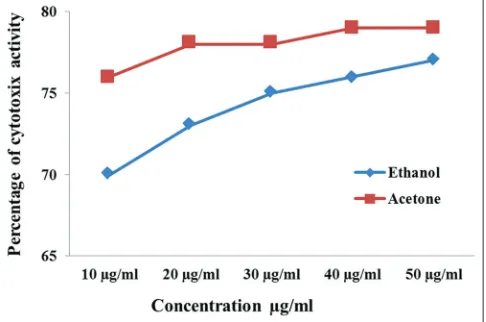
on cancerous cell line, i.e., A549 human cancerous cell line was estimated by MTT assay and Trypan blue dye exclusion test. The aqueous extract leaf of O. sanctum has not shown anticancer activity on A549 cancer cell line. The acetone and ethanol leaf extracts showed a potent anticancer activity on A549 cancerous cell line by MTT assay and Trypan blue dye
Figure 1: (a) Growth chamber (b) callus induction of the
Ocimum sanctum explant
exclusion assay. The results of the MTT assay of acetone and ethanol extracts of O. sanctum on A549 cancerous cell line are represented in Figure 2. The results of Trypan blue exclusion assay of acetone and ethanol extracts of O. sanctum on A549 cancerous cell line are represented in Figure 3.
DISCUSSION
Natural products have received increasing attention over the past 30 years for their potential as a novel cancer preventive and therapeutic agents.[13,14] In parallel, there is increasing
evidence for the potential of plant-derived compounds as inhibitors of various stages of tumorigenesis and associated inflammatory processes, underlining the importance of these products in cancer prevention and therapy. Approximately 60% of drugs currently used for cancer treatment have been isolated from natural products.[15] These include vinca
alkaloids, Taxus diterpenes, Camptotheca alkaloids, and
Podophyllum lignans. Similar results has been reported in the extracts of Bidens pilosa in cervix cancer;[16,17] Citrullus colocynthis in breast cancer;[18,19]Crocus sativus in cervical
epithelioid carcinoma cancer.[20,21] At this time, more than
3000 plants worldwide have been reported to have anticancer properties. Globally, the incidence of plant-derived products for cancer treatment has increased. Hence, an attempt was made to study the cytotoxic activity of various extracts of O. sanctum against A549 human cancerous cell line.
CONCLUSIONS
The plant was initially selected and tested for anticancer activity based on their historical and other traditional uses. The leaf extracts of O. sanctum (acetone, ethanol, and aqueous) were prepared and tested for their potential as anticancer activity by in vitro evaluation methods, i.e., MTT assay and Trypan blue exclusion assay. This was done by closely monitoring the viability of cultured human cells exposed to the plant extracts. More efforts are needed to explore potent anticancer plants from the mother earth and save humans around the world from cancer.
ACKNOWLEDGMENTS
The author would like to thank UGC-Southern Eastern Regional Office (SERO) for funding in the form of Minor Research Project (MRP-5810/15 [SERO/UGC] January 2015).
REFERENCES
1. Veeresham C, Chitti P. Therapeutic agents from tissue cultures of medicinal plants. Nat Prod Chem Res 2013;1:118.
2. Newman DJ, Cragg GM. Natural products as sources of new drugs over the 30 years from 1981 to 2010. J Nat Prod 2012;75:311-35.
3. Rahman M, Khan MA. Anti-cancer potential of South Asian plants. Nat Prod Bioprospect 2013;3:74-88. 4. Amin AR, Kucuk O, Khuri FR, Shin DM. Perspectives
for cancer prevention with natural compounds. J Clin Oncol 2009;27:2712-25.
5. Russo M, Spagnuolo C, Tedesco I, Russo GL. Phytochemicals in cancer prevention and therapy: Truth or dare? Toxins(Basel) 2010;2:517-51.
6. Kolewe ME, Gaurav V, Roberts SC. Pharmaceutically active natural product synthesis and supply via plant cell culture technology. Mol Pharm 2008;5:243-56.
7. Pattanayak P, Behera P, Das D, Panda SK. Ocimum sanctum Linn. A reservoir plant for therapeutic applications: An overview. Pharmacogn Rev 2010;4:95-105.
8. Sankaranarayanan S, Bama P, Sathyabama S, Figure 2: Anticancer activity of various leaf extracts
of Ocimum sanctum on A549 cancer cell line by 3-(4,5-dimethylthiazole- 2-yl)-2,5-diphenyltetrazolium bromide assay
Bhuvaneswari N. Anticancer compound isolated from the leaves of Tridax procumbens against human lung cancer cell a-549. Asian J Pharm Clin Res 2013;6:91-6. 9. Ghosh S, Sarma MD, Patra A, Hazra B.
Anti-inflammatory and anticancer compounds isolated from
Ventilago madraspatana Gaertn, Rubia cordifolia
Linn, and Lantana camara Linn. J Pharm Pharmacol 2010;62:1158-66.
10. Ramamurthy SK, Kotturi R, Devi P, Kumar S. In vitro
cytotoxic activity of methanol and acetone extracts of
Parthenium hysterophorus flower on A549 cell lines. Int J Pharm Sci Rev Res 2011;10:95-9.
11. Priya VP, Radhika K, Rao SA. In vitro anti-cancer activity of aqueous and acetone extracts of Tridax procumbens
leaf on PC 3 cell lines. Int J Pharm Pharm Sci 2011;3:1-4. 12. Priya VP, Rao SA. Evaluation of anticancer activity of
Tridox procumbens leaf extract on A549 and Hep G2 cell lines. Asian J Pharm Clin Res 2015;8:129-32.
13. Saklani A, Kutty SK. Plant-derived compounds in clinical trials. Drug Discov Today 2008;13:161-71. 14. Gordaliza M. Natural products as leads to anticancer
drugs. Clin Transl Oncol 2007;9:767-76.
15. Gerson-Cwilich R, Serrano-Olvera A, Villalobos-Prieto A. Complementary and alternative medicine (CAM) in Mexican patients with cancer. Clin Transl Oncol 2006;8:200-7.
16. Abajo C, Boffill MA, del Campo J, Mendez MA, Gonzalez Y, Mitjans M, et al. In vitro study of the antioxidant and immunomodulatory activity of aqueous infusion of Bidens pilosa. J Ethnopharmacol 2004;93:319-23.
17. Sundararajan P, Dey A, Smith A, Doss AG, Rajappan M, Natarajan S. Studies of anticancer and antipyretic activity of Bidens pilosa whole plant. Afr Health Sci 2006;6:27-30.
18. Tannin-Spitz T, Grossman S, Dovrat S, Gottlieb HE, Bergman M. Growth inhibitory activity of cucurbitacin glucosides isolated from Citrullus colocynthis on human breast cancer cells. Biochem Pharmacol 2007;73:56-67. 19. Tannin-Spitz T, Bergman M, Grossman S. Cucurbitacin
glucosides: Antioxidant and free-radical scavenging activities. Biochem Biophys Res Commun 2007;364:181-6. 20. Abdullaev FI. Cancer chemopreventive and tumoricidal
properties of saffron (Crocus sativus L.). Exp Biol Med 2002;227:20-5.
21. Escribano J, Alonso GL, Coca-Prados M, Fernandez JA. Crocin, safranal and picrocrocin from saffron (Crocus sativus L.) inhibit the growth of human cancer cells
in vitro. Cancer Lett 1996;100:23-30.