www.fm.viamedica.pl
Address for correspondence: J. Wysocki, Laboratory of Clinical Anatomy of the Head and Neck, Institute of Physiology and Pathology of
Topographical anatomy and morphometry
of the temporal bone of the macaque
J. Wysocki
1Clinic of Otolaryngology and Rehabilitation, II Medical Faculty, Warsaw Medical University, Poland, Kajetany,
Nadarzyn, Poland
2Laboratory of Clinical Anatomy of the Head and Neck, Institute of Physiology and Pathology of Hearing,
Poland, Kajetany, Nadarzyn, Poland
[Received 7 July 2008; Accepted 10 October 2008]
Based on the dissections of 24 bones of 12 macaques (Macaca mulatta), a systematic anatomical description was made and measurements of the cho-sen size parameters of the temporal bone as well as the skull were taken. Although there is a small mastoid process, the general arrangement of the macaque’s temporal bone structures is very close to that which is observed in humans. The main differences are a different model of pneumatisation and the presence of subarcuate fossa, which possesses considerable dimensions. The main air space in the middle ear is the mesotympanum, but there are also additional air cells: the epitympanic recess containing the head of malleus and body of incus, the mastoid cavity, and several air spaces on the floor of the tympanic cavity. The vicinity of the carotid canal is also very well pneuma-tised and the walls of the canal are very thin. The semicircular canals are relatively small, very regular in shape, and characterized by almost the same dimensions. The bony walls of the labyrinth are relatively thin. (Folia Morphol 2009; 68, 1: 13–22)
Key words: anatomy, inner ear, macaque, middle ear, temporal bone
INTRODUCTION
The temporal bone containing the vestibuloco-chlear organ is significant in the pathology of hu-mans and animals. The middle and inner ear of many animals, including the macaque [2, 7], are used as laboratory models in various research projects. The anatomical description of the ear structures in ani-mals, as found in veterinary anatomy textbooks, is scarce, schematic, and does not go beyond a rough enumeration of the structures often with no data on topography and variableness. This work is a con-tinuation of a program of investigation of the tem-poral bones of laboratory animals [16]. The aim of undertaking this study has been to come up with a systematic anatomic description of the temporal
bone, including the topographic aspects for clinical needs in the selected animal species.
MATERIAL AND METHODS
measurements of the temporal bone selected size parameters were made. Measurements of the mid-dle and inner ear structures were made using an eyepiece (ocular) with 0.05 mm to 0.1 mm accuracy gauge (depending on the size of the objective used). Neither thorough investigation of the interior of the cochlea nor measurements of its scalae had been performed and previous studies dealing with this problem are available [14]. The dimensions of the skull and the dimensions of the whole temporal bone were made with linear callipers with an accuracy of 1 mm. The full length of the skull was measured as the distance between the opistocranion and pros-thion points. The length of the neurocranial basis was evaluated as the distance between the hormi-on and opisthihormi-on points. The Student-t test was used for the evaluation of differences between genders and body sides.
RESULTS
The morphological description is illustrated with figures, and the measurements are presented in Tables 1–3. The acquired numerical values (data) are presented combined (together) for males and fe-males as well as for right and left side of the body, since no statistically significant differences depend-ing on sex or the side of the body were found.
Morphology of the temporal bone
The temporal bone of the macaque is composed of three parts: squamous, tympanic and petrous, which in adult individuals are combined totally into one part. The temporal bone borders with the oc-cipital bone caudally, rostrally with the sphenoid and zygomatic bones, and with the parietal from the dorsal side (Fig. 1).
The squamous part is flat, low, and elongated in the antero-posterior direction. It has sharp ends in the anterior and posterior part, which intussus-cept between the sphenoid and parietal as well as occipital and parietal bones, respectively. Zy-gomatic and postglenoid processes come out from the squamous part. A not very prominent articu-lar tubercle and a mandibuarticu-lar fossa are located between the epiphyseal parts of the processes. The temporal canal and its external opening, the post-glenoid foramen, are not always found and are rather small.
The tympanic part has the form of a wide, in-complete ring, opened in the dorsal part, comple-mented in this place by a fragment of the squamous part. Then, together with the squamous part, it forms the external acoustic meatus (Fig. 2).
The petrous part of the temporal bone is elon-gated, slender, and has the shape of a regular four
Table 1. Results of measurements of selected size parameters of the macaque’s temporal bone. All values are in millimetres. Arithmetical means with ranging below
Structure Parameter Value
Whole skull Full length of skull 116.17 (105.0–137.0)
Full length of neurocranial basis 62.67 (56.0–73.0)
Whole bone Full length of bone 44.92 (32.0–53.0)
Length of petrous part 29.4 (26.1–32.7) Height of petrous part 11.28 (9.2–14.7) Length of external auditory meatus 9.3 (7.4–12.5)
External auditory pore and its vicinity Height 3.08 (2.2–5.2)
Width 2.97 (2.2–3.6)
Distance between stylomastoid foramen 10.41 (7.4–15.1) and posterior wall of external auditory meatus
Internal auditory meatus Height 2.87 (2.0–3.4)
Width 3.12 (2.2–3.8)
Length 4.64 (4.1–5.8)
Subarcuate fossa Height 3.26 (2.4–4.3)
Width 2.97 (2.3–3.4)
A.
B.
parietal pyramid with sharp edges (Fig. 3). The apex of the pyramid wedges between the basilar part of the occipital bone and the body and greater wing of the sphenoid bone. The base of the pyramid fac-es caudally and laterally and changfac-es into a flat, narrow ending intussuscepted between the occipi-tal and parieoccipi-tal bone. The dorsal part of the pyra-mid, in its caudal part, is smooth, flat, and forms a little mastoid process. In the anterior part, its
sur-face is irregular and develops a short, wide, and sharply tipped styloid process. The external open-ing of the internal carotid artery canal, stylomas-toid foramen, and the opening of the musculotubal canal are situated here as well (Figs. 2, 3).
There are several important structures visible on the walls of the pyramid. The medial wall of the petrous part has two large recesses (depressions) in the central 1/3 (Fig. 3). The rostral recess constitutes
Table 2. Results of measurements of selected size parameters of tympanic cavity of the macaque’s temporal bone. All values are in millimetres. Arithmetical means with the ranging below
Structure Parameter Value
Auditory ossicles Malleus — full length 4.93 (4.35–5.25)
Incus — full length 3.42 (3.25–3.65) Stapes — full height 1.76 (1.15–2.05)
Tympanic cavity Full height of tympanic cavity 8,78 (7.1–10.6) Full length of tympanic cavity 6.63 (5.8–9.3) Height of epitympanic recess 2.28 (1.4–3.8) Length of epitympanic recess 4.18 (3.2–5.8)
Mastoid cavity Length 5.23 (4.2–6.8)
Height 4.86 (3.4–5.8)
Width 2.34 (1.8–3.2)
Distance between round window and 11.48 (9.2–16.5) internal orifice of Eustachian tube
Distance between round window 3.31 (2.8–4.3) and internal carotid artery
Eustachian tube Full length of its bony part 6.92 (6.1–8.9)
Table 3. Results of measurements of selected parameters characterizing magnitude of internal ear of the macaque. All values are in millimetres. Arithmetical means with ranging below
Structure Parameter Value
Semicircular canals Superior Vertical diameter 4.68 (4.25–5.35) Horizontal diameter 4.17 (3.15–5.0)
Posterior Vertical diameter 4.19 (3.25–4.85) Horizontal diameter 4.12 (3.75–4.8)
Lateral Vertical diameter 4.18 (3.8–5.1) Horizontal diameter 4.08 (3.65–4.85)
Cochlea Diameter of base of cochlea 5.28 (4.25–5.85)
Height 2.87 (2.4–3.65)
Oval window Height 0.72 (0.65–0.85)
Width 1.68 (1.45–1.95)
Round window Height 1.29 (1.0–1.65)
the internal acoustic meatus, and the caudal, the subarcuate fossa. The petrous crest does not appear on the superior border of the petrous part as it does in other animals.
The internal acoustic meatus is a short, bony ca-nal. It runs obliquely from the anterior and medial part caudally and sideward, forming about a 45-degree angle with the axis of the pyramid. The subarcuate fossa holding the cerebellum flocculus is a slightly bigger recess surrounded caudally and dorsally by a bony prominence, in which the superi-or semicircular canal runs. Directly inferisuperi-orly and cau-dally from the internal acoustic meatus lies the external opening of the cochlear canaliculus, and
di-rectly inferiorly and caudally from the entrance to the subarcuate fossa is the external opening of the vestibular aqueduct. Both openings are veiled by small bony overhangs. In the antero-inferior and postero-inferior part of the medial wall of the pe-trous part lie rather shallow grooves for the inferior petrosal sinus and the sigmoidal sinus, respectively. The superior petrosal sinus groove occupies the lat-eral 1/3 of the superior border of the petrous part. The groove for the inferior petrosal sinus runs along the facing borders of the basilar part of the occipi-tal and temporal bones. A prominent groove for the sigmoid sinus ends in the considerable jugular fora-men. Sometimes the internal opening of the mas-toid emissary vein is found close to it.
The dorsal wall of the petrous part in the macaque, which is called the anterior wall in human anatomy, has openings of lesser and greater petrosal nerve ca-nals in its central part, and the trigeminal impression can be found rostrally from them. A shallow groove for the petrosquamous sinus runs towards a rather small (and not always found in the macaque) postgle-noid foramen in the posterior wall of the petrous part. The internal opening of the internal carotid canal is visible at the apex of the petrous part.
Middle ear
The tympanic membrane separates the external acoustic meatus from the tympanic cavity. Its shape,
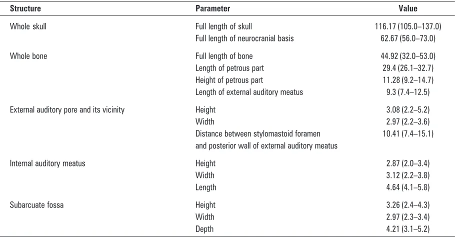
Figure 2. Skull base of the macaque; 1 — vomer; 2 — foramen magnum; 3 — mastoid process; 4 — inferior wall of external auditory meatus; 5 — postglenoid process; 6 — articular tubercle; 7 — external opening of carotid canal; 8 — apex of pyramid; 9 — right opening of choanae.
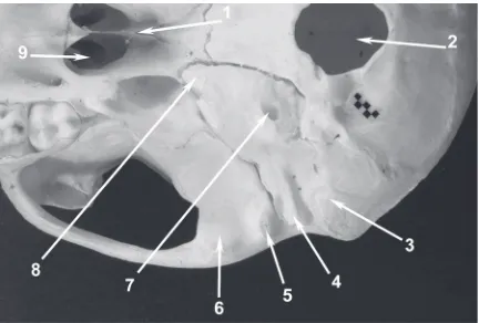
Figure 1. Localization of the macaque’s temporal bone in the neuroc-ranium and viscerocneuroc-ranium. Lateral wall of the skull from the right side. One millimetre gauge on the figure; 1 — external acoustic pore; 2 — squamous part of temporal bone; 3 — zygomatic arch; 4 — articular tubercle; 5 — postglenoid process; 6 — hypoglossal canal; 7 — foramen magnum; 8 — stylomastoid foramen.
viewed from the lateral side, is elliptic, with the long-er diagonal running from the antlong-erior and suplong-erior caudally and inferiorly. The dimensions of the ellipse vary slightly and on average are 4.51 ¥ 5.54 mm. The angle between the tympanic membrane plane and the horizontal plane is about 50–60 degrees, and the angle formed with the sagittal plane is about 50 degrees. Both angles are opened rostrally.
Similarly to humans, the macaque’s middle ear consists of the tympanic cavity, mastoid cavity, and auditory tube. The shape of the tympanic cavity can be described as a biconcave lens embraced in a small cuboid block, in which the wide surfaces form
later-al and medilater-al wlater-alls, and the rest of the wlater-alls are much narrower. The width of the cavity is greatest in the superior part and, unlike in humans, also in the inferior part.
The lateral wall of the tympanic cavity is formed mainly by the tympanic membrane, from the supe-rior by the petrous part and from the infesupe-rior by tympanic part. The quite well-defined superior part of the tympanic cavity is the epitympanic recess (a term drawn from human anatomy, absent in an-imal anatomy). The lateral bony wall of the recess is thick and lies deep under the initial part of the zy-gomatic process. It corresponds to the lateral wall of the “attic” (clinical term) in humans.
The medial wall of the tympanic cavity has the most diversiform carving. The promontory forms a distinct, centrally located prominence of that wall (Figs. 4–6). The caudoventral edge of the promon-tory curves dorsally and caudally, forming a promi-nent lateral labium of the entrance (aditus) to the fossa of the round window. The apex of the prom-ontory is visible by looking in the axis of the exter-nal acoustic meatus.
The oval window has a beanlike shape. The infe-rior border of the oval window is slightly concave; the superior is convex and seems to be hollower, owing to the underlain prominence of the facial ca-nal. The round window is slightly oval and extends caudally and a little laterally. The round window is situated in a rather deep small cavity called the niche or the fossa of the round window.
Figure 6. Macaque’s left tympanic cavity interior with auditory ossicles inside. Widened external acoustic meatus. Macerated bone; 1-milimeter scale bar; 1 — handle of malleus; 2 — long limb of incus; 3 — incudostapedial joint; 4 — bony trabecula on posterior wall of tympanic cavity; 5 — stylomastoid foramen; 6 — round window niche; 7 — promontory; 8 — tympanic opening of auditory tube.
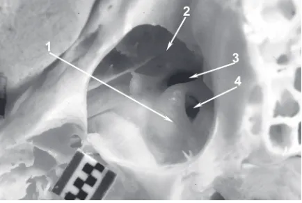
Figure 5. View of macaque’s left tympanic cavity from side of external acoustic pore which has been widened for better insight. Macerated bone; 1-milimeter scale bar; 1 — promontory; 2 — prominence of facial canal; 3 — oval window; 4 — round window niche.
The round window niche has a sphenoidal shape with the apex at the round window membrane and the base at the entrance (aditus) to the niche. The niche is deeper in the posterior and shallower in the anterior. Posteriorly from the round window fossa lies a concavity, which, by analogy with human anat-omy, would be called the tympanic sinus. It under-mines the facial nerve canal and the pyramidal em-inence from the ventral side.
Directly over the oval window lies the horizontal segment of the facial nerve canal, forming a dis-tinct bony eminence on the medial wall of the tym-panic cavity — the prominence of the facial canal. At the level of the canal, but rostrally from the oval window, lies an opening (foramen) with irregular borders, intended for the tendon of the tensor tym-pani. The prominence of the lateral semicircular ca-nal is situated caudally and dorsally from the facial nerve canal, at the place of passage from the tym-panic to the mastoid cavity. It curves superiorly and posteriorly, limiting the fossa of incus dorsally.
The anterior part of medial wall of the tympanic cavity is smooth and elongates into the bony canal of the auditory tube in its superior part. The bony part of the auditory tube lies directly laterally from the internal carotid canal. In some cases the inter-nal carotid artery may be visible in the cainter-nal itself because the canal walls are thin and often incom-plete, having small natural defects called dehiscenc-es. Dorsally from the auditory tube canal runs the semicanal and farther the canal for the tensor tym-pani muscle. Dorsally from these structures, on the medial wall of tympanic cavity and on the border with its superior wall (the tegmental wall), there is a relatively deep bony niche already belonging to the epitympanic recess.
The anterior wall of the tympanic cavity, the wall of the carotid artery, is very thin. It is strongly pneuma-tised in the macaque by a few tympanic cells (Fig. 4). It is perforated by the mentioned tympanic open-ing of the auditory tube and the tensor tympani sem-icanals. In the vicinity of these structures there is a prominence formed by the carotid artery canal, highly visible on all the studied bones, as it has only a thin bony wall around itself. Here, the entrance to a series of pneumatic cells of the anterior part of pyramid is located.
The inferior wall of the tympanic cavity is also pneumatised. Depending on the degree of pneuma-tisation, the space between the capsule of the bony labyrinth and the inferior tympanic wall may have a different thickness. The petrous bulla is the
larg-est of pneumatic cells in this part of the temporal bone. It spreads from the posterior wall of the tym-panic cavity to the top of the pyramid. In the supe-rior wall of the bulla, and then in the infesupe-rior wall of the hypotympanum, runs the tympanic canaliculus with the tympanic nerve inside. The bulla is usually so big that it undermines the labyrinth as well as the carotid and musculo-tubal canals.
The posterior wall (the mastoid wall) is very nar-row, reaches superiorly to the lateral semicircular canal, and inferiorly turns into the inferior wall with-out any visible border. The facial nerve canal forms a distinct prominence on the posterior wall, sur-rounded by an array (range) of pneumatic cells. A small bony process, a pyramidal eminence, lies laterally from the canal, on the level of the oval win-dow. It holds the stapedial muscle. A small opening of the chorda tympani canaliculus lies more lateral-ly and below. The largest of all the pneumatic cells of the pyramid posterior part is the mastoid cavity. It has four walls: anterior, posterior, medial, and inferior, through which it communicates with the rest of the pneumatic cells of the mastoid process and the epitympanum. The prominence of the lat-eral semicircular canal lies on the medial wall. The superior wall of the tympanic cavity is the tegmen (cover) of the tympanic cavity, which also forms part of the middle cranial fossa.
The interior of the macaque’s tympanic cavity, similarly to that of humans, can be divided into three spaces: hypotympanum, mesotympanum and epi-tympanum. The epitympanum, i.e. the epitympanic recess of the tympanic cavity, is located in the supe-rior area of tympanic cavity. It is medially limited by the prominence of the lateral semicircular canal and facial canal, laterally by a bony wall above the tym-panic ring (by analogy with human anatomy or as a clinical term, the wall may be called the “attic”). This space contains the body of incus and the head of malleus together with their ligaments (Fig. 6). The epitympanic recess occupies about 1/3 of the middle ear space. Caudally, it communicates with the mastoid cavity — the largest air cell pneumatis-ing the mastoid process and the posterior part of the temporal bone pyramid.
Auditory ossicles with their structure and pro-portions resemble human ones; however, the malleus possesses a prominent muscular process that is absent in humans (Figs. 6, 7).
The facial nerve canal, also known as the facial canal, originates in the fundus of the internal acous-tic meatus and ends at the stylomastoid foramen. Unlike in humans, its wall does not have any natural defects — dehiscences. Three parts can be distin-guished in the course of the facial nerve canal: laby-rinthine, tympanic, and mastoid. In the labyrinthine part, the nerve runs between the basal turn of the cochlea and the superior semicircular canal, and appears on the anterior wall of the vestibule. The labyrinthine part of the facial canal ends in the place where it approaches the medial wall of the tympan-ic cavity and forms a distinct gentympan-iculum at an acute to almost right angle. Here lies the geniculate gan-glion and here originates the first of the intratem-poral branches of the facial nerve — the greater petrosal nerve. The second part of the facial nerve canal, the tympanic part, lies in the labyrinthine wall of the tympanic cavity, between the prominence of the lateral semicircular canal and the oval window. In this part, the nerve runs obliquely, caudally, and inferiorly, up to the area of the pyramid eminence in the posterior wall of the tympanic cavity. Here the canal passes into its mastoid part, which runs in a gentle arch towards the stylomastoid foramen. In the mastoid part, the facial nerve gives back the sta-pedial nerve and the chorda tympani. The tympanic chord emerges from the facial nerve just before it leaves the stylomastoid foramen. It runs between the handle of malleus and the long limb of incus, to leave
through a small opening in the anterior wall, on the border with the lateral. The opening lies above the tensor tympani canal and is separated from it by a recess on the medial wall. The recess has the shape of an ellipse, elongated in the antero-posterior direction. The internal carotid artery in the macaque runs in a bony canal in the temporal bone at its full length (Fig. 4). The carotid canal participates in the forma-tion of the anterior wall of the tympanic cavity, and is therefore called the carotid wall. The canal runs in a gentle arch, with the convexity heading caudally and slightly to the side. The vicinity of the canal is strongly pneumatised, so the canal is somehow sus-pended in a “cribriform” porous base. The internal carotid artery closely neighbours with other impor-tant structures of the temporal bone. Laterally from it lie the musculotubal canal, and medially, the ex-ternal acoustic meatus and the cochlea.
Bony labyrinth
The bony labyrinth, the location of the vestibu-locochlear organ, is situated in the central part of the pyramid. It is oblong, and its long axis is in line with the axis of the pyramid and equals about 10 mm. The bony labyrinth includes the vestibule, the cochlea, and the semicircular canals. The vestibule is an approximately oviform, 2 ¥ 3 mm cavity, and six walls can be distinguished in it. The lateral wall separates the vestibule from the tympanic cavity, and in its great part is occupied by the oval window (Fig. 5). The medial wall, with the most complicated carving, has recesses including the utricle, the sac-cule and the initial part of the cochlear duct. The anterior wall is narrow and very thin and neighbours the facial canal in its labyrinthine part. In its inferior part, the anterior wall has an opening called the el-liptic foramen, which leads to the scala vestibuli. The semicircular canals enter and leave the vestibule in its superior and posterior walls. The posterior wall has an opening leading to the ampulla of the pos-terior semicircular canal. The superior wall has four openings leading to the semicircular canals: the two anterior openings lead to the ampullar parts of the lateral semicircular canal (which lies more laterally) and to the superior canal (which lies more medial-ly); the two posterior openings lead to the non-am-pullar end of the lateral semicircular canal and to the common limb of the superior and posterior (also non-ampullar) semicircular canals. The inferior wall is formed by the spiral lamina.
The semicircular canals lie caudally and superior-ly from the vestibule. Their geometry is irregular.
All three bony canals have a more or less elliptical shape and they are a little flattened in the trans-verse section. The semicircular canals originate and end in the vestibule. Therefore, each of them has two openings (apertures), but because the anterior and posterior canal have a common limb, there are in total 5 not 6 openings in the vestibule.
The lateral canal is visible in the medial wall of the tympanic cavity where, as mentioned, it forms its own prominence. The prominence is most dis-tinct close to the aditus to the mastoid antrum (Figs. 5, 6). A little farther down, the canal sinks into the surroundings and disappears among the air cells. The plane of the canal runs strongly obliquely from the anterior and superior caudally, and forms an angle of approximately 45 degrees with the long axis of the oval window, thus it is situated more slantwise than in humans. Caudally from the prom-inence of the lateral semicircular canal lies an area deprived of bigger cells, constituting the base of the subarcuate fossa, and caudally from it there is an area corresponding to the sigmoid sinus, located in the cranial cavity. The superior semicircular canal lim-its the entrance (aditus) to the subarcuate fossa on the posterior wall of the pyramid. The posterior ca-nal is difficult to find because it is not distinguish-able by any topographic point either on the surface or inside the bone. It runs slightly arched around the external orifice of the vestibular aqueduct and after joining the superior canal as the common limb embraces inferiorly the entrance (aditus) to the sub-arcuate fossa.
The cochlea looks a little like a conch or the shell of a snail (Fig. 8). The long axis of the cochlea is directed rostrally and laterally and is nearly perpen-dicular to the long axis of the pyramid. The base of the cochlea is directed caudally and medially, pre-cisely towards the internal acoustic meatus. The spi-ral canal forms from 2.5 to 2.75 turns. The cochlea neighbours medially with the internal acoustic me-atus, laterally with the musculotubal canal and the promontory, anteriorly with the carotid canal, and superiorly with the facial canal, particularly with the area of geniculum
The cochlear canaliculus is a perilymphatic duct and the vestibule aqueduct is endolymphatic. The co-chlear canaliculus unites the initial part of the scala tympani with the subarachnoid space of the posterior cranial cavity. The internal opening of the cochlear canaliculus, called the ear orifice, lies in the medial wall of the basal turn of the scala vestibuli near the round window. The external opening, called its cranial
ori-fice, is located on the inferior surface of the temporal bone pyramid, close to the jugular fossa (Fig. 3). The vestibular aqueduct originates in the medial wall of the vestibule, then runs medially, and exits on the medial wall of the pyramid in a depression called the external opening of the vestibular aqueduct.
DISCUSSION
Data concerning the topographic anatomy of the temporal bone in macaques are accessible only in a dozen or so publications [1, 3–6, 9–16].
In the external morphology of the macaque’s temporal bone, the deep subarcuate fossa attracts the most attention, as indicated by Stelmasiak and Wójtowicz [12] and Straus [13].
According to Saad et al. [10], the shape of the middle ear space is similar to that in humans, and, certainly, this should be agreed with. He divided the tympanic cavity into three parts, similarly to how it has been done in this description of inves-tigations. However, Bast [1] divided macaque’s tympanic cavity only into two spaces; the tympa-num and epitympatympa-num, since according to him, the hypotympanum does not appear in the macaque as a distinct (separate) space. Similarly, Kirikae [8] stated that the mastoid antrum is well developed and the hypotympanum is underdevel-oped, in the macaque.
Our own investigations confirm Saad’s et al. ob-servations [10] that the antero-posterior and super-oinferior dimension of the middle ear space is about 9 mm, and including only the tympanic cavity, 6.3 mm. The promontory intussuscepts into the
tympanic cavity, narrowing it down to a width of 1 mm. As far as the shape and dimensions of the tympanic membrane are concerned, there are some controversies indicated by investigators. According to Saad et al. [10], the macaque’s tympanic membrane is almost a round disc with both its diameters equal to about 5.8 mm.
Also according to Saad et al. [10], the narrow external acoustic meatus (diameter 2.5–3 mm) wid-ens medially. One should agree with this statement which indicates that this meatus is relatively very long and potentially makes all surgical interventions through this approach more difficult. On the other hand, the vicinity of the meatus with the mastoid antrum, and through the atrium, with the lateral semicircular canal, allows for relatively easy access to the latter without causing any major surgical in-jury.
According to Saad et al. [10], the oval window is beanlike in shape and measures about 1.8 by 0.5 mm, which is in accordance with our observations. How-ever, the value of the oval window height given by Saad is lower in comparison with our observations. The round window is a little longer in its horizontal diameter than in the vertical, measuring 1.5 mm and 1.2 mm, respectively, which is close to own mea-surements.
Our own observations concerning the vestibular aqueduct and cochlear canaliculus are in accordance with Neiger’s [9] description.
The spiral canal of cochlea, according to previ-ous studies, amounts to an average of 21 mm [14]. The course of the carotid canal is quite similar as in humans. According to Sikorska-Piwowska et al. [11], the external opening of the canal measures 1.11– –2.4 mm (average 2 mm). The carotid canal in the macaque runs in two phases: the ascending part and the horizontal part. The angle between them amounts to about 80–119 degrees (average 89.6 de-grees).
Some authors underline the good pneumatisa-tion of the macaque’s temporal bone. The air cells spread from the mastoid process to the apex of the pyramid including the tegmen of the tympanic cav-ity and its fundus [6, 10].
It is impossible to agree with the statement that there is no subiculum of promontory in the macaque; therefore, the access to the round window is easy [6]. In my opinion, the access is easy because there is no promontory bridge, thus there is no natural limit (border) of the tympanic sinus superiorly: as it flat-tens and gradually declines dorsally.
According to Saad et al. [10], the malleus is about 6 mm, and the author gives separately the dimension of the head (2 mm) and the handle (4 mm). It is a little more than the dimension cal-culated in these studies. The difference probably comes from overlaying of the head and handle di-mensions measured, in a way twofold, by Saad et al. [10]. The size of the body of incus is 3 ¥ 2.5 mm. The long limb is 2 mm long, which gives a higher value, by around 4–5 mm, as the total length of the incus, compared with our measurements. The full length of the stapes given by Saad et al. [10] is about 2 mm, falling into the upper range of values obtained in this investigation.
The mastoid atrium, mastoid cells, petrous bul-la, and apex of the pyramid cells belong to the sys-tem of the middle ear air cells. It is worth noting that none of the investigators mentions the clearly isolated (distinct) mastoid antrum as an element of the middle ear in the macaque. Indirectly related to this is a mention by Saad et al. [10], who noticed that 2/3 of the 9 mm long middle ear is occupied by the tympanic cavity. The pneumatic cells are unusu-ally developed, are a different size and shape, and are surrounding and fixing the carotid canal with its bony walls, which, in a way, is suspended among them like inside a tracery construction designed by an engineer. Saad et al. [10] called one of them the pneumatic cell of the pyramid, because it is particu-larly big and accompanies the auditory tube canal along its full length; however, it was encountered only once during this study.
The macaque’s auditory tube has many similari-ties with that of humans, as indicated earlier [3, 4]. The temporal canal and the postglenoid foramen do not always appear in the macaque, as confirmed earlier in our own investigations [15].
REFERENCES
1. Bast TH (1971) The eye and ear. In: Hartman CG, Straus LW eds. The anatomy of the rhesus monkey (Macaca mu-latta). Hafner Publ Comp, New York, pp. 339–358. 2. Burnett PA, Miller JM, Mangham CA (1984) Intra-aural
reflexes elicited by a cochlear prosthesis in monkeys. Hear Res, 16: 175–180.
3. Doyle WJ, Rood SR (1979) Anatomy of the auditory tube and related structures in the rhesus monkey (Maca-ca mulatta). Acta Anat (Basel), 105: 209–225. 4. Doyle W, Rood SR (1980) Comparison of the anatomy
of the auditory tube in the rhesus monkey (Macaca mu-latta) and man implications for physiologic modeling. Am J Otol, 89: 49–57.
5. Doyle WJ, Webster DB (1991) Neonatal conductive hearing loss does not compromise brainstem auditory function and structure in rhesus monkeys. Hear Res, 54: 145–151. 6. Kelemen G (1968) Non-experimental aural pathology
in rhesus monkeys (Macaca mulatta). Acta Otolaryn-gol, 66: 399–408.
7. Kesser BW, Hashisaki GT, Spindel JH, Ruth RA, Scheld WM (1999) Time course of hearing loss in an animal model of pneumococcal meningitis. Otolaryngol Head Neck Surg, 120: 628–637.
8. Kirikae I (1963) Physiology of the middle ear. Arch Oto-laryngol, 78: 317–323.
9. Neiger M (1968) Zur Morphologie und Physiologie des Aquaeductus cochleae. Experimentelle und morpholo-gische Untersuchungen beim Rhesusaffen. Experimen-telle Untersuchung über die Herkunft der Perilymphe. Forstchr. Hals-Nasen-Ohrenheilk, 15: 113–126. 10. Saad MM, Doyle WJ, Gest TR (1982) Morphology of
the middle ear in the rhesus monkey (Macaca Mulat-ta). Acta Anat, 112: 117–130.
11. Sikorska-Piwowska Z, Kukwa A, Łukaszewska-Otto H, Kozłowski P, Kryst-Widźgowska T, Giza A (1985) The carotid canal in rhesus monkey (Cercopithecidae). Folia Morphol, 44: 33–38.
12. Stelmasiak M, Wójtowicz Z (1972) Doły i jamy czaszki u Macacus rhesus. Folia Morphol, 31: 1–10.
13. Straus WL (1960) The subarcuate fossa in primates. Anat Rec, 138: 93–103.
14. Wysocki J (2001) Dimensions of the vestibular and tym-panic scalae of the cochlea in selected mammals. Hear Res, 161: 1–9.
15. Wysocki J (2002) The morphology of the temporal ca-nal and postglenoid foramen with reference to the size of the jugular foramen in man and selected kinds of animals. Folia Morphol, 61: 199–208.