www.fm.viamedica.pl
Address for correspondence: Dr. A. Ibraheem Zaki, MSc, MD, Faculty of Medicine, King Abdul Aziz University, Jeddah, Saudi Arabia, P.O. Box 42806, Jeddah 21551, Female Section Building 7, tel: +966505931745, e-mail: rafiah-az@hotmail.com
Anatomical study of the carotid bifurcation
and origin variations of the ascending
pharyngeal and superior thyroid arteries
A. Al-Rafiah, A.A. EL-Haggagy, I.H.A. Aal, A.I. Zaki
Faculty of Medicine, King Abdulaziz University Hospital, Jeddah, KSA, Saudi Arabia
[Received 12 October 2010; Accepted 13 January 2011]
Background: Background:Background: Background:
Background: Human anatomy texts in current use have very little precise infor-mation as to the frequency of variations in the bifurcation of the common carotid artery, and a clear description of the relation between external and internal carotid arteries as well as the variation of the origin of the ascending pharyngeal and superior thyroid arteries is limited.
Material and methods: Material and methods:Material and methods: Material and methods:
Material and methods: Sixty common carotid arteries in the sagittal section of the head and neck of 30 human adult cadavers were obtained from the Ana-tomy Department of King Abdulaziz University. The data collected were analy-sed using the Chi square-test.
Results: Results:Results: Results:
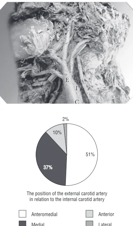
Results: The carotid bifurcation was at the level of the superior border of the thyroid cartilage in 48.3% of cases, 25% were opposite the hyoid bone, and 18.3% were at the level between the thyroid cartilage and the hyoid bone. The bifurcation appeared at a lower level than the superior border of the thyroid cartilage in 5% of cases, while in 3.3% of cases the bifurcation level was seen higher than the hyoid bone. The usual anteromedial position of the external carotid artery to the internal carotid artery was found in 51.7% of cases, whereas it was medial to the internal carotid artery in 36.7% of cases. In 10% it was seen in an anterior position and only in 1.7% the external carotid artery was lateral to the internal carotid artery. In 93.3% of the cases the ascending pharyngeal artery originated from one root, while in the remaining 6.7% of cases it originated from two roots. In 80% of cases the superior thyroid artery arose from the external carotid artery. In 18.3% of cases it originated from the common carotid artery, and in 1.7% it arose from a thyrolingofacial trunk.
Conclusions: Conclusions:Conclusions: Conclusions:
Conclusions: The carotid bifurcation can occur as high as the hyoid bone, or as low as the cricoid cartilage. The anteromedial position of the external caro-tid artery (ECA) in relation to the internal carocaro-tid artery (ICA) was the most common anatomical position. The origins and configurations of the ascending pharyngeal artery and the superior thyroid artery are variable. (Folia Morphol
2011; 70, 1: 47–55)
tery (APA) arises from the external carotid artery and is the only medial branch. The APA ascends on the pharynx deep to the ICA [25]. The superior thyroid artery (STA) is the first branch of the ECA; it arises from the anterior surface of the ECA just below the level of the greater cornu of the hyoid bone [31]. Ozgur et al. [27] emphasised that knowledge of the CCA and its branches are important for vascular sur-gical procedures in the neck region.
Gulsen et al. [12] mentioned that the CCA general-ly bifurcates into the internal and external carotid ar-teries at the level of C3–4. Ito et al. [17] found that the location of the external and internal carotid arteries was reversed. Anil et al. [4] reported a case with the right APA arising from the carotid bifurcation (CB). Bergman et al. [6] found that the APA sometimes arose in common with the occipital artery. Hayashi et al. [14] stated that the APA is frequently said to originate from the back of the ECA near the bifurcation of the CCA. Lucev et al. [23] mentioned that lack of experience re-garding possible variations could lead to fatal errors if one blood vessel should be mistaken for another. For example, the unexpected origin of the STA from the CCA could be a cause of a possible mistake. The great blood vessels of the neck have numerous variations, and their explorations are essential for a better ana-tomic knowledge of the neck. This knowledge is very important in choosing a surgical approach and for diag-nosis in radiology. The aim of the present work is to study the variations of the level of CCA bifurcation as well as the variations of the origin of the ascending pharyngeal and superior thyroid arteries in human adult cadavers.
MATERIAL AND METHODS
The present study was carried on 60 common ca-rotid arteries in the sagittal section of the head and neck of 30 adult cadavers obtained from the dissec-tion room of the Anatomy Department, Faculty of Medicine, King Abdul Aziz University, Saudi Arabia.
Various univariate analyses were used to assess each variation, to provide an understanding of some of the relationships between the variables. All data were entered and analysed using SPSS version 10. Analysis was done using chi-square test with a p va-lue £ 0.05 considered significant.
RESULTS
The bifurcation level of common carotid artery
The level of bifurcation in 29 cases (48.3%) corre-sponded to the superior border of the thyroid cartilage (Fig. 1). In 15 (25%) cases, the level was found opposite to the body of the hyoid bone (Fig. 2). Eleven (18.3%) cases showed the bifurcation at a higher level than the superior border of the thyroid cartilage (between the thyroid cartilage and the hyoid bone) (Fig. 3). The dis-tance between the superior border of the thyroid carti-lage and the site of bifurcation ranged from 0.3 to 1.8 cm. The bifurcation in 3 (5%) cases was found at a low-er level than the suplow-erior bordlow-er of the thyroid cartilage (Fig. 4). The distance between the superior border of the thyroid cartilage and the site of bifurcation ranged from 1.1 to 1.3 cm. In two (3.3%) cases the bifurcation was at a higher level than the hyoid bone (Figs. 5, 6).
The relationship between the external and internal carotid arteries
In 31 (51.7%) cases the ECA was anteromedial to the ICA (Figs. 2, 6). The external carotid artery 22 (36.7%) cases was found medial to the internal carotid artery (Figs. 3, 4, 5, 7).
In six (10%) cases the ECA was situated anterior to the ICA (Fig. 8), and in only one (1.7%) case the exter-nal carotid artery lay lateral to the interexter-nal carotid ar-tery (Fig. 1).
Ascending pharyngeal artery
from the point of the carotid bifurcation (Figs.1, 10). In one (1.7%) case it originated from the ICA (Fig. 6). The following two different patterns of origin of the ascending pharyngeal were found:
— pattern I was observed in 56 (93.3%) cases. The ascending pharyngeal artery originated from one root (Figs. 1, 3, 5, 6, 8);
— pattern II was observed in 4 (6.7%) cases. The artery originated from two roots (Figs. 9, 10).
The distribution of the localization of the ascending pharyngeal artery of pattern I
In 35 cases (58.3%) the APA originated from the medial side of the ECA (Fig. 5). In one (1.7%) case the origin of the APA was from the point of the ca-rotid bifurcation (Fig. 1).
Origin of the artery above the level of bifurcation
In 19 (31.7%) cases the APA originated from the medial side of the external carotid (Figs. 3, 8). The dis-tance of APA from the level of bifurcation was from
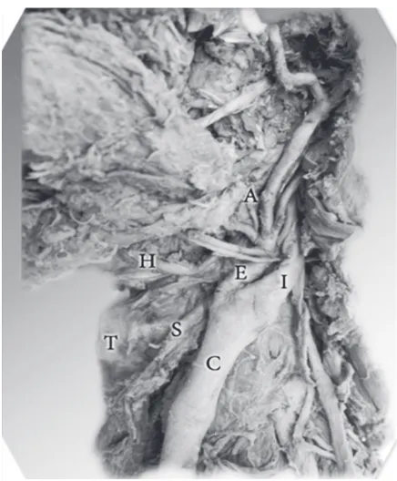
Figure 1. The bifurcation of the left common carotid artery (C) at
the superior border of the thyroid cartilage (T); the external carotid artery (E) lies lateral to the internal carotid artery (I); the origin of the ascending pharyngeal artery (A) from the point of the carotid bifurcation and the origin of the superior thyroid artery (S) from the external carotid artery at the level of the bifurcation.
Figure 2. The bifurcation of the right common carotid artery (C)
opposite the hyoid bone (H); the external carotid artery (E) lies anteromedial to the internal carotid artery (I) and the origin of the superior thyroid artery (S) from the external carotid artery above the level of the bifurcation; T — thyroid cartilage.
Figure 3. The bifurcation of the left common carotid artery (C)
0.2 to 2.4 cm, and in one (1.7%) case it originated from the ICA (Fig. 6). The distance of the origin of the artery from the bifurcation was 1.1 cm.
The distribution of the localization of the ascending pharyngeal artery of pattern II
In three (5%) cases the APA originated from the ECA above the level of bifurcation (Fig. 9). The dis-tance of the artery from the level of bifurcation was from 0.2 to 2.8 cm, and in one (1.7%) case it origi-nated from the ECA from the bifurcation (Fig. 10).
Superior thyroid artery
The distribution of the localization of the superior thyroid artery was observed in 46 cases (76.7%); the superior thyroid artery originated from the ECA at the level of the bifurcation (Figs. 1, 3, 4).
In two (3.3%) cases, it originated from the ECA above the level of the bifurcation (Figs. 2, 7). The distance of the origin from the bifurcation was from 0.9 to 1.1 cm. In 11 (18.3%) cases it originated from the common carotid artery (Fig. 6). The distance of the origin from the bifurcation was from 0.4 to 1.0 cm. In one (1.7 %) case it arose from a thyrolingofacial trunk (Fig. 5). In 59 (98.3%) cases the superior thy-roid artery originated from the anteromedial aspect of the ECA (Figs. 2, 3, 4, 5, 10).
In one (1.7%) case, it arose from the lateral aspect of the ECA and ran in front of the CCA to descend to the thyroid gland (Fig. 1).
Statistical analysis
The statistical analyses between each two vari-ables were done. The che-square test value equaled
0.954, which is a p value > 0.05. No significant results were shown.
DISCUSSION
The carotid bifurcation
Several anatomic textbooks state that the common carotid artery bifurcates into the exter-nal and interexter-nal carotid arteries at the level of the superior border of the thyroid cartilage [1, 20, 31, 34].
The present study showed that normal bifurca-tion of the CCA was found in 48.3% of cases. Lucev et al. [23] found this to be true in 50% of cases and
A B
Figure 4. The bifurcation of the left common carotid artery (C)
below the upper border of the thyroid cartilage (T); the external carotid artery (E) lies medial to the internal carotid artery (I) and the origin of the superior thyroid artery (S) from the external carotid artery at the level of the bifurcation.
Figure 5. The bifurcation of the right common carotid artery (C)
Ilic et al. [16] reported a similar finding in 58% of cases. In addition, Espalieu et al. [9] found it to be true in 65% of cases, as well as Von Poisel and Golth [34] who found it in 67%.
In the present work, the CCA may bifurcate high-er or lowhigh-er than the usual level; a high bifurcation is more common. The bifurcation can occur as high as the hyoid bone or even higher than, or as low as, the lower border of the thyroid cartilage. These varia-tions are of clinical importance for surgical ap-proaches in the head and neck region.
Figure 6. The bifurcation of the left common carotid artery (C)
above the hyoid bone (H); the external carotid artery (E) lies antero-medial to the internal carotid artery (I); the origin of the ascending pharyngeal artery from the internal carotid artery and the origin of the superior thyroid artery (S) from the common carotid artery below the level of the bifurcation; T — thyroid cartilage.
Figure 7. The level of the bifurcation of the right common carotid
artery (C) at the superior border of the thyroid cartilage (T); the external carotid artery lies medial to the internal carotid artery (I) and the origin of the superior thyroid artery (S) from the external carotid artery (E) above the level of the bifurcation; H — hyoid bone; A — ascending pharyngeal artery.
Figure 8. The left common carotid artery (C); the external carotid
artery (E) lies anterior to the internal carotid artery (I) and the ori-gin of the ascending pharyngeal artery (A) from the external caro-tid artery distal to the level of the bifurcation.
Figure 9. The origin of the ascending pharyngeal artery (A) from
Standring [31] mentioned a higher level of bi-furcation opposite the hyoid bone. He reported this level as the most common bifurcation level. In this study, the extent of bifurcation at this level was 25%. This is in agreement with Ito et al. [17], who found this level in 31.2% of cases, and it was detected in 12.5% of the cases examined by Lucev et al. [23]. It was also described by Kipré et al. [19] in 13% of his examined cases.
In 18.3% of cases in this study the bifurcation was observed at a higher level than the superior border of the thyroid cartilage. This finding
corre-sponds to Von Poisel & Golth [34] and Krmpotić--Nemanić et al. [20], who found it in 20% of cases. It also corresponds to Lucev et al. [23], who found it in 25%, whereas Ilić et al. [16] found this higher level in 31% of cases. In contrast to this study, Kipré et al. [19] found it in 75% of cases.
In 5% of cases examined in this study the caro-tid bifurcation was found at the level below the up-per border of the thyroid cartilage. Ilić et al. [16] found this level in 11% of cases, Lucev et al. [23] found it in 12.5% of cases, and Kipré et al. [19] re-corded it in 15% of cases.
Figure 10. The origin of the ascending pharyngeal artery (A) by two roots from the bifurcation of the common carotid artery;
In this study the level of the carotid bifurcation above the hyoid bone was found in 3.3% of cases. This is in agreement with the results of Nikolić [26], who found the bifurcation at this level in 2% of cases, and Ilić et al. [16] who found it in only 1% of cases.
Relation between the external carotid artery and the internal carotid artery
According to the findings of this study, there are different patterns of relationships between these two arteries. The anteromedial position of the ECA was present in 51.7% of cases, which corresponds to the result of Krmpotić-Nemanić et al. [20], who reported this position in 50% of cases, while Lucev et al. [23] found it in 30% of cases.
Ozgur et al. [27] mentioned that the most common position of the ECA in relation to the ICA was the medial position. In this study, the medial position was found in 36.7% of cases, whereas it was in only 10% of the examined cas-es of Lucev et al. [23].
In the current study the anterior position of the ECA was found in only 10% of cases, while Krmpotić-Nemanić et al. [20] reported this position in 21% of cases, and Lucev et al. [23] found it in 47.5% of cases. In this study the lateral position of the ECA in relation to the ICA was found in only 1.7% of cases. Also, three case reports of the same position were reported by Handa et al. [13], Teal et al. [33], and Prendes et al. [28]. Lucev et al. [23] found the later-al position in 10% of cases.
Anangwe et al. [3] found that the ECA was situ-ated anterolateral to the ICA in 30% of cases. This is in contrast to Lucev et al. [23], who reported it in only 2.5% of cases. This position was not encoun-tered in the current study.
The ascending pharyngeal artery
Knowledge of variations in the origin of the APA is vital for the exact identification of the neck ves-sels during surgery, to avoid a fatal mix-up with the ICA [5, 11, 20].
In the present study, the APA arose from the ECA at the level of carotid bifurcation in 56.7% of cases; it is found to be 65% in study of Hollinshead [15], and Czerwinski [8] who found it in 74.1% of cases. In the current work, the APA originated from the ICA in 1.7% of cases. This finding was in agreement with the study of Zumre et al. [35], who detected it in 2.5% of cases, and Hayashi et al. [14], who found it in 2% of cases. This was also in accordance with the findings in the case report of Chitra [7].
In the present work, the APA originated from the ECA above the level of bifurcation in 36.7% of cases. This is in accordance with the study of Anil et al. [4], who found it in 30% of cases. In 5% of the cases of this study the APA originated from the ca-rotid bifurcation. This is in agreement with Anil et al. [4] who detected the same origin.
In this study, 6.7% of cases of the APA originat-ed by two roots from the ECA, it either arose above the level of bifurcation or from the point of bifur-cation. Chitra [7] observed two ascending pharyn-geal arteries: one from the occipital artery and an-other from the ICA. Czerwinski [8] stated that the APA originated from the CCA in 2% of cases. Berg-man et al. [6] reported that it might arise from the CCA in 7–9% of cases, while Chitra [7] found it to be a branch of the occipital artery. These variations were not encountered in the current study.
Finally, the statistical results show that the place of origin of the APA depends significantly on the position of the ECA in relation to the ICA. This corre-lation between the origin of the APA and the posi-tion of the ECA in relaposi-tion to the ICA was not dis-cussed in the available literature.
The superior thyroid artery
In the current study, the superior thyroid artery (STA) originated from the ECA at the carotid bifur-cation in most of the examined cases (76.7%). This finding was consistent with the findings of Czer-winski [8], who found this origin in 62% of cases and Hayashi et al. [14], who found it in 70% of cases; however, Lucev et al. [23] found this to be true only in 30% of cases and Ozgur et al. [27] de-tected it in 40% of cases. Akyol et al. [2], Kaneko et al. [18], Matsumoto et al. [24] and Smith and Lars-en [29], showed that the STA originated from the common carotid artery. The incidence of this varia-tion ranged from 1% to 18% of cases in the stud-ies of Akyol et al. [2] and Lemaire et al. [21]. In the current study, the STA originated from the CCA in 18.3% of cases. This result was identical to the re-sults of Faller and Scharer [10] and Czerwinski [8], but it differed from those of Hayashi et al. [14], who found this origin in 30% of cases, Lucev et al. [23] who found it in 47.5% of cases, and Lo et al. [22] who found it in 52.3% of cases. This finding was also recorded by Von Poisel and Golth [34], who found it only in 6.41% of cases.
who reported it in 25% of cases.
In this study, thyrolingofacial trunk was seen in 1.7% of the cases. This finding was in agreement with Zumre et al. [35], who observed a thyrolin-guofacial trunk in 2.5% of cases.
The present study results showed some differ-ences with respect to similar previous study results from the available literature, which suggests that the vessels show great variability. Thus, physicians must be very cautious and take all possibilities into consideration.
CONCLUSIONS
AND RECOMMENDATIONS
The present work shows that carotid bifurcation can occur as high as the hyoid bone, or as low as the cricoid cartilage. The anteromedial position of the ECA in relation to the ICA was the most com-mon anatomical position, although other positions were also encountered. The origin and configura-tion of the APA are quite variable. The superior thy-roid artery can arise at the level of the bifurcation, from the common carotid artery, or from the thyro-lingofactial trunk. Anatomical knowledge of the varia-bility of carotid bifurcation levels, the position of the ECA in relation to ICA, as well as the variations of the origin of the APA and STA will be useful for surgeons, to avoid unnecessary complications.
ACKNOWLEDGEMENTS
I would like to thank my family for the support they provided me through my entire life, and in par-ticular, I must acknowledge my husband and my children, without whose love, encouragement, and patience I would not have finished this research.
My endless thanks and appreciation to my dear-est mother for her support.
In conclusion, I recognize that this research would not have been possible without the assis-tance of the University and my supervisors, Prof.
tomical variations of the carotid arteries in adult ken-yans. Est Afr Med J, 85: 244–247.
4. Anil A, Turgut HB, Peker T, Pelin C (2000) Variations of the branches of the external carotid artery. Gazi Med J, 11: 81–83.
5. Anu VR, Pai MM, Rajalakshmi R, Latha VP, Rajanigandha V, D’Costa S (2007) Clinically-relevant variations of the carotid arterial system. Singapore Med J, 48: 566. 6. Bergman RA, Thompson SA, Afifi AK, Saadeh FA (1988)
Compendium of human anatomic variations. Urben and Schwarzenberg, Baltimore.
7. Chitra R (2008) Trifurcation of the right common ca-rotid artery. Indian J Plast Surg, 41: 85–88.
8. Czerwinski F (1981) Variability of the course of exter-nal carotid artery and its rami in man in the light of anatomical and radiological studies. Folia Morphol, 4: 449–453.
9. Espalieu P, Cottier M, Relave M, Youvarlakis P, Cuill-eret J (1986) Radio-anatomic study of the carotid axis with regard to the implantation of microsurgical vas-cular anastomoses. Surg Radiol Anat, 8: 257–263. 10. Faller A, Scharer O (1947) Uber die Variabilitat der
Ar-teriae Thyroideae. Acta Anat, 4: 119–122.
11. Gluncic V, Petanjek Z, Marusic A, Gluncic I (2001) High bifurcation of common carotid artery, anomalous ori-gin of ascending pharyngeal artery and anomalous branching of external carotid artery. Surg Radiol Anat, 23: 123–125.
12. Gulsen S, Caner H, Altinors N (2009) An anatomical variant: low-lying bifurcation of the common carotid artery and its surgical implications in anterior cervical discectomy. J Korean Neurosurg Soc, 45: 32–34. 13. Handa J, Matsuda M, Handa H (1972) Lateral position
of the external carotid artery. Report of a case. Radiol-ogy, 102: 361–362.
14. Hayashi N, Hori E, Ohtani Y, Ohtani O, Kuwayama N, Endo S (2005) Surgical anatomy of the cervical carotid artery for carotid endarterectomy. Neurol Med Chir (To-kyo), 45: 25–29.
15. Hollinshead WH (1954) Anatomy for surgeons. Vol. 1. The Head and Neck, Hoeber Harper, New York, pp. 553–557.
17. Ito H, Mataga I, Kageyama I, Kobayashi K (2006) Clini-cal anatomy in the neck region the position of external and internal carotid arteries may be reversed. Okaji-mas Folia Anat Jpn, 82: 157–167.
18. Kaneko K, Akita M, Murata E, Imai M, Sowa K (1996) Unilateral anomalous left common carotid artery; a case report. Ann Anat, 178: 477–480.
19. Kipré YZ, Ouattara D, Broalet E, Kouakou F, Gotta FS, Kakou M, Yangni-Angaté H, Kassanyou S, BiN’guessan G (2008) Investigation of the collateral branches of the external carotid artery in population from west Africa. Morphologie: Bulletin de‘Association des Anatomists, 92: 176–180.
20. Krmpotić-Nemanić J (1978) Anatomie Variationen und Mißbildungen der Gefäßge im Kopf-und Halsbereich. Arch Oto-RhinoLaryingol, 219: 1–91.
21. Lemaire V, Jacquemin G, Medot M, Fissette J (2001) Thyrolingual trunk arising from the common caro-tid artery: a case report. Surg Radiol Anat, 23: 135– –137.
22. Lo A, Oehley M, Bartlett A, Adams D, Blyth P, Al-Ali S (2006) Anatomical variations of the common carotid artery bifurcation. ANZ J Surg, 76: 970–972.
23. Lucev N, Bobinac D, Maric I, Drescik I (2000) Variations of the great arteries in the carotid triangle. Otolaryn-gol Head Neck Surg, 122: 590–591.
24. Matsumoto M, Okuda H, Ishidoh E, Mitsui H (1986) An anomalous case of the common carotid artery giving off several branches and high division of the internal carotid artery. Okajimas Folia Anat Jpn, 63: 37–44. 25. Moore KL (2006) Clinically oriented anatomy. 5th Ed.
Lippincott, Williams and Wilkins, pp. 1067–1068.
26. Nikolić VO (1961) Kolateralnim granama gornjeg dijela zajednicke karotide. Rad Med Fak, 3: 269–278. 27. Ozgur Z, Govsa F, Ozgur T (2008) Anatomic evaluation
of the carotid artery bifurcation in cadavers: implica-tions for open and endovascular therapy. Surg Radiol Anat, 30: 475–480.
28. Prendes JL, Mckinney WM, Buonanno FS, Jones AM (1980) Anatomic variations of the carotid bifurcation affecting Doppler scan interpretation. J Clin Ultrasound, 8: 147–150.
29. Smith D, Larsen JL (1979) On the symmetry and asym-metry of the bifurcation of the common carotid artery: a study of bilateral carotid angiograms in 100 adults. Neuroradiology, 17: 245–247.
30. Snell RS (2008) Clinical anatomy by regions. 8th Ed.
Williams and Wilkins, Lippincott.
31. Standring S (2005) Gray’s anatomy: the anatomical basis of clinical practice. 39th Ed. Elsevier, Churchill Livingstone, Edinburgh, London, New York, Oxford, Philadelphia, Sydney, Toronto.
32. Smith RS, Benton SD (1978) A rare origin of the supe-rior thyroid artery. Acta Anatomica, 101: 91–93. 33. Teal JS, Rumbaugh, CL, Bergeron RT, Segall HD (1973)
Lateral position of the external carotid artery: a rare anomaly? Radiology, 108: 77–81.
34. Von Poisel S, Golth DZ (1974) Variabilität der großen Arterien im Trigonum caroticum. Wien Med Wochen-schr, 15: 229–232.