www.fm.viamedica.pl O R I G I N A L A R T I C L E
Address for correspondence: Prof. Janusz Moryś, Department of Anatomy & Neurobiology, Medical University of Gdańsk, ul. Dębinki 1,
Morphometric analysis of the small intestine
in wild type mice C57BL/6J — a developmental
study
Monika Gulbinowicz
1, Bożena Berdel
1, 2, Sławomir Wójcik
1, Jerzy Dziewiątkowski
1,
Seija Oikarinen
3, Marja Mutanen
3, Veli-Matti Kosma
4, Hannu Mykkänen
2, Janusz Moryś
11Department of Anatomy & Neurobiology, Medical University of Gdansk, Poland 2Department of Clinical Nutrition, University of Kuopio, Finland
3Department of Applied Chemistry and Microbiology, Division of Nutrition, University of Helsinki, Finland 4Department of Pathology, University of Kuopio, Finland
[Received 12 August 2004; Accepted 24 September 2004]
Recently the increasing prevalence of gastrointestinal diseases, including neo-plasm, has resulted in the necessity of characterising not only the tumours, but also healthy mucosa. Research into the morphological changes of healthy mu-cosa under different experimental conditions, including drugs, special diets and the use of probiotic bacteria, is greatly facilitated by the availability of animal models. In spite of the widespread use of mice in gastrointestinal research, there is a lack of information on the qualitative and quantitative histological charac-teristics of the intestinal mucosa of the mouse.
The aim of this study was to assess the morphological characteristics and the postnatal development of the small intestine of wild type mice — C57BL/6J. The mice were aged either 5 weeks or 12 weeks. The 12-week-old mice had been weaned at the age of 5 weeks. After dissection the small intestine was divided into 5 equal portions and randomly chosen microscopical sections from each were stained with haematoxylin and eosin. The parameters describing the morphology of the small intestine (villus height, depth of the crypt, villus width near the crypt, width of the villus connective tissue near the crypt, thickness of the muscular layer and the height of the enterocytes and their nuclei) were evaluated under a light microscope.
In both age groups the height and width of the villi decreased, while the thick-ness of the muscular layer increased in the distal direction. The height of the enterocytes decreased and the height of the enterocyte nucleus increased to-wards the colon in both age groups. The depth of the crypts was greater in the younger animals than in the older ones.
Our data provides the baseline morphological description of the small intestinal mucosa in wild type mice, strain C57BL/6J, which can be used as a reference for testing the influence of drugs, toxins, nutrients and inborn mutations on the mouse intestine.
INTRODUCTION
Morphometric analysis is widely used in gtrointestinal research, since it is a quantitative as-sessment and thus more reliable and reproducible than a subjective assessment, which is especially important in the diagnosis of different pathological conditions not readily apparent during routine his-tological assessment. For example, computerised image analysis has been used to discriminate be-tween various types of inflammatory bowel disease [42] and has enabled various childhood enteropa-thies to be differentiated, showing that villous atro-phy and crypt hyperplasia were most severe in pa-tients with coeliac disease [20]. In addition, mor-phometry has been used to evaluate the condition of the intestinal mucosa after antibiotic treatment in patients with small intestinal bacterial overgrowth [12], the effectiveness of treatment and the progno-sis in patients with coeliac disease [7], and the influ-ence of environmental factors on mucosal morphol-ogy [19].
Morphometric analysis is also of great importance in cancer research where early diagnosis determines successful treatment. This method has been shown to be reliable in making a distinction between nor-mal mucosa, early adenoma and adenocarcinoma and has assisted in distinguishing regenerative hy-perplasia from dysplasia and separating high grade dysplasia from adenocarcinoma [8].
A combination of morphometric analysis and animal experiments is increasingly providing impor-tant information relevant to cancer and its genetic background. Most animal work has focused on the familial cancer genes because of their clear mecha-nistic relationship with cancer. In this respect the mouse is most commonly used to obtain transgenic animals which mimic hereditary human syndromes. In colon cancer research mice with mutation in the APC gene (APC mouse models) are most commonly used. The inactivating mutations of this gene in hu-mans are responsible for familial adenomatous poly-posis, an autosomal dominant predisposition to the development of hundreds to thousands of adeno-matous polyps in the colon and rectum [3, 4]. There are several APC mouse models for intestinal cancer produced by introducing specific germline mutations into the mouse Apc gene (ApcMin
, Apc716
, Apc1638N
,
Apc1638T) on the same inbred genetic background
— C57BL/6J [9]. The molecular and phenotypic anal-yses of these mice have provided very important clues to the understanding of the function of the APC gene
in homeostasis and tumorigenesis. The close pheno-typic resemblance to the human disease makes these mice unique pre-clinical models for intestinal can-cer, providing highly informative examples of the efficacy of the combination of genetics and phar-macology in assessing the chemopreventive and/or therapeutic effects of experimental drugs and dif-ferent diets on intestinal cancer [1, 16, 17, 21, 24, 29–32, 36, 37, 39]
Although transgenic mice are commonly used in intestinal cancer research, data regarding the mor-phology of healthy intestinal mucosa in mice are still scanty. The existing morphological studies of the intenstine of ApcMin mice are limited only to
assess-ment of the tumours that occur [10]. The character-istics of macroscopical healthy tissue would appear to be an important element in describing the patho-logical processes of the intestine, since the morphol-ogy of the mucosa reflects the influence of the cer-tain pathogenic factors and is a valuable diagnostic element when taken as a biopsy. Moreover, the pre-vention of cancer is mainly directed to the tissue that is still healthy in order to protect it from the deve-lopment of a lesion. In view of this, we decided to describe the morphological characteristics and the postnatal development of the small intestine of wild type mice — C57BL/6J.
MATERIAL AND METHODS
The experiment was performed on the small in-testines of 15 wild type mice — C57BL/6J of both sexes. The animals were divided into 2 groups. The first consisted of 7 suckling 5-week-old mice, while the second consisted of 8 12-week-old mice, weaned at the age of 5 weeks and fed on a high-fat fibre-free semi-synthetic diet and tap water. The animals were cared for and treated in accordance with the guidelines for laboratory animals established by the National Institute of Health as well as those laid down by the local ethical committee. The material was collected at the University of Helsinki. The mice were terminated by CO2 asphyxiation. Immediately afterHistochemical and morphometrical analysis In each case a minimum of 5 transverse sections were stained from each portion of the small intes-tine using the haematoxylin and eosin technique. The sections for staining were selected in a systematic random manner.
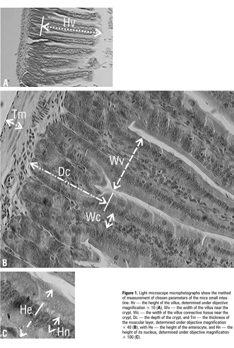
The stained sections were examined using the light microscope DMLS (Leica, Germany) connected to a computer equipped with a QWIN morphomet-ric system (Leica, Germany). All measurements were performed on images grabbed on a SONY 19” moni-tor with no prior knowledge of the age of the animal. The following parameters were established: the height of the villus (Hv) under magnification × 10, the width of the villus near the crypt (Wv), the width of the villus connective tissue near the crypt (Wc), the depth of the crypt (Dc), and the thickness of the muscular layer (Tm) under magnification × 40. The parameters concerning the enterocytes were the height of the enterocyte (He) and the height of its nucleus (Hn) established under magnification × 100 in two characteristic places, at the beginning of the villus and at the mid-point of its length (Fig. 1).
In order to obtain valuable data we evaluated variation coefficients (SD/mean) and on the basis of these we sampled each portion of the intestine in each animal according to the following proto-col: at least 25 villi for measurement of their height and at least 50 for measurement of their width. Additionally, in each portion of the intestine we measured the depth of 30 crypts and the height of at least 100 enterocytes.
The mean values of each measured parameter for each portion of the intestine in each animal were evaluated and entered as raw data on the database. The analysis of variance using Statistica v.6.0 (Statsoft, USA) was performed to test whether the values of each measured parameter were dependent on the portion of the intestine and the age of ani-mal. The interaction effect between these main fac-tors was also tested. The relation between the por-tion of the intestine and a given parameter was test-ed by regression analysis. In all statistical analyses p = 0.05 was the level of significance.
RESULTS
RESULTS
RESULTS
RESULTS
RESULTS
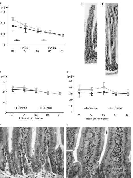
The height of the villus decreased from the prox-imal to the distal end of the small intestine in the case of both the 5-week and the 12-week-old mice (r = –0.76 and r = –0.93, respectively; Fig. 2A). An interaction was present between the age of the
an-imal and the portion of the small intestine. Age in-fluenced the villus height predominantly in the prox-imal portions (D5, D4) of the intestine and older mice had longer villi than younger mice. The mean value of villus height in D5 was 436.7 µm in younger mice and 586.4 µm in older mice, while in D1 it was 229.4 µm and 202.6 µm in younger and older mice, respec-tively (Fig. 2B, C).
The width of the villus near the crypt diminished on approaching to the colon in both groups of ani-mals studied (Fig. 2D). This effect was stronger in the 12-week-old group than in the 5-week-old group (r = –0.79 and r = –0.5, respectively). There was no interaction between the age of the animals and the portion of the small intestine.
The width of the villus connective tissue near the crypt was greater in the 12-week-old than in the 5-week-old mice (Fig. 2E–G). The mean value of this parameter in D5 was 34% and in D1 11% higher in the older than in the younger mice. Only in the 12-week-old animals did the mean value of this pa-rameter decrease from the proximal towards the dis-tal part of the intestine (r = –0.37). There was no interaction between the age of the animals and the portion of the small intestine.
The thickness of the muscular layer (Tm) in both age groups was greater in the distal part of the small intestine (5 weeks r = +0.44, 12 weeks r = +0.36, Fig. 3A). No significant differences between the age groups were observed. There was also no interac-tion between the age of the animals and the por-tion of the small intestine.
The depth of the crypts was greater in the young-er animals than in the oldyoung-er ones, especially in D2 (Fig. 3B). There was no interaction between the age of the animals and the portion of the small intestine. The height of the enterocytes decreased to-wards the distal end of the small intestine in both groups (5 weeks r = –0.51, 12 weeks r = –0.68). The mean values for the height of the enterocytes were greater in the 5-week then in the 12-week--old mice (Fig. 3C). There was no interaction between the age of the animals and the portion of the small intestine.
DISCUSSION
The present study is the first to present a mor-phometric analysis of the chosen parameters of the small intestine in the C57BL/6J mouse strain. Two age groups were selected as a result of their key role during the maturation of the gastrointes-tinal tract. The 5th week of C57BL/6J mouse life is
usually the weaning period [13, 34]. This is later than in the case of other mouse strains [25]. Wean-ing is a crucial factor and generally has a crucial influence on the morphology of the mucosal layer in the small intestine [5, 11, 40, 41]. The effect of weaning is at its most significant during the first week following. Later, approximately 2 weeks af-ter weaning, the morphological parameaf-ters have stabilised and resemble the morphology charac-teristic for an adult animal [5, 14, 22]. After 12 weeks of life the gastrointestinal tract is indisput-ably mature.
Figure 3. The graphs present the mean values [mm] and standard deviation of the parameters evaluated in particular portions of the small intestine: the muscular layer (A), the depth of the crypts (B), the height of the enterocyte (C), and the height of enterocyte nucleus (D).
The research conducted showed that in the mouse small intestine both the height and the width of the villi decreased on approaching the colon. This accords with the characteristics commonly consid-ered valid for the histological structure of the small intestine mucosa. Similar results have been described in rat, human, mouse, guinea pig, rabbit and piglet intestines [6, 18, 23, 26, 27, 33].
proximal villi increased in successively older rats, while the height of the villi at the two distal sites decreased with age. Wang et al. [38] reported that the height of jejunal villi increased after birth and peaked in the 3rd
postnatal month.
The widening of the villi near the crypt during ageing has previously been described in piglets [26]. In the present study no differences in this parameter were observed between the two age groups, al-though the higher values for the width of the villus connective tissue and the slightly lower values of the height of the enterocytes observed in the 12-week--old mice indicate the transformations which take place with ageing. In contrast, ageing had no effect on the size of rabbit enterocytes [18].
The lack of distinction in the thickness of the muscular layer between the 5-week and 12-week--old animals is in accord with the changes described by Wang et al. [38]. In rats the thickness of the je-junal muscular layer increased twofold until the 3rd
week and between the 3rd
and the 12th
months. In our material the thickness of the muscular layer in-creased in the distal direction of the small intes-tine, which is in accord with the result of Ogiolda et al. [28].
The fact that the mice crypts become shallower with age is in agreement with the results obtained by Penna et al. [33] on the human intestine. How-ever, Nunez et al. [26] observed that the crypts in 35-day-old piglets were deeper than in 5-day-old ones. The assumption that increased crypt depth is an indication of increased cell production is widely accepted. More intensive proliferation cannot be observed in younger transgenic animals of 5 weeks old, which can be explained by the lower metabol-ic activity of the cells.
The depth of the crypt in piglets decreases as it approaches the colon between the 3rd and 9th day
after weaning, the pigs being weaned at 4 weeks of age [15]. The piglet’s crypts described by Nunez et al. [26] were deeper in the duodenum than in ileum, but in the jejunum they achieved their high-est value. The same differences were seen in the group of rabbits fed diets supplemented with 12% lignin. However, supplementation by cellulose, pec-tin or alfalfa respectively caused the crypts in the jejunum to reach their lowest depth.
In adult rats the crypts in the jejunum are deeper than in the ileum [2].
The depth of the crypt in rats fed a 2% or 20% casein diet as described by Qu et al. [35] also
de-The enterocyte nuclei diminish with age. This decrease can be explained by the lower metabolic activity of the cells.
When taken together, our data provide the base-line morphological description of the small intesti-nal mucosa in wild type mice, strain C57BL/6J, which can be used as a reference for testing the influence of drugs, toxins, nutrients and inborn mutations on the mouse intestine.
REFERENCES
1. Ahn B, Ohshima H (2001) Suppression of intestinal polyposis in Apc(Min/+) mice by inhibiting nitric oxide production. Cancer Res, 61: 8357–8360. 2. Banwell JG, Howard R, Kabir I, Adrian TE, Diamond RH,
Abramowsky C (1993) Small intestinal growth caused by feeding red kidney bean phytohemagglutinin lectin to rats. Gastroenterology, 104: 1669–1677.
3. Bussey HJ (1987) Historical developments in familial polyposis coli. Semin Surg Oncol, 3: 67–70.
4. Bussey HJ (1990) Familial polyposis coli and hepato-cellular neoplasms. Hepatology, 12: 175–176. 5. Cera KR, Mahan DC, Cross RF, Reinhart GA,
Whitmoy-er RE (1988) Effect of age, weaning and postweaning diet on small intestinal growth and jejunal morpholo-gy in young swine. J Anim Sci, 66: 574–584.
6. Clarke RM (1977) The effects of age on mucosal mor-phology and epithelial cell production in rat small in-testine. J Anat, 123: 805–811.
7. Cummins AG, Thompson FM, Butler RN, Cassidy JC, Gillis D, Lorenzetti M, Southcott EK, Wilson PC (2001) Improvement in intestinal permeability precedes mor-phometric recovery of the small intestine in coeliac disease. Clin Sci (London), 100: 379–386.
8. Deans GT, Hamilton PW, Watt PC, Heatley M, William-son K, PatterWilliam-son CC, Rowlands, BJ, Parks G, Spence R (1993) Morphometric analysis of colorectal cancer. Dis Colon Rectum, 36: 450–456.
9. Fodde R, Smits R (2001) Disease model: familial ade-nomatous polyposis. Trends Mol Med, 7: 369–373. 10. Fodde R (2002) The APC gene in colorectal cancer. Eur
J Cancer, 38: 867–871.
11. Gu X, Li D, She R (2002) Effect of weaning on small intestinal structure and function in the piglet. Arch Tierernahr, 56: 275–286.
12. Haboubi NY, Lee GS, Montgomery RD (1991) Duode-nal mucosal morphometry of elderly patients with small intestinal bacterial overgrowth: response to an-tibiotic treatment. Age Ageing, 20: 29–32.
13. Halberg RB, Katzung DS, Hoff PD, Moser AR, Cole CE, Lubet RA, Donehower LA, Jacoby RF, Dove WF (2000) Tumorigenesis in the multiple intestinal neoplasia mouse: redundancy of negative regulators and specificity of modifiers. Proc Natl Acad Sci USA, 97: 3461–3466. 14. Hampson DJ (1986) Alterations in piglet small
intesti-nal structure at weaning. Res Vet Sci, 40: 32–40. 15. Hedemann MS, Hojsgaard S, Jensen BB (2003) Small
16. Hioki K, Shivapurkar N, Oshima H, Alabaster O, Oshi-ma M, Taketo MM (1997) Suppression of intestinal polyp development by low-fat and high-fiber diet in Apc(Delta 716) knockout mice. Carcinogenesis, 18: 1863–1865.
17. Huerta S, Irwin RW, Heber D, Go VL, Koeffler HP, Usko-kovic MR, Harris DM (2002) 1alpha,25-(OH)(2)-D(3) and its synthetic analogue decrease tumor load in the Apc(min) Mouse. Cancer Res, 62: 741–746.
18. Keelan M, Walker K, Thomson AB (1985) Intestinal morphology, marker enzymes and lipid content of brush border membranes from rabbit jejunum and ileum: effect of aging. Mech Ageing Dev, 31: 49–68. 19. Kelly P, Menzies I, Crane R, Zulu I, Nickols C, Feakins R,
Mwansa J, Mudenda V, Katubulushi M, Greenwald S, Farthing M (2004) Responses of small intestinal archi-tecture and function over time to environmental fac-tors in a tropical population. Am J Trop Med Hyg, 70: 412–419.
20. Kuitunen P, Kosnai I, Savilahti E (1982) Morphometric study of the jejunal mucosa in various childhood en-teropathies with special reference to intraepithelial lymphocytes. J Pediatr Gastroenterol Nutr, 1: 525–531. 21. Mahmoud NN, Boolbol SK, Dannenberg AJ, Mestre JR, Bilinski RT, Martucci C, Newmark HL, Chadburn A, Bertagnolli MM (1998) The sulfide metabolite of sulin-dac prevents tumors and restores enterocyte apopto-sis in a murine model of familial adenomatous poly-posis. Carcinogenesis, 19: 87–91.
22. Marion J, Biernat M, Thomas F, Savary G, Le Breton Y, Zabielski R, Le Huerou-Luron I, Le Dividich J (2002) Small intestine growth and morphometry in piglets weaned at 7 days of age. Effects of level of energy intake. Re-prod Nutr Dev, 42: 339–354.
23. Mitjans M, Ferrer R (2004) Morphometric study of the guinea pig small intestine during development. Mi-crosc Res Tech, 63: 206–214.
24. Mutanen M, Pajari AM, Oikarinen SI (2000) Beef in-duces and rye bran prevents the formation of intesti-nal polyps in ApcMin mice: relation to beta-catenin and PKC isozymes. Carcinogenesis, 21: 1167–1173. 25. Nakamura K, Kikusui T, Takeuchi Y, Mori Y (2003) The
influence of early weaning on aggressive behavior in mice. J Vet Med Sci, 65: 1347–1349.
26. Nunez MC, Bueno JD, Ayudarte MV, Almendros A, Rios A, Suarez MD, Gil A (1996) Dietary restriction induces biochemical and morphometric changes in the small intestine of nursing piglets. J Nutr, 126: 933–944. 27. O’Connor TM (1966) Cell dynamics in the intestine of
the mouse from late fetal life to maturity. Am J Anat, 118: 525–536.
28. Ogiolda L, Wanke R, Rottmann O, Hermanns W, Wolf E (1998) Intestinal dimensions of mice divergently se-lected for body weight. Anat Rec, 250: 292–299. 29. Oikarinen SI, Pajari AM, Mutanen M (2000)
Chemopre-ventative activity of crude hydroxymatairesinol (HMR) extract in Apc(Min) mice. Cancer Lett, 159: 183–187.
30. Orner GA, Dashwood WM, Blum CA, Diaz GD, Li Q, Al-Fageeh M, Tebbutt N, Heath JK, Ernst M, Dashwood RH (2002) Response of Apc(min) and A33 (delta N beta-cat) mutant mice to treatment with tea, sulindac, and 2-amino-1-methyl-6-phenylimidazo[4,5-b]pyridine (PhIP). Mutat Res, 506–507: 121–127.
31. Paulsen JE, Alexander J (2001) Growth stimulation of intestinal tumours in Apc(Min/+) mice by dietary L-methionine supplementation. Anticancer Res, 21: 3281–3284.
32. Paulsen JE, Elvsaas IKO, Steffensen IL, Alexander J (1997) A fish oil derived concentrate enriched in eicos-apentaenoic and docosahexaenoic acid as ethyl ester suppresses the formation and growth of intestinal polyps in the Min mouse. Carcinogenesis, 18: 1905– –1910.
33. Penna FJ, Hill ID, Kingston D, Robertson K, Slavin G, Shiner M (1981) Jejunal mucosal morphometry in chil-dren with and without gut symptoms and in normal adults. J Clin Pathol, 34: 386–392.
34. Perkins S, Clarke AR, Steward W, Gescher A (2003) Age--related difference in susceptibility of Apc(Min/+) mice towards the chemopreventive efficacy of dietary aspi-rin and curcumin. Br J Cancer, 88: 1480–1483. 35. Qu Z, Ling PR, Tahan SR, Sierra P, Onderdonk AB,
Bis-trian BR (1996) Protein and lipid refeeding changes protein metabolism and colonic but not small intesti-nal morphology in protein-depleted rats. J Nutr, 126: 906–912.
36. Song J, Medline A, Mason JB, Gallinger S, Kim YI (2000) Effects of dietary folate on intestinal tumorigenesis in the apcMin mouse. Cancer Res, 60: 5434–5440. 37. Song J, Sohn KJ, Medline A, Ash C, Gallinger S, Kim YI
(2000) Chemopreventive effects of dietary folate on intestinal polyps in Apc+/-Msh2-/- mice. Cancer Res, 60: 3191–3199.
38. Wang L, Li J, Li Q, Zhang J, Duan XL (2003) Morpho-logical changes of cell proliferation and apoptosis in rat jejunal mucosa at different ages. World J Gastro-enterol, 9: 2060–2064.
39. Wechter WJ, Murray ED Jr., Kantoci D, Quiggle DD, Leipold DD, Gibson KM, McCracken JD (2000) Treat-ment and survival study in the C57BL/6J-APC(Min)/ +(Min) mouse with R-flurbiprofen. Life Sci, 66: 745– 753.
40. Wiese F, Simon O, Weyrauch KD (2003) Morphology of the small intestine of weaned piglets and a novel method for morphometric evaluation. Anat Histol Embryol, 32: 102–109.
41. Yu B, Chiou WS (1997) The morphological changes of intestinal mucosa in growing rabbits. Lab Anim, 31: 254–263.
![Figure 3. The graphs present the mean values [mm] and standard deviation of the parameters evaluated in particular portions of the smallintestine: the muscular layer (A), the depth of the crypts (B), the height of the enterocyte (C), and the height of enterocyte nucleus (D).](https://thumb-us.123doks.com/thumbv2/123dok_us/9949170.1982753/6.581.64.525.70.431/deviation-parameters-evaluated-particular-smallintestine-muscular-enterocyte-enterocyte.webp)
