Vol. 3, No. 4 (2013): 303-308 Review Article
Open Access
I
ISSSSNN::22332200--66881100
Magic Bullets - Nanocapsules in Future Medicine
Vishnu Prabhakar*, Tapan, Aarti Yadav, Rachna Ratan
Nanotechnology Research center, DAVIET, Jalandhar 144008, India
* Corresponding author: Vishnu Prabhakar; e-mail: [email protected]
ABSTRACT
Advanced drug delivery systems are preferred for less administration frequency, fewer side effects, higher drug concentrations at pathological sites, and longer drug bioavailability. A general method to achieve better delivery is through encapsulation of drug inside carriers that are made of biocompatible and biodegradable materials. Self-assembled polymer based nanocapsules, polymer micelles, polymer–drug conjugate, and polymer implants are commonly used drug delivery systems either in under clinical trials or in the market. The encapsulation of the drug within the nanocapsules affects the absorption, distribution, and metabolism of the drug and can result in better treatment protocols that are more familiar to the patient and have shown significant outcomes in treating cancers and immunological diseases on molecular level. The paper highlights that polymeric nanocapsules made up by self-assembly process opening the new ways to enhance quality of life.
Keywords:
Nanopharmacology, Polymers,Nanocapsule, Drug Delivery, Layer by Layer Self-AssemblyINTRODUCTION
Nanopharmacology, is a new branch of pharmacology which gradually emerging with the application of nanotechnology in the field of nanomedicine, a potential step towards curing & prevention of disease using molecular tools & molecular knowledge about human body. Nanopharmacology studies the interactions between nanoscale drugs and proteins, DNA and RNA, cells, tissues or organs that construct human body as structural materials, or the interactions between traditional drugs and physiological systems at nanoscale level.
The hot spot of Nanopharmacology at present is synthesis of self-assembled polymeric nano sized drugs carriers by bottom up assembly & its applications in health care, including controlled & targeted drug delivery, which reduces the risks of drug toxicity, drug concentration while increasing the drug bioavailability at the therapeutic sites [1].
Nanocapsules [2] are drug delivery agents with in the size range of 10-1000 nm which are normally composed of a shell and a space in which desired substance or drug molecules can be placed, and have been made from many years following the example of nature, using molecules called phospholipids, like
liposomes, which comprises the biological membrane and are hydrophobic (water repellent) on one end and hydrophilic (water loving) on the other hand when such molecules are placed in an aqueous environment, they can spontaneously form a capsule in which hydrophobic portions hide inside, protecting them from contact with water, represent thebest example of nature made self-assembly [2]. Self-assembly allow spontaneous association of certain molecules in defined & desired structures without any guidance or management from external sources, and generates defect free, complicated, hierarchical supramolecular architectures from relatively simple molecular components. Recently many other materials such as variety of polymers have been used to make nanocapsules by self-assembly process. Polymeric nanocapsules have been extensively studied as particulate carriers in the pharmaceutical and medical fields, because they show promise as drug delivery systems as a result of their controlled and sustained release properties, subcellular size, biocompatibility with tissue and cells [3].
WHAT MAKES NANOCAPSULES SPECIAL?
one of the useful function, the finer drug carrier are apt to be absorbed more easily through biological systems, called enhanced permeation & retention (EPR effect - the diffusion and flow of nanoparticles through narrow channels), and can manipulate diseased cells by releasing drug at molecular level [4]. The special features of nanocapsules designs in drug delivery system are as follows [5]:
Slow release - The capsule releases drug molecules slowly over a longer period of time (e.g., for slow delivery of a substance in the body).
Quick-release - The capsule shell breaks upon contact with a surface.
Specific release - The shell is designed to break open when a molecular receptor binds to a specific chemical (e.g., upon encountering a tumor or protein in the body).
Moisture release - The shell breaks down and releases drug in the presence of water (e.g. in soil).
Heat-release - The shell releases drug only when the environment warms above a certain temperature;
pH release - Nanocapsule breaks up only in specific acid or alkaline environment (e.g., in the stomach or inside a cell).
Ultrasound release - The capsule is ruptured by an external ultrasound frequency.
Magnetic release - A magnetic particle in the capsule ruptures the shell when exposed to a magnetic field.
DNA nanocapsule - The capsule smuggles a short strand of foreign DNA into a living cell which, once released, hijacks cell machinery to express a specific protein (used for DNA vaccines).
WHY LAYER BY LAYER SELF-ASSEMBLING?
Polymers have found many important applicationsin biomedical, pharmaceutical, electronic, andmolecular diagnostic fields. The recent introduction ofelectrostatic layer-by-layer (LbL) self-assembly techniqueenables the design and development of polymer/polymer and polymer/nanoparticle complexes atnanometer scale for advanced drug delivery & therapeutic purpose.Layer by layer (LBL) self-assembled polyelectrolyte capsules, recognized as one of the nanotechnologies that advanced the field of drug delivery[(Cruz et al. 2006; Amaral et al. 2007)7], may serve as one of the alternatives to solve the problems associated with formulation of complex drug design like chlorinated organic solvents are usually involved in synthesis and this may lead to organic residues in the system and damage encapsulated materials or may alter the properties of chemical drugs [6].Polyelectrolyte nanocapsules present unique advantages such as:
Easy fabrication process.
No necessity of chlorinated organic solvent.
Fine control of permeability (EPR effect) through membrane.
The thickness of the capsule wall can be precisely tuned in the range of a few nanometers by choosing coating materials and number of layers. The pore size on shell wall membrane can be controlled through different polyelectrolyte pairs and assembly conditions.
Wide varieties of Shell materials, includes charged polymers, lipids, proteins, and magnetic nanoparticles can be used during shell assembly. Shells can be switched between “open” and “closed” states for trigger release.
PREPARING NANOCAPSULE
There are two major methods for the encapsulation of therapeutic agents inside polyelectrolyte shells: (a) Layer by layer self-assembled nanocapsule, and (b) loading of drug molecules into hollow polyelectrolyte shell.
Layer by Layer Self-Assembled Nanocapsules
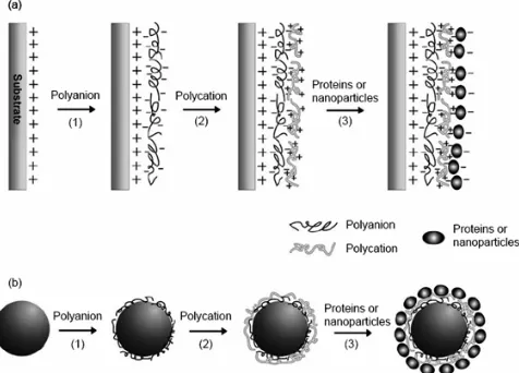
This principle of LBL self-assembly [6] utilizes the electrostatic attraction between oppositely charged groups on different polyelectrolytes polymers/nanoparticles as the driving force to achieve encapsulation of micro/nano drug molecules. The building blocks are naturally occurring charged polymers, such as pectins, gelatins, polyglutamic acid, chitosan and hyaluronic acid. These are adsorbed around the drug particle in alternating layers of oppositely charged material. The supramolecular structure is held together electrostatically through the formation of complexes between the polycations and polyanions. The resulting capsules have a wall thickness of 10-40 nm and range from 20-20 mm in diameter [7].
The encapsulation process is as follows: First, for adsorption of a polyanion layer, a solid support (e.g. slide or drug) with positive surface charge is incubated in a solution containing polyanion for a certain amount of time, usually 30 min. Next, solid supports are rinsed with pure water two or three times to remove excess free polyelectrolyte. Then, the slide is immersed in a solution of cationic polyelectrolytes and a layer is adsorbed. The original surface charge (positive) is restored, and the surface is ready for washing. Generally, the above-mentioned steps are repeated alternately until a film of desired thickness is obtained [8-11].
core. Increasing the number of layers decreased the shell permeability and resulted in prolonged dye-core dissolution, which solves the problem of quick photo bleaching in case of general fluorescent imaging dyes. Hence enhance molecular imaging as well as visible pharmacokinetics of drugs in molecular level has been achieved [12].
Figure 1. (a) Schematic illustration of film deposition process through LbL self-assembly on a flat substrate.
(b) Encapsulation of drug molecule by LBL self-assembled polyelectrolyte polymers [12].
Loading Drug into Hollow Polyelectrolyte Shell This method is based on preparing a multilayer of polyelectrolyte polymeric shell using a core template (weakly cross linked melamine formaldehyde particles/bio colloids) and further removal of this core template using short time exposure of acidic solution, which leaving behind a hollow shell and can be further loaded by suitable drug molecules[13].This is done by dispersing the particle in the respective polymer solution and subsequent centrifugation to settle and separate the coated particles. Each adsorption step is followed by repeated centrifugation and washing to remove weakly adsorbed polymer. Subjecting the polyelectrolyte coated particles to either strong acidic conditions or oxidative degradation results in decomposition of core template thus formation of hollow shell obtains [8].
Polyelectrolyte shells are potential candidates for drug and DNA delivery and may be applied for bio sensing when loaded with molecular probes. The interior environment of a polyelectrolyte shell is aqueous, similar to that of liposome and polymer vesicles. Generally, hydrophilic materials are favorable candidates for loading. In comparison, the hydrophobic core of a polymer micelle is good for encapsulation of hydrophobic drugs or diagnostic agents.
Figure 2. Hollow shell fabrication
Loading Efficiency
There are mainly two ways of performing drug loading: (1) incorporation of the drug into the system before the reaction has started; &(2) introduction of the drug after termination of the reaction or after some time when initiation took place (partly adsorption). The loading efficiency of the drug into the polymeric matrix is determined by the time of introduction of the drug into the reaction medium. A high binding degree can be achieved by dissolving the drug in the aqueous medium before adding the monomer into the system.
Harivardhan L. Reddy et al. synthesized nanoparticles of
polybutyl cyanoacrylate (PBCA) loaded with methotrexate by emulsion polymerization and dispersion polymerization techniques by anionic polymerization. The drug was dissolved in thepolymerization medium prior to addition of butyl cyanoacrylate (BCA) [14].
POLYMERS USED
The polymers should be compatible with the body in the terms of adaptability toxicity) and (non-antigenicity) and should be biodegradable and biocompatible. The most commonly used natural polymers in preparation of polymeric nanoparticles are: Chitosan, Gelatin, Sodium alginate, Albumin [17-20] etc. Also there are many synthetic polymers used in encapsulation of drug like as:polylactides (PLA), polyglycolides (PGA), poly(lactide co-glycolides) (PLGA), polyanhydrides, polyalkylcyanoacrylates (PACA), polycaprolactone, poly glutamic acid, poly malic acid, poly(N-vinyl pyrrolidone), poly (methyl methacrylate), poly(vinyl alcohol), poly (acrylic acid), poly acrylamide, poly(ethylene glycol), poly (methacrylic acid), poly(styrene sulfonate), poly(allylamine hydrochloride) [21-31] etc.
NANOTRANSPORTER
The nanotransporter is built up by hyper branched polymers network like dendrimers and also based on the electrostatic adhesion between two opposite charged molecules. Polymer network covers up whole drug molecule or metal ions having opposite charge and shape it in the form of nanocapsule.The functional groups present on nanotransporters are soluble in blood, serum, PBS buffer, water, ethylene glycol, methanol, ethanol, acetonitrile, DMSO, DMF, acetone, chloroform, ethyl acetate, and Toluene etc.
Several chemical molecules, metal ions and metal nanoparticles have already been encapsulated with nanotransporters like as Nimopidin, Methotrexate, Doxorubicin, congo red, vitamin A, vitamin B2, vitamin B6, vitamin D3, carotene, gold, silver, ruthenium, platinum, palladium and copper etc. The encapsulation of the substrates is done in two different ways either through the choice of functional groups presents in polymer network or by the agglomeration of several polymer units. The unique properties & ease of fabrication of nanotransporters has shown practically significant results to deliver therapeutic and medical imaging agents on molecular level [15].
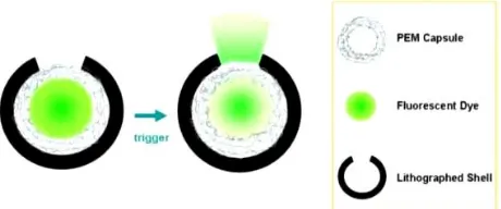
NANOCAPSULE DESIGNED BY LITHOGRAPHY&SA A tiny nanocapsule composed of polymer layers and nanoparticles may provide drug delivery that targets diseased cells without harming the rest of the body. This delivery system could be robust and flexible enough to deliver a variety of substances.The outer spheres are fabricated by lithography technique with leaving about 5 percent of the surface as an escape area for the drugs and biocompatible with biological organism. Thealternating positive and negative layers of self-assembles polymer form the capsules inside the spheres.
Figure 4. Lithographically made nanocapsule
Targeted drug delivery systems release their drug from the tiny hole in their surface, when they encounter with diseased area, this will concentrate the drug at the target and reduce the amount of toxins circulating in the body. In these spheres variety of substance can be filled. Drugs can be either solid & incorporated when the capsules are made or liquid which is filled later. Chemicals that target the diseased cells can be attached in a variety of ways.The robustness of the nanocapsules is measured by the dehydrating and then reconstituting them. Their ability to withstand long periods dried out and then successfully rehydrate is important for capsule life and because that is the way that liquid medications will be inserted in the microcapsules as needed [16].
NANOCAPSULES IN RECENT MEDICINE
The task of creation of polymeric nanocapsules is especially important in a treatment of diseases which require intensive and long term therapy with high dosage of drugs as in chemotherapy of cancer diseases. The use of potent drugs for a long period of time often leads to expressed side reactions (toxic and allergic effects) thus considerably decreasing the treatment efficacy. Polymeric nanoparticles and nanocapsules are
used in targeted drug delivery into the site of inflammation such as tumors and tuberculosis, therefore developing of novel forms of drugs for the delivery of antituberculosis and antitumor preparations on the basis of well-known biocompatible polymers would help to increase essentially the efficacy of chemotherapy of such diseases.
Delivery of Antitumor Drugs
The results of numerous investigations have shown obvious potentials of the use of polymeric nanocapsule for the treatment of such chronic diseases as tuberculosis, tumors, arterial hypertension and other diseases which demand long-term therapy with multiple doses of potent drugs.Nowadays there are several research groups working basically on synthesizing polymeric nanocapsules systems for controlled drug release. One of the founders in the field of creation of nanoparticles for controlled release purposes was Prof. Peter Speiser (ETH, Zurich) [26].
The most important ideas of Prof. Speiser were the development of nanoparticles for targeted drug delivery and the idea of using nanoparticles for the delivery of drugs which have to pass through the blood–brain barrier and to be delivered directly to the brain. This direction was continued by the Prof. Kreuter J. (Institute of Pharmaceutical Technology, Goethe University, Frankfurt, Germany). The research group under the supervision of Prof. Kreuter has been working on developing polymeric systems for controlled release of antitumor drug doxorubicin, apoliprotein, DNA, RNA, etc. on the basis of different natural and synthetic polymers including human serum albumin and polyalkyl cyanoacrylates [14, 26].A number of antitumor drugs have been successfully encapsulated into PLGA nanoparticles. These are some specific examples:
9-Nitrocamptothecin was encapsulated in PLGA through the nano-precipitation method; as a result the encapsulation efficiency was more than 30 % [32].
Cisplastin was encapsulated in PLG Amethoxy PEG [33].
Paclitaxel-loaded nanoparticles were prepared by the solvent evaporation/extraction method; in this case the loading efficiency reached 100 % with full antitumor activity [34].
The poorly water-soluble Xanthones was incorporated into PLGA nanoparticles [35].
Dexamethazone was incorporated into PLGA nanoparticles by the solvent evaporation method. The drug released completely after 4 h of incubation at 370C in vitro [36].
Nanocapsule in Gene Therapy
Gene therapy is an emerging technology that aims to permanently or temporarily correct a gene defect by the intracellular delivery of nucleic acids [38]. Gene defects can either arise during cell division processes or be due to external agents (such as ultraviolet or other radiation, chemical substances). The introduction of a plasmid DNA, encoding the native form of the gene can be a way to treat these gene defects. Since naked plasmid DNA is quickly degraded by blood nucleases and in general shows no relevant therapeutic effects when administered systemically [38], vectors are necessary to transport plasmid DNA into the cell nucleus. In general, as with any drug, gene transfer complexes must reach their intended site of action to induce therapeutic effects Basic challenge for nonviral gene therapy is to develop an approach that delivers a therapeutic gene into selected cells. Two different types of promising nanocarriers were developed for gene therapy purpose: lipid nanocapsules (DNA LNCs) and multimodular systems (DNA MMS).
DNA LNCs consist of a lipophilic lipid core, containing a mixture of triglycerides and polyglyceryl-6 dioleate surrounded by a shell composed of free polyethylene glycol (PEG) and hydroxystearate-PEG [39]. To encapsulate hydrophilic DNA in the lipophilic lipid core, the first step consists of complexing the anionic DNA with cationic lipids, to form lipoplexes which are then introduced into the formulation process of DNA LNCs, based on phase-inversions of an emulsion [40].DNA LNCs encapsulating a luciferase-coding plasmid DNA proved their transfection efficacy in vitro [41].
DNA MMS also exhibit a dual structure, a core composed of lipoplexes and an external corona of steric stabilizerscapable of carrying the ligands necessary for active targeting. The first DNA MMS that were developed consisted of the cholesterol derivate BGTC (bis-guanidinium-tren-cholesterol) as a cationic lipid, the plasmid pCMV luciferase, and the steric stabilizer F108, a block copolymer of poly(ethylene oxide) and poly(propylene oxide), with or without galactose (GAL) as a ligand. They had already been tested in vitro on primary hepatocytes and demonstrated a specific transfection for galactosylated DNA MMS due to the recognition of the GAL ligands by ASGP-R, present on hepatocytes [42].
Future Nanocapsule Bandages
The conventional dressings require to be taken out if the skin becomes affected or it slows the healing [(Radhikaet al.2011)43]. In contrast, nanocapsule dressings trigger automatically to discharge antibiotics when the wound becomes infected. They do not require to be removed, thereby enhancing the chances of healing wound without scarring. Nanocapsule bandages can also be used for additional types of wounds like ulcers and most consistently by the military people on the battlefield. These medicinal
dressings release antibiotics from the nanocapsules activated by the presence of disease causing pathogenic or causative bacterial organism, targeting the treatment prior to the infection aggravates. The bacterial toxins burst the capsules comprising the antibiotics, which cover as the dressings. In this way, antibiotics are produced when needed, thus it reduces the risk of the evolution of antibiotic resistant microbes such as Methicillin resistant Staphylococcus Aureus(MRSA).
CONCLUSION
There has been a considerable research interest in the area of drug delivery using particulate delivery systems as carriers for small and large molecules. Particulate systems like nanocapsules have been used as a physical approach to alter and improve the pharmacokinetic and pharmacodynamic properties of various types of drug molecules. Nanocapsules also have the efficient applications in various fields of the agrochemicals, wastewater treatments, genetic engineering, cosmetics, cleaning products, as well as in adhesive component. They are also used in encapsulation of enzymes, adhesives, catalysts, polymers, oils, inorganic micro and nanoparticles, latex particles, and even the biological cells. The enhanced delivery of bio-active molecules &active pharmaceutical ingredients (APIs) through the targeted delivery in the form of nanocapsules, opens numerous challenges and opportunities for the research and future development of novel improved therapeutic systems.
REFERENCES
1. Yingge Zhang, Institute of Pharmacology and Toxicology and Key laboratory of Nanopharmacology and Nanotoxicology, Beijjing Academy of Medical Sciences, 27 Taiping Road, Haidian District, Beijing 100850, China.
2. Drug delivery capsules, courtesy of capsulation nanoscience, Cientifica, oct 2003.
3. Different techniques for preparation of polymeric nanoparticles, Asian journal of pharmaceutical & clinical research, Nagavarma, HemantYadav, Department of Pharmaceutics, JSS College of Pharmacy, JSS University, Mysore,Karnataka – 570015. 4. Nanoparticle technology handbook, H. Maeda: J. Control.
Release, 19, 315–324 (1992).
5. www.azonano.com/Drug encapsulation at nano level.
6. Sukhorukov, G.B. et al. (1998) Layer-by-layer self-assembly of polyelectrolytes on colloidal particles. Colloids and Surfaces A: physicochemical and engineering aspects. 137, 253–266. 7. Size-controlled polyelectrolyte nanocapsules via layer-by-layer
self-assembly, H. AI, J. GAO, Department of Biomedical Engineering, Case Western Reserve University, Cleveland, OH 44106.
8. Self-assembly & Self-Organization, Roy Shenhar, Tyler B. Norsten, Department of chemistry, University of Massachusetts. 9. Yuri M. LVOV, Nanofabrication of ordered multilayers by
alternate adsorption of polyions, nanoparticles and proteins: From planar films to microtemplates. Institute for Micromanufacturing, LaTech, Ruston, LA 71272
10. Decher, G. (1997) Fuzzy nanoassemblies:Toward layered polymeric multicomposites.Science 227, 1232–1237.
11. Bertrand, P., Jonas, A., Laschevsky, A., andLegras, R. (2000) Ultrathin polymer coating bycomplexation of polyelectrolytes, Macromol, 21,319–348.
13. F. Caruso, R. A. Caruso, H. Möhwald, Nanoengineering of inorganic and hybrid hollow spheres by colloidaltemplating. Science, 282, 1111, 1998.
14. Kreuter. J. Colloidal Drug Delivery Systems.-New York, Marcel Dekker,1994.
15. www.nanopartica.com/Nanotransporter
16. www.sciencedaily.com/releases/2009/01/090112121815.htm 17. Farrugia C.A, M.J. Grover, Gelatin behavior in dilute J. Kreuter,
Nanoparticles, in: J. Kreuter (Ed.), Colloidal Drug aqueous solutions: Designing a nanoparticulate formulations, Delivery Systems, Marcel Dekker, New York, 1994, pp. J. Pharm. Pharmacol. 51 (1999) 643–649.
18. Fernandez-Urrusuno.R, P. Calvo, C. Remunan-Lopez, J.L Villa-Jato, M.J Alonso, Enhancement of nasal absorption of insulin using chitosan nanopartilces, Pharm. Res. 16 (1999) - 1576– 1581.
19. Aynie I.C, C.Vauthier, E. Fattal, M. Foulquier, P. Couvreur, Alginate nanoparticles as a novel carrier for antisense oligonucleotide, in: J.E. Diederichs, R. Muler (Eds.), Future Strategies of Drug Delivery With Particulate Systems, Med- 405– 427. Pharm Scientific Publisher, Stuttgart, 1998, 5–10.
20. Barbara Luppi, Federica Bigucci, Giuseppe Corace, Alice Delucca, Teresa Cerchiara, Milena Sorrenti et al. Albumin nanoparticles carrying cyclodextrins for nasal delivery of the anti-Alzheimer drug tarcine. Eur J Pharm Sci 44(2011) 559-565.
21. Shokri N, AkbariJavar H, FouladdelSh, Khalaj A, Khoshayand MR., Dinarvand. R et al. Preparation and evaluation of poly (caprolactonefumurate) nanoparticles containing Doxorubicin Hcl. DARU (19) 1, 2011.
22. Ghosh. PK Hydrophilic polymeric nanoparticles as drug carriers. Indian J BiochemBiophys 2000 (37), 273-282.
23. Heidi M. Mansour, MinJiSohn, Abeer Al-Ghananeem and Patrick P. DeLuca materials for pharmaceutical dosage forms: molecular pharmaceutics and controlled release drug delivery aspects. Int. J. Mol. Sci. 2010 (11), 3298-3322.
24. SushmithaSundar, JoydipKundu and Subhas C Kundu. Biopolymeric nanoparticles Sci. Technol. Adv. Mater. 11 (2010) 014104 (13pp).
25. Birrenbach G and Speiser P 1976 J. Pharm. Sci. 65 1763. 26. Kreuter J and Speiser P P 1976 Infect. Immun. 13 204.
27. Couvreur P, Kante B, Roland M, Guiot P, Bauduin P and Speiser P 1979 J. Pharm. Pharmacol. 31 311.
28. Gurny R 1981 Drug Dev. Ind. Pharm. 7 1.
29. Vauthier-Holtzscherer C, Benabbou S, Spenlehauer G, Veillard M and Couvreur P 1991 STP Pharm. Sci. 1 109.
30. Allemann E, Leroux J C, Gurny R and Doelker E 1993 Pharm. Res. 10 1732.
31. Annick Ludwig .The use of mucoadhesive polymers in ocular drug delivery Advanced Drug Delivery Reviews 57 (2005) 1595– 163.
32. Derakhshandeh K., Erfan M., Dadashzadeh S., Encapsulation of 9-nitrocamptothecin, a novel anticancer drug, in biodegradable nanoparticles, Eur. J. Pharm. Biopharm. 2007. 66. 1. 34–41. 33. Avgoustakis K., et al., PLGA–mPEG nanoparticles of cisplatin: in
vitro nanoparticle degradation, J Control, 2002. 79 (1–3). - P. 123–135.
34. Fonseca C., Simoes S., Gaspar R., Paclitaxel-loaded PLGA nanoparticles: preparation, physicochemical characterization and in vitro anti-tumor activity, J. Control, 2002. 83 (2) 273– 286.
35. Pinto M.M., Sousa E.P., Natural and synthetic xanthonolignoids: chemistry and biological activities, Curr. Med. Chem. 2003. 10 (1). P. 1–12.
36. Release of dexamethasone from PLGA nanoparticles, Cascone MG, Pot PM, Lazzeri L, Zhu Z., Department of Chemical Engineering, University of Pisa, Via Diotisalvi 2, 56126 Pisa, Italy.
37. Gao H., et al., Synthesis of a biodegradable tadpole-shaped polymer via the coupling reaction of polylactide, J. Control. Release. 2005. 107 (1). P. 158–173.
38. Viola, JR, El-Andaloussi, S, Oprea, II and Smith, CI (2010). Non-viral nanovectors for gene delivery, Opin Drug Deliv7: 721–735. 39. Vonarbourg, A, Passirani, C, Desigaux, L, Allard, E, Saulnier, P,
Lambert, O et al. (2009). The encapsulation of DNA molecules within biomimetic lipid nanocapsules. Biomaterials30: 3197– 3204.
40. Heurtault, B, Saulnier, P, Pech, B, Proust, JE and Benoit, JP (2002). A novel phase inversion-based process for the preparation of lipid nanocarriers. Pharm Res19: 875–880. 41. Morille, M, Passirani, C, Letrou-Bonneval, E, Benoit, JP and
Pitard, B (2009). Galactosylated DNA lipid nanocapsules for efficient hepatocyte targeting. Int J Pharm379: 293–300. 42. Letrou-Bonneval, E, Chèvre, R, Lambert, O, Costet, P, André, C,
Tellier, C et al. (2008). Galactosylatedmultimodularlipoplexes for specific gene transfer into primary hepatocytes. J Gene Med10: 1198–1209.
43. Radhika PR, Sasikanth and Sivakumar T. 2011. Nanocapsules: A new approach for drug delivery. Int J PharmaSci Res, 2(6), 1426-29.
*****
© 2013; AIZEON Publishers; All Rights Reserved