Vol. 6, No. 2 (2016): 1459-1466 Research Article
Open Access
I
ISSSSNN::22332200--66881100
Protective and Therapeutic Effects of Cranberry
extract against Paracetamol-induced Liver toxicity
Abdel-Maksoud A. Hussien
1, Mohammed Abdalla Hussein
2,* and Saieda Mohamed
11
Department of Biochemistry, Faculty of Veterinary Medicine, Benha University, 13736 Moshtohor, Qalioubeya, Egypt. 2
Biochemistry Department, Faculty of Pharmacy, October 6 University, October 6 city, Egypt.
* Corresponding author: Mohammed Abdalla Hussein, e-mail: [email protected]
ABSTRACT
The analgesic paracetamol causes a potentially fatal, hepatic centrilobular necrosis when taken in overdose. We aimed to assess the hepatoprotective effects of cranberry extract against paracetamol-induced hepatotoxicity in rats. Oral administration of cranberry extract at a concentration of 75 and 150 mg/kg b.w daily for 15 days showed a significant protection against-induced alteration in aspartate amino transferase (AST), alanine amino transferase (ALT), alkaline phosphatase (ALP), lactate dehydrogenase (LDH), superoxide dismutase (SOD) and catalase (CAT) activities and concentrations of thiobarbituric acid reactive substances (TBARS), protein thiols (Pr-SHs), reduced glutathione (GSH), tumor nicrosis factor alpha (TNF-α) and nitric oxide (NO), by paracetamol. The extract of cranberry significantly reduced the plasma levels of these elevated liver enzyme markers in a dose-dependent manner. Histopathological examination of liver tissues also showed hepatoprotective effects of cranberry in restoring normal functional ability of the liver. The results of our study strongly suggest that the extract of cranberry has strong hepatoprotective effects against paracetamol-induced liver injury, thereby scientifically affirming its traditional therapeutic role in liver injury.
Keywords:
Antioxidants, paracetamol, Cranberry extract and liver enzymes.1. INTRODUCTION
The liver, being the center of metabolic functions, plays a crucial role in metabolizing a variety of xenobiotics; it is therefore more vulnerable to the toxicity of these chemicals [1]. Overdoses of the analgesic and antipyretic acetaminophen represent one of the most common pharmaceutical product poisonings in the United States today [2]. Although considered safe at therapeutic doses, in overdose, acetaminophen produces a centrilobular hepatic necrosis that can be fatal [3]. Whereas the initial biochemical and metabolic events that occur in the early stages of toxicity have been well described, the precise mechanisms of hepatocyte death are poorly understood. Necrosis is recognized as the mode of cell death and apoptosis has been ruled out [4].
Normally, paracetamol is metabolized by cytochrome P450 enzymes into an active intermediate, i.e. N-acetyl-p-benzoquinone imine (NAPQI), which is rapidly detoxified by conjugation with glutathione [5]. Excess
NAPQI binds to the mitochondrial proteins and also damages the mitochondria in hepatocytes, leading to extreme generation of free radicals followed by lipid peroxidation and finally hepatic cell death [5].
of quercetin [15]. Myricetin is the second most abundant flavonol, followed by kaempferol [14 and 15]. These compounds are yellow in color, and there are 20 different flavonol glycosides in cranberry, as confirmed by another article [12 and 13]. No reports about antioxidant of cranberry extract against paracetamol induced liver toxicity in rats. As a continuation of our interested research in pharmaceutical and medical importance of natural products [6-11]. We report herein, a facile route to explain antioxidant and hepatoprotective effects of cranberry extract in rat’s model of paractamol-induced liver toxicity.
2. MATERIALS AND METHODS
2.1 Chemicals
- Paracetamol was provided as gift from El-Nile
Pharmaceutical Company (Cairo, Egypt). When intended to be used in vivo experiments, paracetamol was suspended in 0.5 % tween 80 and orally administrated in dose of 1g/kg.B.W.(19).
- Tween 80 was produced by Prolabo, Farance. - Cranberry extract was purchased it from Virgin
Extracts (TM), China. Cranberry was given to female mice with 1/150 LD50 (75mg/kg.b.w.) and 1/75 LD50 (150mg/kg.b.w.) daily for 3weeks by oral gastric gavage tube.
2.2 Animals
Adult albino rats weighing around 200-220gms were purchased from Faculty of Veterinary Medicine, Cairo University. They were acclimatized to animal house conditions. Animals were provided with standard diet and water adlibtum. Rats were kept under constant environmental condition and observed daily throughout the experimental work.
2.3 Experimental set up
This experiment was carried out to examine the prophylactic potential of ethanolic and aqueous extracts of cranberry extract, gaven repeatedly for 2 weeks, against paracetamol hepatotoxicity in vivo.
Groups of animals each consisting of 8 rats were treated daily for 14 days as follows. A suspended solution of 3g% was prepared for intragastric intubation of rats.
Group I: Normal (was given similar volume of tween 80, 1% in saline orally)
Group II: Control (was given similar volume of tween 80, 1% in saline orally)
Group III: Was treated with cranberry extract (75mg/kg b.w.) suspended in tween 80 orally in a single daily dose [11].
Group IV: Was treated with cranberry extract (150 mg/kg b.w.) suspended in saline orally in a single daily dose [11].
Group V: Was treated with vitamin C (1g/kg b.w.) suspended in tween 80 orally in a single daily dose [16].
At day 13, i.e. one day before the last treatment, animals of all groups were fasted for 18 h. At day 14, one hour after the last dose of drug treatment, all animals in groups II, III, IV and V recived paracetamol (1 g/kg.b.w.) [8].
2.3.1 Treatment of blood samples
After 15 days of treatment blood samples were withdrawn from the retro-orbital vein of each animal and each sample was collected into 2 tubes, heparinized and non-heparinized. The heparinized blood samples were centrifuged at 1000 xg for 20 min. The separated plasma were used for the estimation of plasma activity of ALT, AST, ALP, LDH as well as levels of TNF-α, NO, TBARs, Pr-SHs and total protein. The heparinized blood samples were divided into 2 aliquots. The first aliquot was used for determination of CAT activity.
The second aliquot was haemolyzed using bidistilled water and the haemolysate of each sample was divided into two portions was treated with chloroform/ethanol (3:5 V/V) mixture to precipitate and the resultant supernatant was used for the determination of SOD activity. The second portion was deproteinized with meta-phosphoric acid and the clear supernatant was used for the estimation of GSH level. Haemoglobin levels were determined in the heparinized blood samples and used in the calculation of the enzyme activity.
2.3.2 Preparation of liver samples
Animals were killed by cervical dislocation, and then livers were rapidly removed. A part of each liver was weighed and homogenized, using glass homogenizer (Universal Lab. Aid MPW-309, mechanika precyzyjna, Poland), with ice-cooled saline to prepare 25% W/V homogenate. The homogenate was divided into three aliquots. The first one was deproteinized with ice-cooled 12% trichloroacetic acid and the obtained supernatant, after centrifugation at 1000 xg, was used for the estimation of GSH. The second aliquot was centrifuged at 1000 xg and the resultant supernatant was used for estimation of TBARS, Pr-SHs, total protein and albumin levels. The third aliquot of homogenate was used to prepare a cytosolic fraction of the liver by centrifugation at 10500 xg for 15 min at 4 OC using a cooling ultra-centrifuge (Sorvall comiplus T-880, Du Pont, USA), and the clear supernatant (cytosolic fraction) was used for the determination of SOD and CAT activities.
2.4 Biochemical assays
Superoxide dismutase (SOD) and catalase (CAT) activities were carried out Sinha [25], Marklund and Marklund [26], respectively. Liver GSH were estimated according to the method of Sedlak and Lindsay [27]. Blood haemoglobin was determined according to the method of Van Kampen and Zijlstra [28]. The protein content of liver tissue was measured by applying the method of Lowry et al. [29].
2.5 Histopathology
The liver tissues isolated from the test animals were fixed in formaline-saline for 48 hours. The fixed tissue were processed manually through graded ethanol, cleared in xylene, impregnated and embedded in paraffin wax. Thin sections were cut with a rotary microtome, stained by haematoxylin and eosin technique, examined microscopically for pathological changes according to the method of Bancroft and Steven[30].
2.6 Statistical Analysis
Statistical analysis of the obtained results was carried out using T-test and student’s F-test according to [31].
P values of less than 0.05 were considered to indicate statistical significance. All the results were expressed as mean ± SD for eight separate determinations.
3. RESULTS AND DISCUSSION
Table 1 showed that oral administration of paracetamol at 1g/kg.b.w. resulted in a significant increase in plasma ALT, AST, ALP and LDH compared to the normal control group (p< 0.01). Supplementation of cranberry extract at 75 and 150mg/k.g.b.w. resulted in a significant decrease in plasma ALT, AST, ALP and LDH compared to the group that received paracetamol (p< 0.05). The effect of cranberry 150mg/kg. is more pronounced than vitamin C (p< 0.05).
Table 1: Activity of alanine transaminase (ALT), aspartate transaminase (AST), alkaline phosphatase (ALP) and lactate dehydrogenase (LDH) in plasma of normal and experimental groups of rats.
No. Groups ALT
(U/L) (U/L) AST (U/L)ALP (U/l) LDH
(I) Normal 1 % tween 80
27.45 ± 3.65 36.9 ± 5.32 92.5 ± 6.88 121.33 ± 9.52
(II) Control
(Paracetamol 1 g/kg.b.w) 87.4 ± 6.11* 108.01 ±15.49* 216.83 ± 21.75* 300.33 ±16.59
*
(III) Cranberry extract
75 mg/kg.b.w. 56.66 ± 7.46
@ 61.5 ± 6.77@ 177.33 ± 13.90@ 246.5 ± 22.70@
(IV) Cranberry extract
150 mg/kg b.w. 32.6 ± 5.98
@ 34.33 ± 3.16@ 136.5 ± 11.82@ 170.66± 17.63@
(V) Vitamin C
1 g/kg,b.w 46.5 ± 4.65
@ 50.33 ± 4.96@ 153.66 ± 16.35@ 196.16 ± 10.55@
Paracetamol was given orally as a single dose of 1g/kg.b.w. to 18 h fasted animals. It was given to all groups except the normal one. Cranberry extract and vitamin C were orally given daily for 2 weeks and the last dose of each was given 1 h before Paracetamol administration. Blood
samples were collected 24 h after paracetamol administration. Values are given as mean ± SD for groups of eight animals each. * Significantly different from normal group at p< 0.01 @ Significantly different from control group at p< 0.05.
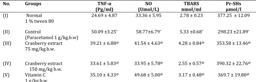
Table 2: Level of plasma tumor necroses factor -α (TNF-α), nitric oxide (NO), lipid peroxides (TBARS) and protein thiols (Pr-SHs) in normal and experimental groups of rats.
No. Groups TNF-α
(Pg/ml) (Umol/L)NO nmol/mlTBARS µmol/l Pr-SHs
(I) Normal
1 % tween 80 24.69 ± 4.87 33.36 ± 5.95 2.78 ± 0.23 377.25 ± 12.09
(II) Control
(Paracetamol 1 g/kg.b.w) 50.09 ±3.25
* 58.77±6.79* 5.33 ±0.68* 298.23 ±21.89*
(III) Cranberry extract
75 mg/kg.b.w. 39.21 ± 6.88
@ 41.54 ± 4.63@ 4.28 ± 0.84@ 353.58 ± 13.46@
(IV) Cranberry extract
150 mg/kg b.w. 33.61 ± 5.83
@ 33.95 ± 5.78@ 2.55 ± 0.57@ 390.32 ± 22.76@
(V) Vitamin C
1 g/kg,b.w 35.10 ± 4.33
@ 49.68 ± 5.00@ 3.17 ± 0.48@ 369.7 ± 19.80@
Paracetamol was given orally as a single dose of 1g/kg.b.w. to 18h fasted animals. It was given to all groups except the normal one. Cranberry extract and vitamin C were orally given daily for 2 weeks and the last dose of each was given 1 h before paracetamol administration. Values are
given as mean ± SD for groups of eight animals each.
* Significantly different from normal group at p< 0.01. @ Significantly different from control group at p< 0.05.
Table 2 showed that oral administration of paracetamol at 1g/kg.b.w. resulted in a significant increase in plasma tumor necroses factor -α (TNF-α), nitric oxide (NO) and lipid peroxides (TBARS) compared to the normal control group (p< 0.01). Supplementation of
administration of paracetamol at 1g/kg.b.w. resulted in a significant decrease in plasma protein thiols (Pr-SHs) compared to the normal control group (p< 0.01). Supplementation of cranberry extract at 75 and 150mg/k.g.b.w. resulted in a significant increase in Pr-SHs compared to the group that received paracetamol (p< 0.05).
Table 3 showed that oral administration of paracetamol at 1g/kg.b.w. resulted in a significant increase in liver lipid peroxides (TBARS) compared to the normal control group (p< 0.01). Supplementation of cranberry extract at 75 and 150mg/k.g.b.w. resulted in a significant decrease in liver TBARS compared to the group that received paracetamol (p< 0.05). Also, oral administration of paracetamol at 1g/kg.b.w. resulted in a significant decrease in liver protein thiols (Pr-SHs) compared to the normal control group (p< 0.01). Supplementation of cranberry extract at 75 and 150mg/k.g.b.w. resulted in a significant increase in Pr-SHs compared to the group that received paracetamol (p< 0.05). The effect of cranberry 150mg/kg. is more pronounced than vitamin C (p< 0.05).
Table 4 showed that oral administration of paracetamol at 1g/kg.b.w. resulted in a significant decrease in blood reduced glutathione (GSH) and activities of superoxide dismutase (SOD) and catalase (CAT) compared to the normal control group (p< 0.01). Supplementation of cranberry extract at 75 and 150mg/k.g.b.w. resulted in a significant increase in blood GSH, SOD and CAT compared to the group that received paracetamol (p< 0.05). The effect of cranberry 150mg/kg. is more pronounced than vitamin C (p< 0.05).
Table 5 showed that oral administration of paracetamol at 1g/kg.b.w. resulted in a significant decrease in liver reduced glutathione (GSH) and activities of superoxide dismutase (SOD) and catalase (CAT) compared to the normal control group (p< 0.01). Supplementation of cranberry extract at 75 and 150mg/k.g.b.w. resulted in a significant increase in liver GSH, SOD and CAT compared to the group that received paracetamol (p< 0.05). The effect of cranberry 150mg/kg. is more pronounced than vitamin C (p< 0.05).
Table 3: level of liver adenosine triphosphate (ATP), lipid peroxides (TBARS) and protein thiols (Pr-SHs) in normal and experimental groups of rats.
No. Groups TBARS
(nmol/g protein) (nmol/ mg protein) Pr-SHs
(I) Normal
1 % tween 80 0.69 ± 0.22 111.59 ± 13.25 (II) Control
(Paracetamol 1 g/kg.b.w)
0.86 ±0.09* 53.63 ±6.47*
(III) Cranberry extract
75 mg/kg.b.w. 0.67 ± 0.15
@ 93.12 ± 10.94@
(IV) Cranberry extract
150 mg/kg b.w. 0.5 ± 0.32
@ 112.84 ± 12.87@
(V) Vitamin C (1 g/kg,b.w) 0.62 ± 0.11@ 107.02 ± 10.96@
Paracetamol was given orally as a single dose of 1g/kg.b.w. to 18h fasted animals. It was given to all groups except the normal one. Cranberry extract and vitamin C were orally given daily for 2 weeks and the last dose of each was given 1 h before paracetamol administration. Values are
given as mean ± SD for groups of eight animals each.
* Significantly different from normal group at p< 0.01. @ Significantly different from control group at p< 0.05.
Table 4: Level of blood reduced glutathione (GSH) and activities of superoxide dismutase (SOD) and catalase (CAT) in normal and experimental groups of rats.
No. Groups GSH
(mg %) (U/mL) SOD (U/mL)CAT
(I) Normal
1 % tween 80 40.71 ± 3.46 214.74 ± 11.25 74.18 ± 6.57 (II) Control
(Paracetamol 1 g/kg.b.w) 19.28 ± 5.15* 147.58 ±17.60* 32.88 ± 4.77* (III) Cranberry extract
75 mg/kg.b.w. 29.37 ± 4.38
@ 174.14 ± 13.45@ 41.73 ± 3.85@
(IV) Cranberry extract
150 mg/kg b.w. 38.74 ± 5.00
@ 206.90 ± 16.54@ 72.75 ± 5.98@
(V) Vitamin C
1 g/kg,b.w 35.66 ± 3.86
@ 178.04 ± 13.90@ 66.27 ± 5.70@
Paracetamol was given orally as a single dose of 1g/kg.b.w. to 18 h fasted animals. It was given to all groups except the normal one. Cranberry extract and vitamin C were orally given daily for 2 weeks and the last dose of each was given 1h before Paracetamol administration. Blood samples were collected 24h after paracetamol administration. Activity is expressed as: 50% of inhibition of pyrogallol autooxidation per min for
SOD. Values are given as mean ± SD for groups of eight animals each.
Table 5: Level of liver reduced glutathione (GSH) and activities of superoxide dismutase (SOD) and catalase (CAT) in normal and experimental groups of rats.
No. Groups GSH
(mg/g tissue) (U/mg protein) SOD (U/mg protein)CAT
(I) Normal
1 % tween 80 2.49 ± 6.10 4.67 ± 3.57 17.52 ± 11.69 (II) Control
(Paracetamol 1 g/kg.b.w) 1.15 ± 4.40* 3.18 ±2.71* 6.63 ± 9.06* (III) Cranberry extract
75 mg/kg.b.w. 2.39 ± 6.08
@ 3.68 ± 4.00@ 10.84 ± 13.42@
(IV) Cranberry extract
150 mg/kg b.w. 2.94 ± 5.10
@ 4.51 ± 3.84@ 15.63 ± 8.43@
(V) Vitamin C
1 g/kg,b.w 2.64 ± 2.94
@ 4.21 ± 2.11@ 13.83 ± 12.72@
Paracetamol was given orally as a single dose of 1g/kg.b.w. to 18 h fasted animals. It was given to all groups except the normal one. Cranberry extract and vitamin C were orally given daily for 2 weeks and the last dose of each was given 1h before Paracetamol administration. Blood samples were collected 24h after paracetamol administration. Activity is expressed as: 50% of inhibition of pyrogallol autooxidation per min for
SOD and the obtained values were divided by the protein concentration. Values are given as mean ± SD for groups of eight animals each. * *Significantly different from normal group at p< 0.01. @ Significantly different from control group at p< 0.05
3.1 Histopathology of the liver
Histopathological examination of the liver sections from normal rats showed normal parenchymal architecture; no significant lesions were observed (Fig. 1a). In the rats treated with paracetamol alone, cloudy swelling, fatty degeneration, hepatocellular necrosis,
heavy haemorrhage and irregular appearance due to cell death were seen (Fig. 1b). The above changes were reduced in the liver of rats treated with cranberry extract (75 and 150mg/kg) + paracetamol together (Fig. 1c&d). The histological pattern was almost normal in rats treated with vitamin C + paracetamol (Fig. 1e).
Figure1: Representative photographs from the liver showing the protective effect of Cranberry extract on paracetamol-induced hepatic injury in rats.
(A), Control rat liver. Normal hepatic parenchyma; (B), Paracetamol-treated rat liver showing cloudy swelling, fatty degeneration of hepatocytes, with necrosis, heavy haemorrhage, irregular appearance and damaged central vein; (C and D)
cranberry extract (75 and 150mg/kg) + paracetamol-treated rat liver. Normal appearance of hepatocytes with mild sinusoidal dilation; (E) Vitamin C (1g/ kg) + paracetamol treated rat liver showing near-normal appearance of hepatocyte saround the
central vein.
Paracetamol (4'-Hydroxyacetanilide) is oral analgesic and antipyretic drug [32]. It is metabolized extensively by the liver via three main pathways; sulfonation, glucuronidation and oxidation [33]. The first two pathways are quantitatively more important than the last, but the oxidative pathway is the culprit as far as toxicity is concerned [34]. Oxidation of paracetamol occurs in the hepatic microsomes and is primarily catalyzed by cytochrome P-450 [35]. The process produces a highly reactive arylating compound called N-acetyl-p-benzoquinoneimine (NAPQI) [36]. In human
liver microsome P-4501A2, were shown to be principal catalysts of paracetamol activation [37].When more NAPQI if formed than can be conjugated to GSH, the unbound NAPQI becomes toxic by binding to macromolecules, including cellular proteins [37].
In the present study administration of paracetamol treated rats showed an increase in the activities of AST, ALT, ALP, LDH and TBARs levels, however there was a decrease of Pr-SHs levels besides GSH, SOD and GPx activities when compared with control rats. Oral administration of etanolic and aqueous extracts of
cranberry extract (75 and 150mg/kg body weight) and vitamin C to paracetamol treated rats showed an inhibition in activities of plasma AST, ALT, ALP and LDH as well as reduction of Pr-SHs, GSH, SOD and CAT levels than paracetamol alone treated rats. Mitra et al., [40] have reported that administration of paracetamol caused significantly increased plasma AST and ALT activities.
Since cranberry has shown antioxidant and free radical scavenging activity [41], the present study primarily ameliorating the effect of cranberry’ polyphenols on Diclofenac induced liver toxicity in rat is studied. Oral administration of cranberry extract significantly inverse the Diclofenac sodium induced peroxidative damage in liver which is evidenced from the lowered levels of thiobarbituric acid reactive substances and lipid hydroperoxides.
This may be due to the antioxidative effect of polyphenols [42]. An antioxidant is a molecule capable of slowing or preventing the oxidation of other molecules. Oxidation is a chemical reaction that transfers electrons from a substance to an oxidizing agent. paracetamol induces cellular injury and functional abnormalities in hepatocytes by the process of lipid peroxidation [7]. Because the liver has a central role in the maintenance of lipid homeostasis, excess iron may alter the concentration of serum lipids, which could reduce or increase the risk of atherosclerosis. The preliminary studies conducted by this work revealed the non-toxic nature of cranberry extract on normal rats. Hepatic necrosis following massive paracetamol administration is well documented [40]. Drastic elevation in the activity of liver ALT, AST, ALP and LDH were shown in the current study after administration of paracetamol (1g/kg.b.w) due to the intracellular accumulation of Ca2+, which results in activation of phosphofructokinase and anaerobic glycolysis leading to lactate formation [43]. Loss of Ca2+ homeostasis as a result of oxidative damage and increase in intracellular Ca2+ has been reported to be late and perhaps irreversible final stage in the process of cell death for paracetamol [44]. Cranberry extract
administration controlled plasma and hepatic LDH activities.
In the present study, plasma and hepatic TBARS levels were significantly lower in the cranberry extract– treated groups compared to the paracetamol treated group. The above result suggests that the cranberry exrtact may exert antioxidant activities and protect the tissues from lipid peroxidation. The protective effect due to treatment with cranberry extract strongly indicated the possibility of the extracts being able to prevent and/or mitigate any leakages of marker
enzymes into circulation, conditions the hepatocytes to accelerate regeneration of parenchymal cells, and preserve the integrity of the plasma membranes and hence restores these enzymes activities [45].
Plasma and hepatic protein thiols (Pr-SHs) contents were markedly decreased after paracetamol administration, as shown in the current investigation. The loss of Pr-SHs is held to be a critical event in the genesis of lethal injury by an acute oxidative stress [46]. Such depletion is presumed to be a direct oxidation of the thiol groups of contiguous amino acids with the formation of protein-protein disulphides [47]. The cytotoxic effects of paracetamol have been attributed to depletion of Pr-SHs level [48]. In addition, the metabolism of paracetamol also generates GSSG, a product that reacts with Pr-SHs to form GSH mixed disulphides [49]. In the present study, the elevation of Pr-SHs levels in plasma and liver was observed in the
cranberry extract-treated rats. This indicates that the
cranberry extract can either increase the biosynthesis of Pr-SHs or reduce the oxidative stress.
GSH has a multifactorial role in antioxidant defense. It is a direct scavenger of free radicals as well as a co-substrate for peroxide detoxification by glutathione peroxidases [50]. Liu et al., [51], suggested that the decrease in blood and liver GSH level could be the result of decreased synthesis or increased degradation of GSH by oxidative stress and tissue injury. Increased oxidative stress, resulting from significant increase in aldehydic products of lipid peroxidation has probably decreased hepatic GSH content. In the present study, the elevation of GSH levels in blood and liver was observed in the Cranberry extract - treated rats. This indicates that the Cranberry extract can either increase the biosynthesis of GSH or reduce the oxidative stress leading to less degradation of GSH, or have both effects.
SOD has been postulated as one of the most important enzymes in the enzymatic antioxidant defense system which catalyses the dismutation of superoxide radicals to produce H2O2 and molecular oxygen [52], hence diminishing the toxic effects caused by their radical. The observed decrease in SOD activity could result from inactivation by H2O2 or by glycation of enzymes [53]. The superoxide anion has been known to inactivate CAT, which involved in the detoxification of hydrogen peroxide [54]. Thus, the increase in SOD activity may indirectly play an important role in the activity of catalase.
GPx plays a primary role in minimizing oxidative damage. Glutathione peroxidase (GPx), an enzyme with selenium and Glutathione-s-transferase (GST) works together with glutathione in the decomposition of H2O2 or other organic hydroperoxides to non-toxic products at the expense of reduced glutathione [55].
Several authors reported the decrease in SOD and GPx activities in paracetamol-treated animals [56& 57].
The results indicates that the cranberry extract can either increase the biosynthesis of SOD and GPx or reduce the oxidative stress leading to less degradation of SOD and GPx, or have both effects.
The histopathological study of liver tissues of animals is related to their function. The paracetamol hepatotoxicity presents as centrilobular necrosis [58]. The histopathological studies support the biochemical findings. The paracetamol treated rats showed fatty changes, necrosis, vacuoles, space formation and loss of cell boundaries in liver. Oral administration of
cranberry extract (75 and 150 mg/kg body weight) and vitamin C to paracetamol treated rats brought back the above-mentioned changes in liver to near normal Coen
et al., [59] reported that marked changes in liver such as vacuolated hepatocytes, necrosis and congested sinusoids in paracetamol treated rats. Oliveira et al., [60] have reported that administration of α- and β-amyrin to paracetamol treated rats liver showed normal histoarchitecture.
The preliminary phytochemical screening of cranberry extract revealed the presence of flavonoids. Flavonoids (or bioflavonoids) are natural products, they are capable of modulating the activity of enzymes (SOD and GPx) and affecting the behavior of many cell systems and possess a significant antihepatotoxic, antiallergic, anti-inflammatory, antiosteoporotic, and even antitumor and antioxidant activities [61 and 62].
In conclusion, the results of this study demonstrated that Cranberry extract possesses a potent hepatoprotective action upon paracetamol-induced hepatic damage in rats. This may be due to its antioxidative activity with its ability to scavenge free radicals and inhibit lipid peroxidation, all of which are capable of hepatocellular injury.
5. REFERENCES
1. Liaqat H, Javaria I, Kanwal R, Muhammad T, Muhammad I and Muhammad S. (2014). Hepatoprotective effects of Malva sylvestris L. against paracetamol-induced hepatotoxicity. Turk J Biol (2014) 38: 396-402.
2. Litovitz TL, Klein-Schwartz W, Rodgers GC Jr, Cobaugh DJ, Youniss J, Omslaer JC, May ME, Woolf AD, and Benson BE (2002). Annual report of the American Association of Poison Control Centers Toxic Exposure Surveillance System. Am J Emerg Med 20:391–452.
3. Prescott LF (1980). Hepatotoxicity of mild analgesics Br J Clin Pharmacol 10 (Suppl 2):373S–379S.
4. Gujral JS, Knight TR, Farhood A, Bajt ML, and Jaeschke H (2002). Mode of cell death after acetaminophen overdose in mice: apoptosis or oncotic necrosis? Toxicol Sci 67:322–328. 5. McGill MR, Sharpe MR, Williams CD, Taha M, Curry SC,
Jaeschke H (2012). The mechanism underlying acetaminophen-induced hepatotoxicity in humans and mice involves mitochondrial damage and nuclear DNA fragmentation. J Clin Invest 122: 1574–1583.
6. Hussein, MA. (2008). Antidiabetic and antioxidant activity of Jasonia montana extract in Streptozotocin – induced diabetic rats. JSP., 16:214-221.
7. Hussein, MA. (2010). Purslane Extract Effects on
Obesity-Induced Diabetic Rats Fed a High-Fat Diet. Mal J Nut., 3:419-429.
8. Hussein, MA. and Abdelgwad SM. (2010). In vivo Hepato-protective Properties of Purslane Extracts on Paracetamol-Induced Liver Damage. Mal J Nutr., 1: 161–170.
9. Hussein, MA, Abdel-Khalek NG. and El Bishbishy, MH. (2014). Composition, in vitro antioxidant and antitumor properties of essential oil from the seeds of Moringa oleifera. International Journal of Pharma Sciences, 3: 532-540. 10.Boshra, S.A. and Hussein, M.A. (2014). The protective role of
Colchicine on Diclofenac sodium induced hepato-renal toxicity in albino rats model. International Journal of Pharmaceutical Sciences and Research. 12: 5136-5144. 11.Abdel-Maksoud, H.A., Hussein MA and Barakat M (2015).
Cranberry extract as a functional food in treatment of myocardial toxicity induced by nicotine in Rats. 5; 4: 1174-1180
12.Aherne, S.A. and O’Brien, M. (2002). Dietary flavonols: Chemistry, food content, and metabolism. Nutr., 18:75–81 13.Mullen W., Marks, SC., Crozier, A. (2007). Evaluation of
phenolic compounds in commercial fruit juices and fruit drinks. J Agric Food Chem.; 55:3148–57.
14.Vvedenskaya, IO., Rosen, RT. And Guido, JE., (2004). Characterization of flavonols in cranberry (Vaccinium macrocarpon) powder. J Agric Food Chem., 52:188–95. 15.Manach, C., Scalbert, A. and Morand, C., (2004). Polyphenols:
Food sources and bioavailability. Am J ClinNutr., 79:727–47. 16.Luo Z, Harada T, London S, Gajdusek C, Mayberg M.
Antioxidant and iron chelating agents in cerebral vasospasm. Neurosurgery, 1995; 37: 1054.
17.Reitman S, Frankel S. A colorimetric method for the determination of plasma oxaloacetic acid and glutamic pyruvic transaminases. Am. j. Clin. Pathol., 1957; 28: 56 – 63. 18.Kind PRN, King EJ. Estimation of plasma phosphatase by
determination of hydrolysed phenol with amino-antipyrine. J. Clin. Pathol 1954; 7: 322 – 326.
19.Buhl SN, Jackson KY. Optimal conditions and comparison of lactate dehydrogenase catalysis of the lactate to pyruvate to lactate reactions in human plasma at 25, 30 and 37 0C. Clin. Chem. 1978; 2415: 828.
20.Uchiyama M, Mihara M. Determination of malondialdehyde precursor in tissues by thiobarbituric acid test. Anal. Biochem., 1978; 86:271.
21.Koster JF, Biemond P, Swaak AJ. Intracellular and extracellular sulphydryl levels in rheumatoid arthritis. Ann. Rheum. Dis., 1986; 45: 44.
22.Chanarin I . Text book of Laboratory Haematology: An Account of Laboratory techniques, Churchill Livingstone, New York PP. 1989; 107.
23.Beyaert, R. and Fiers, W. (1998). Tumor Necrosis Factor and Lymphotoxin. In Cytokines, A.R.M.-S. a. R. Thorpe, eds. Academic Press, San Diego, p. 335-360.
24.Miranda, K.M.; Espey, M.G.; Wink. D.A. (2001). A rapid, simple spectrophotometric method for simultaneous detection of nitrate and nitrite. Nitric Oxide. 5: 62-71.
25.Sinha, AK. (1972). Colorimetric assay of catalase. J. Anal Biochem.47 (2): 389-94.
26.Marklund S, Marklund D. Involvement of the superoxide anion radical in the autoxidation of pyrogallol and a convenient assay for superoxide dismutase. Eur. J. Biochem., 1974; 47:469.
27.Sedlak J, Lindsay RH. Estimation of total, protein bound and non-protein sulfhydryl groups in tissue with Ellman’s reagent. Anal Biochem., 1968; 25: 192.
28.Van Kampen, E, Zijlstra, W. Standardization of hemoglobinometry.II. The hemiglobin-cyanide method. Clin. Chim. Acta, 1961; 6:538.
29.Lowry OH, Rosenbrough NJ, Farr AL, Randall RJ. Protein measurement with folin-phenol reagent. J Biol Chem., 1951; 193:265.
30.Bancroft GD, Steven A: Theory and Practice of Histological Technique, 4th ed. Churchill Livingstone, New York, 1983, pp. 99–112.
31.Snedecor and Cochran (1969): Statistical methode 6th ed. The Iowa State Univ., Press, Iowa, USA.
Pharmacol. Ther., 1974; 16:676.
33.Jollow DJ, Mitchell JR, Potter WZ, Davis DC, Gillette JR, Brodie BB. Acetaminphen-induced hepatic necrosis. II.Role of covalent binding in vivo. J. Pharmacol Exp. Ther., 1973; 187: 195.
34.Potter WZ, Davis DC, Mitchell JR, Jollow GJ, Gillette JR, Brodie BB. Aetaminphen-induced hepatic necrosis. III. Cytochrome p450-mediated covalent binding in vitro. J. Pharmacol. Exp. Ther., 1973; 187: 203.
35.Dahlin DC, Miwa GT, Lu AY, Nelson SD. N-acetyl-p-benzoquinoneimine: a cytochrome p-450 mediated oxidation product of acetaminophen. Proc. Natl. Acad. Sci. USA., 1984; 81:1327.
36.Raucy JL, Lasker HM, Lieber CS, Black M. Acetaminophen activation by human liver cytochromes p-450 II EI and p-450 IA2. Arch. Biochem. Biophys., 1989; 27: 270.
37.Vermeulen NPE, Bassems JGM, Van de Straat R. Molecular aspects of paracetamol-induced hepatotoxicity and its mechanism based prevention. Drug Metab. Rev., 1992; 24: 367.
38.Zayova E, Stancheva I, Geneva M, Petrova M, Dimitrova L. Antioxidant activity of in vitro propagated Stevia rebaudiana
Bertoni plants of different origins. Turk J Biol 2013, 37: 106– 113.
39.Zia-Ul-Haq M, Ahmad S, Qayum M, Ercişli S. Compositional studies and antioxidant potential of Albizia lebbeck (L.) Benth. pods and seeds. Turk J Biol 2013, 37: 25–32.
40.Mitra SK, Venkataranganna MV, Sundaram R, Gopumadhavan S, J Ethnopharmacol 1998; 63: 181-186.
41.Sies H. (1997). Oxidative stress, oxidants and antioxidants, Exp. Physiol. 82: 291–295.
42.Veerappan, R.M.; Senthil, S.; Rao, M. andRavikumar, M. (2004). Redox status and lipid peroxidation in alcoholic hypertensive patients and alcoholic hypertensive patients with diabetes, Clin. Chem. Acta 340: 207–212.
43.Kumar G, Sharmila Banu G, Kannan V, Rajasekara Pandian M, Ind J Exp Biol 2005; 43: 351-355.
44.Waters E, Wang JH, Redmond HP, Wu QD, Kay E, Bouchier Hayes D. Role of taurine in preventing acetaminophen-induced hepatic injury in the rat. Am. J. Physiol. Gastrointest. Liver Phsiol., 2001; 280: G1274.
45.Landowne D, Ritchie J. On the control of glycogenolysis in mammalian nervous tissue by calcium. J. Physiol., 1971; 212:503.
46.Strubelt O, Younes M. The toxicological relevance of paracetamol-induced inhibition of hepatic respiration and ATP depletion. Biochem. Pharmacol., 1992; 44: 163. 47.Al-Howiriny T, Al-Sohaibani M, Al-Said M, Al-Yahya K,
ElTahir S, Rafatullah. Hepatoprotective properties of
Commiphora opobalsamum (“Balessan”), a traditional medicinal plant of Saudi Arabia. Drug Exptl. Clin. Res. 2004; 30: 213.
48.Di Monte D, Ross D, Bellomo G, Eklow L, Orrenius S.
Alterations in intracellular thiol homeostasis during the metabolism of menadione by isolated rat hepatocytes, Arch. Biochem. Biophys., 1984; 235:334.
49.Kyle ME, Sakaida I, Serroni A, Farber JL. Metabolism of acetaminophen by cultured rat hepatocytes. Depletion of protein thiol groups without any loss of viability. Biochem. Pharmacol., 1990; 40:1211.
50.Wohaieb S, Godin DV. Alterations in free radical tissue - defense mechanisms in streptozotocin diabetes in rats: Effect of insulin treatment. Diabetes, 1987; 36: 1014.
51.Liu PT, Ioannides C, Symons AM, Parke DV. Role of tissue glutathione in prevention of surgical trauma. Xenobiotica, 1993; 23: 899.
52.Mc Crod JM, Keele BB, Fridovich I. An enzyme based theory of obigate anaerobiosis, the physiological functions of superoxide dismutase. Pro Nati Acad Sci. USA, 1976; 68: 1024.
53.Sozmen BY, Sozmen B, Delen Y, Onat T. Catalase/superoxide dismutase (SOD) and catalase/paraoxonase (PON) ratios may implicate poor glycemic control. Ara Med Res., 2001; 32: 283.
54.Chance B, Green Stein DS, Roughton RJ.The mechanism of catalase action 1-steady state analysis. Arch Biochem Biophys, 1952; 37: 301.
55.Bruce A, Freeman D, James C. Biology of disease. Free radicals and tissue injury. Lab Invest, 1982; 47: 412. 56.O'Brien P, Slaughter M, Swain A, Birmingham J, Greenhill R,
Elcock F, Bugelski P. Repeated acetaminophen dosing in rats: adaptation of hepatic antioxidant system. Hum. Exp. Toxicol., 2000; 19:277.
57.Ahmed MB, Khater MR. Evaluation of the protective potential of ambrosia maritime extract on acetaminophen-induced liver damage, J. Ethnopharmacol., 2001; 75: 169.
58.Curry R, Robinson JD, Sughrue MJ. JAMA 1982; 247:1012-1014.
59.Coen M, Ruepp SU, Lindon CJ, Nicholson JK, Pognan F, Lenz, EM, Wilson ID. J Pharm Biomed Anal 2004; 35:93-105. 60.Oliveira FA, Chaves MH, Almeida FRC, Lima JC, et al., J
Ethnopharmacol 2005; 98: 103-108.
61.Carlo DG, Mascolo N, Izzo AA, Capasso F. Flavonoids: Old and new aspects of a class of natural therapeutic drugs. Life Sci., 1991; 65: 337.
62.Rathee JS, Hassarajani SA, Chattopadhyay S. Antioxidant activity of Mammea longifolia bud extract. Food Chem., 2006; 99: 436.
*****
© 2016; AIZEON Publishers; All Rights Reserved