Himanshu et al. World Journal of Pharmaceutical and Life Sciences
DIFFERENT METHODS USED TO ACCESS THE DENTAL IMPLANT STABILITY - A
REVIEW
KAPIL ARORA1, NITISHA SHARMA2, RAHUL KUKREJA3, BHAWNA YADAV4, RAJRATNA SONONE5,
HIMANSHU THUKRAL6, VINITA DAHIYA7
1MDS, Periodontist & Oral Implantologist, Mumbai 2
BDS, Mumbai
3,4MDS, Oral Medicine and Radiology, Ghaziabad 5Oral & Maxillofacial Surgeon, Pune
6Oral & Maxillofacial Surgeon, CEO Sarita dental Care, Delhi, India 7
Post Graduate Scholar, I.T.S Dental College, Ghaziabad.
Article Received on 13/06/2017 Article Revised on 04/07/2017 Article Accepted on 02/08/2017
INTRODUCTION
The use of dental implants in the rehabilitation of partially and completely edentulous patients has been significantly increased in dentistry since 1980.[1] Although high survival rates of implants supporting prosthesis have been reported,[2-4] failure still happens due to bone loss as a results of primary and secondary implant stability. Primary stability of an implant is the absence of mobility in the bone bed upon insertion of the implant and mostly comes from mechanical interaction with cortical bone. It is also named as ―Mechanical Stability‖ which is the result of compressed bone holding the implant tightly in the bone. Secondary stability, named as ―Biological Stability‖, happens through bone regeneration and remodelling at the implant/bone interface.[5-6] It is the result of new bone cells forming at the site of the implant and osseointegration. The primary stability is the requirement for successful secondary stability.[6] Secondary stability orders the time of functional loading.[7] Following the placement of an endosseous implant, primary mechanical stability gradually decreases and secondary stability (biologic) gradually increases.
Bone quantity and quality, surgical techniques including the skill of the surgeon, implant (geometry, length, diameter, and surface characteristics) are major factors affecting primary stability.[8] Primary stability, bone modelling and remodelling, and implant surface conditions are the main parameters influencing secondary stability.[8]
Osseointegration is an important factor in specifying a series of criteria that identifies success or failure of an implant. Osseointegration is, however, a patient-dependent wound healing process that happens at two different stages: primary stability and secondary stability.
Dental implant stability measurement, an indirect indication of osseointegration, is a measurement of implant’s resistance to movement.[9] Objective measurement of implant stability is a valuable tool for achieving consistently good results first and foremost because implant stability plays such an important role in achieving a successful outcome. The advantages of measuring implant stability are to make more accurate decisions about the time of crown loading or unloading, select the protocol of choice for implant loading, and
World Journal of Pharmaceutical and Life Sciences
WJPLS
www.wjpls.org SJIF Impact Factor: 4.223
*Corresponding Author: Dr. Himanshu Thukral
Oral and Maxillofacial Surgeon, CEO Sarita Dental Care, Delhi, India.
ABSRACT
Achieving primary stability of greatest importance, at the time of dental implant placement. A rigid fixation of implant within the host bone, in absence of micromotion is the most critical factor for successful osseointegration. Various methods are reported by the authors over the years, in literature to monitor implant stability, which include, tapping the abutment with a metallic instrument, histomorphometry test, removal torque test, cutting torque test, radiography, periotest, and resonance frequency analysis. Resonance frequency analysis (RFA) is most recent advancement in dentistry which offers a clinical, objective way to measure stability and presumed osseointegration of implants. The review focuses on different methods used to assess implant stability and recent advances in this field implant dentistry.
increase trust between patient and practitioner. It is, therefore important to be able to quantify implant stability at various times and have in place a long-term prognosis based on implant stability measurement tool. Although various diagnosis analyses have been employed and several research and development projects have been already made in this field, measuring implant stability remains a challenge in dentistry.
Available Methods Currently used to Assess Implant Stability
The methods for studying stability can be categorized as invasive, which interfere with the osseointegration process of the implant, and non-invasive, which do not. Some of the most famous methods in analyzing dental implant stability are histologic analysis, percussion test, radiographs, reverse torque, cutting resistance, and resonance frequency analysis (RFA). Since histologic analysis is not feasible for daily practice it is not discussed in this artical.
A) Radiographic analysis
Radiographic analysis was one of the first methods applied to evaluate the condition of implants after they had been placed. Radiographic evaluation is a non-invasive method that can be performed at any stage of healing process. Bitewing radiographs are used to measure crestal bone level, defined as the distance from the top of the implant to the position of the bone on the implant surface, because it has been suggested as an indicator for implant success.[10] However, other studies recommended that the resolution of bitewing radiographs cannot be used as the only tool to evaluate either primary or secondary stability.[8] Moreover, crestal bone changes can be only reliably measured if there is no distortion in the radiographic pictures. In those pictures, the distortion happens when the central x-ray tube is not positioned parallel to the implant. Furthermore, panoramic view (a dental X-ray scanning of the upper and lower jaw that shows a two-dimensional view of a half circle from ear to ear) does not provide information on a facial bone level, and bone loss. Finally, regular radiographs cannot be used to quantify neither bone quality nor density. They can be used to assess changes in bone mineral only when there are decreases that exceed 40% of the initial mineralization.[11] Moreover, because of X-radiation hazards other methods with fewer side effects are preferred.
B) Cutting Torque Resistance Analysis (CRA) This method was originally developed in 1994 by Johansson and Strid[12] and later improved in vitro and
in vivo human models. In this method the energy required in cutting off a unit volume of bone during implant surgery is measured. This energy has been shown to be significantly correlated with bone density, which has been suggested as one factor that significantly influences implant stability.[13] The advantages of this method are detecting bone density and its quality during surgery. The major limitation of CRA is that it does not
give any information on bone quality until the osteotomy site (a surgical operation for bone shortening or realignment) is prepared. In addition, this information cannot be used to assess bone quality changes after implant insertion.
C) Reverse Torque Test (RTT)
The Reverse Torque Test (RTT), which is proposed in 1984 by Roberts et al.,[14] measures the critical torque threshold when bone-implant contact is broken. This indirectly provides information on the degree of bone-implant contact in a given bone-implant. Removal Torque Value (RTV) as an indirect measurement of bone-implant contact was reported to range from 45 to 48 N.cm.[15] The disadvantage of this method is the risk of irreparable plastic deformation within implant bone integration and the implant failure when unnecessary load is applied to an implant that is still undergoing osseointegration. In addition, applying torque on implants placed in bone of low quality may result in a shearing of bone-to-implant contact and cause implants to irretrievably fail.
D) Insertion torque analysis
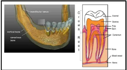
Insertion torque analysis, as an invasive method, expresses the amount of force that is applied to the implant as it is inserted. Implant placement insertion torque is initially minimal, and increases quickly until the cortical layer (see Figure 1) in a jawbone is fully engaged. As the implant is driven into the bone, repeated measurements are taken and a graph is often produced. The maximum value is obtained when the head of the screw makes contact with the cortical plate (the hard, outer shell of alveolar). Insertion torque measurement includes finding the maximum insertion torque value when the screw head contacts the cortical plate. This test has been generally well accepted and has been used for evaluating various implant designs.[16] Insertion torque has been found to correlate with bone density and consequently implant stability.[17] The application of insertion torque has been shown to be limited since estimating the quality of the bone is impossible until the implant insertion is actually started. So, insertion torque measurements cannot be used for the selection of implant sites. This method also cannot be used to follow implant healing and osseointegration procedures.
E) Percussion test
Percussion test is the simplest method for testing implant stability. This test is based upon vibrational acoustic science and impact-response theory. In this method, clinical judgment about osseointegration is made based on the sound heard from the percussion of the implant with a metallic instrument. A ―crystal‖ sound indicates successful osseointegration, while a ―dull‖ sound means weak or failing osseointegration. This method heavily relies on the clinician’s experience level and subjective belief. Therefore, it cannot be used experimentally as a standard testing method.
F) Pulsed oscillation waveform
Kaneko et al.,[18-19] used a Pulsed Oscillation Wave Form (POWF) to analyze the mechanical vibrational characteristics of the implant-bone interface using forced excitation of a steady-state wave.
Pulsed oscillation waveform works based on the frequency and amplitude of the implant vibration induced by a small-pulsed force. This system consists of an acoustoelectric driver, an acoustoelectric receiver, a pulse generator and an oscilloscope. Both the acoustoelectric driver and the acoustoelectric receiver consist of a piezoelectric element and a puncture needle. A multifrequency pulsed force of about 1 KHz is applied to the implant by lightly touching it with two fine needles connected to piezoelectric elements. Resonance and vibration generated from the bone-implant interface of an excited implant are picked up and displayed on an oscilloscope. The sensitivity of this method depended on load directions and positions. The sensitivity of this method is low for the assessment of implant rigidity.[18]
G) Impact hammer method
Impact hammer is an example of transient force as a source of excitement. This method is an improved version of the percussion test except that sound generated from contact between hammer and object is processed through fast Fourier transform (FFT).
The DMC was originally developed by Aoki and Hirakawa.[20,21] It detected the level of tooth mobility by converting the integration of teeth and alveolar bone into acoustic signals. Contac time between the tapping impact hammer and the object is measured. In this theory, the width of the first peak on the time axis of the spectrum generated by transient impulse is inversely proportional to the time axis of the impulse.[22] The lower rigidity of implant-bone integration results in a longer time response. In this device, a microphone is used as a receiver. The response signal of the microphone is processed in the time domain. Some problems were addressed to this method such as difficulties of double tapping and difficulty in obtaining constant excitation.
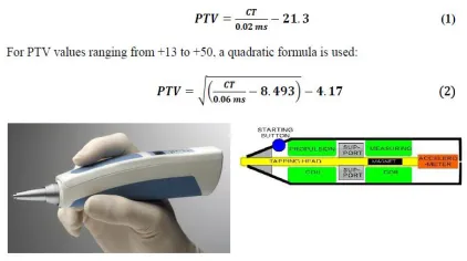
Unlike the DMC, which applies impact force with a hammer, Periotest uses an electromagnetically driven and electrically controlled metallic tapping rod in a
headpiece (see Figure 2). Response to striking is then measured by a small accelerometer incorporated into the head of the device. Similar to DMC, the contact time (CT) between the test object and tapping rod is measured in time domain and then converted to a value called PTV, which is related to the damping characteristics of tissue surrounding teeth or implant. For PTV units in the range of -8 to +13, a linear formula is used:
Figure 2: Periotest device.
The lack of sensitivity is reported as one of the shortcomings of this device.[23] This is because PTV has a very wide dynamic range (PTV is -8 to +50) of possible responses, and the PTV of an osseointegration implant falls only in a relatively narrow zone (-5 to +5) inside the range. Moreover, values measured from Periotest can be affected by excitation conditions such as position and direction. PTV also cannot be used to identify a ―borderline implant‖ which may or may not be considered as a successful osseointegration. Finally, Periotest measurements are limited because they are strongly dependent to the orientation of the excitation source and the striking point. As a result, despite some positive claims for the Periotest, the prognostic accuracy of the Periotest for implant stability has been criticized for a lack of resolution, poor sensitivity and susceptibility to operator variables.[24]
H) Resonance Frequency Analysis (RFA)
In resonance frequency analysis, implants are forced to oscillate and the frequency at which they oscillate at maximum amplitude is registered as their resonance frequency. Similar to all distributed system, an implant can have many resonance frequencies, each called a harmonic. The resonance frequencies are dependent on the material, length and the quality of the supporting mechanism. Since the material and length of the implant are constants, variations of the resonance frequency highly correlate to the quality of the support (osseointegration).
attracted considerable scientific interest in recent years and an increasing number of prominent journal papers are published about it since its first introduction.
The RFA of an implant, as it was briefly mentioned in this section, can be influenced by some factors including implant length, implant diameter, implant geometry, implant surface characteristic and placement position, as well as bone quality, bone quantity, damping effect of marginal mucosa, bone implant contact, effective implant length and connection to transducer.
Currently, there are two commercially available devices used to evaluate the resonance frequency of implants placed into the bone, Implomates (Bio Tech One) and Osstell (Integration Diagnostics). Their main difference is in the way they excite implants.
The Implomates device (Taipei, Taiwan) has been studied extensively by Huang et al.[32,34-37] This device utilizes an impact force to excite an implant. There is a small electrically driven rod inside the device that produces impact force. The received time response signal is then transferred in frequency spectrum for analysis (range 2 to 20 KHz). The first peak in the frequency spectrum (distinguishable from the noise) indicates the primary resonance frequency of implant. Higher frequency for the primary resonance and the sharpness of that peak indicates a more stable implant.
Osstell measures RF by attaching a metal rod to an implant with screw connection and exciting the rod doing a frequency sweep. The rod is excited by a small magnet that is attached to its top that can be stimulated by magnetic pulses from a handled electronic device (see Figure 3). The rod can vibrate in two directions (perpendicular to each other) and thus it has two fundamental resonance frequencies. Implant Stability Quotient (ISQ) is a scale developed by Osstell for implant stability. It converts the resonance frequency values ranging from 3,500 to 8,500 Hz into an ISQ of 0 to 100. A high value indicates greater stability, while a low value indicates instability. Values greater than 65 are recommended as successful implant stability. Even though Osstell is clinically used but there are not much convincing data on the relation between bone-implant interface and ISQ values.[8]
Figure 3: Osstell Device.
I) Finite Element Analysis (FEA)
Finite Element Method (FEM) is a numerical technique, which facilitates the RF analysis by providing an interface where a 3D model of an object and its support can be developed and studied. FEM approximates the real structure with a finite number of elements and assigns mechanical properties of objects such as Young’s Modulus, the Poisson ratio and density. This method can simulate complex geometric shapes, material properties, and generate various boundary conditions of the real situation, which are difficult to produce in the laboratory. FEM simulation method has the advantage of allowing independent control of each parameter in the Finite Element (FE) models.
The first person who used modal analysis, the study of the dynamic properties of structures (will be explained in more details in chapter 2) together with Finite Element Method in analysis of implant stability was Williams & Williams.[39] Since then, FEM has gradually become an important tool in biomedical research. Wang et al.[40]
used FEM for calculating RFA to determine the identifiable stiffness range of interfacial tissue (a thin layer surrounding the implant) of dental implants. They found that when the Young’s modulus of the interfacial tissue is less than 15 MPa, the resonance frequencies are significantly affected by the interfacial tissue and the influence of other parameters such as geometry, boundary constraint, and material property of the bone are negligible. One limitation of finite element modelling is that it is a numerical approach based on many assumptions, which might not necessarily realistic to simulate real cases.
J) Ultrasonic wave propagation
An alternative method to assess implant stability is quantitative ultrasound (QUS), as suggested initially by de Almeida et al.[41] They used the implant as a waveguide and showed a significant correlation between the experimental 1 MHz ultrasonic responses of an aluminum threaded piece and the screwing depth in an aluminum block. They concluded that Ultrasonic waves are sensitive to bone-implant interface properties. In the same study, finite difference numerical simulations depicted an agreement between the 1 MHz ultrasonic response of titanium wave guides and the elastic properties of tissues surrounding the guides. Furthermore, in a recent experimental study by Mathieu et al.,[42] a 10 MHz ultrasonic device was validated with implants placed in rabbit bone. The amount of bone surrounding prototype cylindrical titanium implants was shown to be significantly correlated with a quantitative indicator deduced from the ultrasonic response to a 10 MHz excitation.
CONCULUSION
analysis with fair amount of predictability. The theoretical basis of resonance frequency analysis is based on sound foundation; still there are uncertain issues such as critical value that can suggest success or failure of a particular implant system. Hence, further research is needed to establish higher reliability of the currently discussed methods.
REFERENCE
1. Capek L, Simunek A, Slezak R, Dzan L. Influence of the orientation of the Osstell® transducer during measurement of dental implant stability using resonance frequency analysis: A numerical approach. Med Eng Phys, 2009; 31: 764–9.
2. Poul Holm-Pedersen, Niklaus P.Lang, Frauke Muller. What are the longevities of teeth and oral implants? Clin. Oral Impl. Res, 2007; 18: 15-19. 3. Koller B, Att W, Strub JR. Survival rates of teeth
implants and double crown retained removable dental prostheses systematic literature review. Int J Prosthodont, 2011; 24: 109-17.
4. Chung WE, Rubestien JE, Phillips KM, Raigrodski AJ. Outcomes assessment of patients treated with osseointegrated dental implants at the university Washington Graduate Prosthodontic Program,1988 to 2000. Int J Oral maxillofac Implants, 2009; 24: 927-35.
5. Brunski JB. Biomechanical factors affecting the bone-dental implant interface. Clin Mater, 1992; 10: 153-201.
6. Sennerby L, Roos J.Surgical determinants of clinical success of osseointegrated oral implants: A review of the literature. Int J Prosthodont, 1998; 11: 408-20. 7. Jensen O. (The carter hypothesis. In: Burser D, dahlin C, Schenk RK (eds). Guided bone regeneration in implant dentistry. Hong Kong:Quintessence, 1994; 238: 239.
8. Mihoko Atsumi, Sang-Hoon Park, Hom-Lay Wang. Methods used to assess implant stability status. The International Journal of Oral & Maxillofacial Implants, 2007; 22: 743-754.
9. Meredith N, Shagaldi F, Alleyne D, Sennerby L, Cawley P.The application of resonance frequency measurements to study the stability of titanium implants during healing in the rabbit tibia. Clinical Oral Implants Research, 1997; 8: 234–243.
10. Hermann JS. , Schoolfiled JD, Nummikoski PV, Buser D, Schenk RK, Cichran DL. Crestal bone changes around titanium implants: a methodology study comparing linear radiographic with histometric measurements. Int J Oral Maxillofac Implants, 2001; 16: 475-85.
11. Goodson JM,Haffajee AD, Socransky SS.The relationship between attachment level loss and alveolar bone loss. J Clin Periodontol, 1984; 11: 384-359.
12. Johansson P, Strid K. Assessment of bone quality from cutting resistance during implant surgery. Int J Oral Maxillofac Implants, 1994; 9: 279-288.
13. Friberg B, Sennerby L, Roos J, Johansson O, Strid CG, Lekholm U. Evaluation of bone density using cutting resistance measuremnts and microradiography: An in vitro study in pig ribs. Clin Oral Implants Res, 1995; 6: 164-71.
14. Roberts WE, Smith RK, Zilberman Y, Mozsary PG, Smith RS.Osseous adaption to continuous loading of rigid endosseous implants. Am J Orthod, 1984; 86: 95-111.
15. Sullivan DY, Sherwood RL, Collins TA, Krogh PH. (1996) The reverse torque test: A clinical report. Int J Oral Maxillofac Implants, 1996; 11: 179-85. 16. Lim S, Cha J, Hwang C. Insertion Torque of
Orthodontic Miniscrews According to Changes in Shape, Diameter and Length. Angle Orthod, 2008; 78: 234-40.
17. Turkyilmaz I, Tumer C, Ozbek E, Tozum T. (2007) Relations between the bone density values from computerized tomography, and implant stability parameters: a clinical study of 230 regular platform implants. J Clin Periodontol, 2007; 34: 716-22. 18. Kaneko T. (1991) Pulsed oscillation technique for
assessing the mechanical state of the dental implant-bone interface. Bio-materials, 1991; 12: 555-60. 19. Kaneko T, Nagai Y, Ogino M, Futami T, Ichimura
T. Acoustoelastic technique for assessing the mechanical state of the dental implant-bone interface. J Biomed Mater Res, 1986; 20: 169-76. 20. Aoki H. The mobility of healthy teeth as measured
with the impact hammer method. Kanagawa Shigaku, 1987; 22: 13-31.
21. Hirakawa W. An attempt to measure tooth mobility in terms of time domain wave forms. Kanagawa Shigaku, 1986; 21: 529-43.
22. Matsuo E, Hirakawa K, Hamada S.Tooth Mobility measuremet technique using ECM impact hammer method. Bull Kanagawa Dent Coll, 1989; 17: 9-19. 23. Manz M, Morris H, Ochi S. An evaluation of the
Periotest system. Part I: Examiner reliability and repeatability of readings. Imp Dent, 1992; 1: 142-46. 24. Salvi G, Lang N. Diagnostic parameters for monitoring peri-implant conditions. Int J Oral Maxillofac Implants, 2004; 19: 116-27.
25. N. Meredith, Alleyne D, Cawley P. Quantitative determination of the stability of the implant-tissue interface using resonance frequency analysis. Clin Oral Impl Res, 1996; 7: 261-67.
26. Huwiler MA, Pjetursson BE, Bosshardt DD, Salvi GE, Lang NP. Resonance frequency analysis in relation to jawbone characteristics and during early healing of implant installation. Clin Oral Impl Res, 2007; 18: 275–80.
27. Tozum TF, Turkyilmaz I, Yamalik N, Karabulut E, Eratalay K . Analysis of the potential association of implant stability, laboratory, and image-based measures used to assess osteotomy sites: early versus delayed loading. J Periodontol, 2007; 78: 1675-82.
measurement of implant stability in vivo. A cross-sectional and longitudinal study of resonance frequency measurements on implants in the edentulous and partially dentate maxilla. Clin Oral Impl Res, 1997; 8: 226-233.
29. Bischof M, Nedir R., Szmukler-Moncler S, Bernard JP, Samson J. Implant stability measurement of delayed and immediately loaded implants during healing. Clin. Oral Impl Res, 2004; 5: 529-39. 30. Glauser R., Sennerby L, Meredith N, Ree A,
Lundgren A, Gottlow J, Ha¨ mmerle. Resonance frequency analysis of implants subjected to immediate or early functional occlusal loading. Clin Oral Impl Res, 2004; 15: 428-34.
31. Rasmusson L, Meredith N, Cho HI. The influence of simultaneous versus delayed placement on the stability of titanium implants in onlay bone grafts: a histologic and biomechanics study in the rabbit. International Journal of Oral and maxillofacial Surgery, 1999; 28: 224-31.
32. Huang HM, Chiu CL, Yeh CY, Lin CT, Lin LH, Lee SY. Early detection of implant healing process using resonance frequency analysis. Clin Oral Impl Res, 2003; 14: 437–43.
33. Huang, H.M., Cheng, K.Y., Chen, C.H., Lin, C.T., Lee, S.Y. Design of a stability-detecting device for dental implants. Proceedings of the Institution of Mechanical Engineering—Part H—Journal of Engineering in Medicine, 2005; 219: 203–211. 34. Huang, H.M., Pan, L.C., Lee, S.Y., Chiu, C.L., Fan,
K.H., Ho, K.N. Assessing the implant/bone interface by using natural frequency analysis. Oral Surgery, Oral Medicine, Oral Pathology, Oral Radiology, and Endodontics, 2000; 90: 285–291.
35. Lee SY, haung HM, Lin CY,Shih YH, In vivo and in vitro natural frequency analysis of peridontal conditions: An innovative method. J Periodontol, 2000; 71: 632-640.
36. Huang HM, Chiu CL,Yeh CY, Lee SY, Factors influencing the resonnace frequency of dental implants. J Oral Maxillofac Surg, 2003; 61: 1184-1188.
37. Huang HM, Lee SY, Yeh CY, LIn CT, Resonnace frequency assessemnt of dental implant stability with various bone qualities: A numerical approach. Clin Oral Implants Res, 2002; 13: 65-74.
38. Zix J, Hug S, Kessler-Liechti G, Mericske-Stern R, Measurement of dental implant stability by resonance frequency analysis and damping capacity assessment: comparison of both techniques in a clinical trial. Int J Oral Maxillofac Implants, 2008; 23: 525-530.
39. Williams, K.R. & Williams, A.D.C., Impulse response of a dental implant in bone by numerical analysis. Biomaterials, 1997; 18: 715–719.
40. S. Wang, G.R. Liu, K.C. Hoang, Y. Guo, Identifiable range of osseointegration of dental implants through resonance frequency analysis. Med Eng Phys, 2011; 32: 1094–1106. 87
41. De Almeida, M. S., Maciel, C. D., and Pereira, J. C. Proposal for an ultrasonic tool to monitor the osseointegration of dental implants. Sensors, 2007; 7: 1224–1237.