R E S E A R C H
Open Access
Effect of bone-marrow-derived mesenchymal
stem cells on high-potential hepatocellular
carcinoma in mouse models: an intervention
study
Tianran Li
1*, Bin Song
1, Xiangke Du
2, Zhengmao Wei
2and Tianlong Huo
2Abstract
Background:There are two completely contradictory views regarding the impact of human bone-marrow-derived mesenchymal stem cells (hMSCs) on hepatocellular carcinomas (HCCs). The aim of this study was to investigate the effect of hMSC engraftment on HCC tissues in nude mouse models, and assess the effect on metastatic potential of HCC. Methods:hMSCs were engrafted into the nude mouse models of high metastatic HCC via the tail vein. The mice in the experimental group were engrafted with hMSCs (5 × 10⁵cells per mouse) via the tail vein 15 days after inoculation of tumor cells, twice a week, while the animals in the control group were injected with hMSC culture medium (0.2 mL per mouse) via the tail vein. The subcutaneous tumor size was measured using an electronic digital caliper once every 4 days after hMSC engraftment. After 2, 3, 4, 5 and 6 weeks of tumor cell inoculation, the mice were killed and the tumors were collected in their entirety. The tumor weights and body weights of mice were measured, and the tumor inhibition rate was calculated. Quantitative real-time polymerase chain reaction (RT-PCR) was used to determine the expression of metastasis-related genes including osteopontin (OPN), bone sialoprotein (BSP) and integrinα5 subunit (α-V) in the mouse models of high-metastatic HCC, and the expression of apoptosis-related genes including B cell lymphoma/leukemia-2 (Bcl2), Bcl-2 associated X protein (Bax) and caspase 3 in tumor samples. Results:The tumor weight inhibition rate was 26.62% at 2 weeks, 52.00% at 3 weeks, 38.20% at 4 weeks, 31.98% at 5 weeks, and 30.23% at 6 weeks. Tumor tissue weight comparison results were significantly lower in the hMSC engraftment groups than in the control group at the second and third weeks. The expression of metastasis-related factors OPN, BSP andα-V gene was downregulated with time. The expression of
antiapoptotic gene Bcl2 exhibited an obvious declining tendency, while the expression of apoptotic genes Bax and caspase 3 showed an obvious rising tendency. The expression ofα-V and BSP significantly correlated positively with the expression of Bcl2, and negatively correlated with the expression of Bax and caspase 3. The tumor inhibition rate was not significantly correlated with the expression of antiapoptotic and apoptotic factors, andα-V and BSP factors, though it exhibited a significantly negative correlation with the expression of OPN. Conclusions:The highest tumor inhibition rate was observed 3 weeks after hMSCs engraftment, and the tumor inhibition rate gradually reduced with the progression of time. The metastatic potential of tumor cells was downregulated after hMSC engraftment and hMSCs induce further tumor cells apoptosis. The decrease in the proliferation ability of tumor cells may induce a decline in metastatic potential in tumor cells.
Keywords:Bone-marrow-derived mesenchymal stem cells, Osteopontin, Bone sialoprotein, Integrins, High metastatic potential cell, Animal model
* Correspondence:[email protected]
1
Department of Radiology, The 95th Hospital of PLA, PuTian, FuJian Province 351100, China
Full list of author information is available at the end of the article
metastatic recurrence is the major obstacle to improve the prognosis of HCC patients. Recently, the biological treatment of HCC has become a hot research area [1]. Human bone-marrow-derived mesenchymal stem cells (hMSCs) are a type of adult stem cell with multilineage differentiation potential. hMSCs are closely associated with multiple cells in liver tissues, and can differentiate into oval cells, fibroblasts and myofibroblasts in the liver, which exhibit functions of repairing injured liver tissues [2]. Autologous bone-marrow stem-cell transplantation has been used for treatment of decompensated hepatic cirrhosis, which proves that stem cell transplantation is a novel, safe and effective approach [3]. hMSCs exhibit chemotaxis and homing functions and present a high ef-ficacy of promoting the reconstruction of the immune system, removing residual lesions and preventing recur-rence and metastasis [4]. However, the opposite view also exists. Researchers believe that hMSCs can promote the growth and metastasis of hepatocellular carcinoma cells [5]. In order to investigate intervention effects of hMSCs with regard to hepatocellular carcinoma tissues, in the present study hMSCs were engrafted into animal models of hepatocellular carcinoma (HCC) with high-metastatic potential. The changes in metastasis potential were then observed.
Methods
The work for this study was undertaken in the Gynecology Key Laboratory of Peking University People’s Hospital from May 2010 to May 2012.
Cell lines
hMSCs were purchased from Cyagen Biosciences (Guangzhou) Inc. (Guangzhou, China). Human HCC cells with high metastatic potential (high-metastatic hepatocellular carcinoma cell 97 (MHCC97-H)) were supplied by the Liver Cancer Institute of Fudan University (Shanghai, China).
Experimental animals
A total of 36 6-week-old specific-pathogen-free (SPF) grade nude mice of the Balb/c strain (female:male ratio of 1:1), each weighing 14 to 17 g, were purchased from the Laboratory Animal Center of the National Institutes for Food and Drug Control (Beijing, China). Animals were kept within the animal care facility of the Peking University Health Science Center. The experiments con-form to the Guide for the Care and Use of Laboratory Animals published by the US National Institutes of Health (NIH Publication No. 85–23, revised 1996). The housing and care procedures in the study were
Center and approved by the Institutional Animal Care and Use Committee of the Peking University Health Science Center, China.
Establishment of nude mouse model of HCC with high metastatic potential
Normally cultured MHCC97-H cells were prepared into a single-cell suspension at a concentration of 2 × 10⁵cells/mL using a routine technique. Approximately 0.8 mL of the cell suspension was sampled under aseptic conditions and was subcutaneously seeded onto the armpit of the upper extrem-ities of nude mice.
Grouping and intervention methods
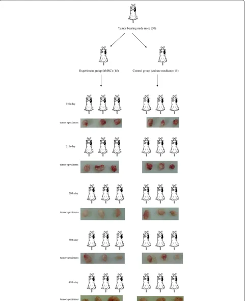
Mice were randomly assigned into one of two groups (n = 15 animals per group), and all mice were weighed and numbered. The mice in the experimental group were engrafted with hMSCs (5 × 10⁵cells per mouse) via the tail vein 15 days after inoculation of tumor cells, twice a week for 6 weeks successively, while the animals in the control group were injected with hMSC culture medium (0.2 mL per mouse) via the tail vein at the same time. The subcutaneous tumor size was mea-sured using an electronic digital caliper once every 4 days after hMSC engraftment. After 2, 3, 4, 5 and 6 weeks of tumor cell inoculation, the mice were killed and the tumors were collected in their entirety. The tumor weight and body weight of mice were measured (see Figure 1).
Data processing method
The tumor inhibition rate was calculated using the fol-lowing formula:
Tumor inhibition rateð Þ ¼% 1‐E gð Þ C gð Þ
100%
E(g): mean tumor weight in the experiment group; C(g): mean tumor weight in the control group.
Determination of expression of osteopontin (OPN), bone sialoprotein (BSP), integrinα5 subunit (α-V), B cell lymphoma/leukemia-2 (Bcl2), Bcl-2 associated X protein (Bax) and caspase 3 in tumor samples from animal models of high metastatic HCC using quantitative real-time polymerase chain reaction (RT-PCR)
Tumor bearing nude mice (30)
Experiment group (hMSC) (15) Control group (culture medium) (15)
14th day
21th day tumor specimens
tumor specimens
35th day
42th day 28th day
tumor specimens tumor specimens tumor specimens
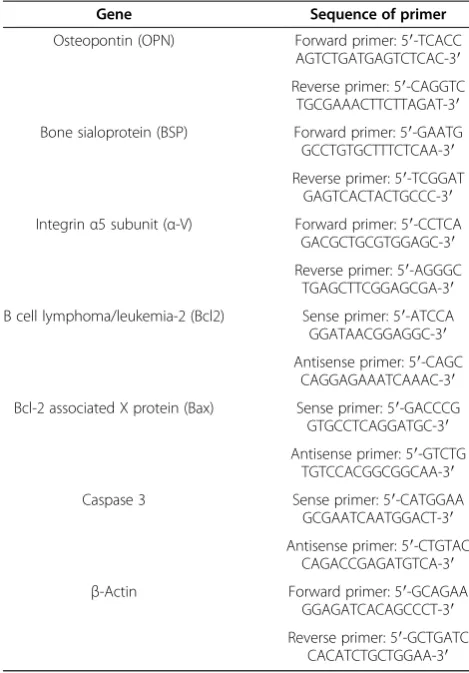
tumor samples, and then reversely transcribed into cDNA using RevertAid™ M-MLV reverse transcriptase and Oligo (dT). The corresponding cDNA was amplified using a RT-PCR technique based on specific primers (Table 1), with predegeneration at 95°C for 5 minutes, followed by 30 cycles of degeneration at 94°C for 30 s, annealing at 58°C for 30 s, extension at 72°C for 30 s, and the final elongation at 72°C for 10 minutes. The PCR amplification product was electrophoresed, and semi-quantitative analysis of the PCR amplification product was performed using a gel electrophoresis processing system.
Statistical analysis
Tumor inhibition rates were calculated, and a graph of the tumor inhibition rates change over time was drawn. The tumor weights were compared between groups at different timepoints.
All statistical analyses were performed using the SPSS statistical software version 16.0 (SPSS Inc., Chicago, IL, USA). The Student t test was used to compare the
Results
Effect of hMSCs on transplanted tumors in nude mice produced by inoculation of MHCC97-H cells
The tumor inhibition rate was calculated according to the tumor tissue specimen weights in vitro before and after hMSC intervention. The experimental results show that the tumor weight inhibition rate was 26.62% at 2 weeks, 52.00% at 3 weeks, 38.20% at 4 weeks, 31.98% at 5 weeks and 30.23% at 6 weeks.
The changes in transplanted tumors in nude mice pro-duced by inoculation of MHCC97-H cells following en-graftment of hMSCs for successive 6 weeks are shown in Figure 2. It was observed that the highest tumor inhib-ition rate was seen 3 weeks after hMSC engraftment, and the tumor inhibition rate gradually reduced with time.
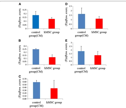
Tumor tissuein vitroweight comparison results were sig-nificantly lower in the hMSC engraftment groups than in the control group for the second and third weeks (P<0.01), but differences were not statistically significant in the fourth, fifth and sixth weeks between groups (P >0.01) (Figure 3).
RT-PCR determines the expression of OPN, BSP,α-V, Bcl2, Bax and caspase 3 genes in animal models of high-metastatic HCC before and after hMSCs engraftment Following hMSC engraftment, the expression of metastasis-related factors OPN, BSP andα-V gene was downregulated with time. Also, in the fourth week, OPN gene expression in hMSC engraftment groups was significantly lower than in control groups (P<0.05). Additionally, in the fourth and fifth week, BSP gene andα-V gene expression in hMSC engraft-ment groups was significantly lower than in control groups (P<0.05). As can be seen from the results, the metastatic
Osteopontin (OPN) Forward primer: 5′-TCACC
AGTCTGATGAGTCTCAC-3′
Reverse primer: 5′-CAGGTC TGCGAAACTTCTTAGAT-3′
Bone sialoprotein (BSP) Forward primer: 5′-GAATG GCCTGTGCTTTCTCAA-3′
Reverse primer: 5′-TCGGAT GAGTCACTACTGCCC-3′
Integrinα5 subunit (α-V) Forward primer: 5′-CCTCA GACGCTGCGTGGAGC-3′
Reverse primer: 5′-AGGGC TGAGCTTCGGAGCGA-3′
B cell lymphoma/leukemia-2 (Bcl2) Sense primer: 5′-ATCCA GGATAACGGAGGC-3′
Antisense primer: 5′-CAGC CAGGAGAAATCAAAC-3′
Bcl-2 associated X protein (Bax) Sense primer: 5′-GACCCG GTGCCTCAGGATGC-3′
Antisense primer: 5′-GTCTG TGTCCACGGCGGCAA-3′
Caspase 3 Sense primer: 5′-CATGGAA
GCGAATCAATGGACT-3′
Antisense primer: 5′-CTGTAC CAGACCGAGATGTCA-3′
β-Actin Forward primer: 5′-GCAGAA
GGAGATCACAGCCCT-3′
Reverse primer: 5′-GCTGATC CACATCTGCTGGAA-3′
potential of tumors was also downregulated after hMSC engraftment, especially in the fourth and fifth weeks (Figure 4A-C).
Following hMSC engraftment, the expression of antiapoptotic gene Bcl2 exhibited an obvious declin-ing tendency, while the expression of apoptotic genes Bax and caspase 3 showed an obvious rising tendency (Figure 4D-F). In the fourth and fifth week, Bcl2 gene expression in hMSC engraftment groups was significantly lower than in negative (control) groups (P<0.05). Also in the fourth and fifth week, Bax and caspase 3 gene expres-sion in hMSC engraftment groups was significantly higher than in negative (control) groups (P<0.05). As can be seen from these results, the proliferative ability of tumor cells decreased and the apoptotic ability of tumor cells in-creased after hMSC engraftment.
The results show that the metastatic potential of tumor cells was downregulated after hMSC engraftment, and hMSCs induced more tumor cells apoptosis.
Correlation analysis between metastasis-related factor expression and apoptotic gene expression in animal models inoculated with MHCC97-H cells after hMSCs engraftment
The expression of metastasis-related factorα-V was sig-nificantly positively correlated with the expression of antiapoptotic factor Bcl2 (r = 0.908,P <0.01), and nega-tively correlated with the expression of apoptotic factors Bax (r =−0.803,P<0.01) and caspase 3 (r =−0.902,P<0.01).
The expression of metastasis-related factor BSP was sig-nificantly positively correlated with the expression of antiapoptotic factor Bcl2 (r = 0.890,P<0.01), and negatively
Figure 3Tumor tissuein vitroweight comparison histogram. (A,B)In the second and third weeks, the tumor weight of human
correlated with the expression of apoptotic factors Bax (r = −0.779, P <0.05) and caspase 3 (r = −0.910, P<0.01).
The expression of metastasis-related factor OPN was not significantly correlated with the expression of antiapoptotic factor Bcl2 (r = 0.553, P >0.05), and was negatively correlated with the expression of apoptotic factors Bax (r =−0.368,P>0.05) and caspase 3 (r =−0.586,P>0.05) (Figure 5).
The results suggested that the decrease of proliferation ability of tumor cells can induce the decline of meta-static potential of tumor cells.
Correlation analysis between apoptotic and metastasis-related gene expression and tumor inhibition rate in animal models inoculated with MHCC97-H cells after hMSC engraftment
The tumor inhibition rate was not significantly correlated with the expression of antiapoptotic factor Bcl2 (r =−0.095,
P >0.05), and apoptotic factors Bax (r = −0.087, P >0.05) and caspase 3 (r = 0.144,P>0.05).
The tumor inhibition rate was not significantly corre-lated with the expression of metastasis-recorre-lated factorsα-V (r =−0.411,P>0.05) and BSP (r =−0.521,P>0.05) factors, but it exhibited a significant negative correlation with the expression of metastasis-related factor OPN (r =−0.766, P<0.05) (Figure 6).
These results suggest that the changes in tumor apop-totic or antiapopapop-totic genes were not the main factors affecting the tumor inhibition rate. Also, the changes in tumor inhibition rate were not the main factors affecting the metastatic potential of tumor cells.
Discussion
Liver cancer is one of the most common malignant tu-mors, and it is the second most common cause of death from cancer in China. Currently, surgical resection re-mains the most effective approach for treatment of the
Figure 4Gene expression in animal models inoculated with high-metastatic hepatocellular carcinoma cell 97 (MHCC97-H) cells before and after human mesenchymal stem cell (hMSC) engraftment. (A)The expression of metastasis-related factor osteopontin (OPN) by the tumor tissue specimen over time.(B)The expression of metastasis-related factor bone sialoprotein (BSP) by the tumor tissue specimen over time.
malignancy. However, the treatment has a high inci-dence of post-surgical recurrence and metastasis. An es-timated 40% to 50% 5-year recurrence rate after surgery is observed in small liver cancers, which is widely recog-nized to have a good prognosis in clinical practice [6]. Therefore, post-surgical early recurrence and metastasis has become critical as the main hindrance to the long-term survival of patients with liver cancer [6]. It has been found that bone-marrow-derived stem cells are a major extrahepatic source of hepatic stem cells, and bone-marrow-derived stem cells engrafted into the liver tissues of patients can differentiate into liver tissues and stem cells under a specific environment, thereby becom-ing involvbecom-ing in liver repair and reconstruction [2,3,7]. Autologous bone-marrow-derived stem cells have been engrafted into the injured liver for treatment of decom-pensated hepatic cirrhosis, which proves that stem cell transplantation is a novel, safe and effective treatment method [8,9]. Stem cell therapy has chemotaxis and homing functions in the treatment of liver cancer, and it presents a high efficacy for promoting the reconstruction of the immune system, removing residual lesions, and preventing recurrence and metastasis [10]. However, there are two completely contradictory views concerning the impact of human bone-marrow-derived mesenchy-mal stem cells (hMSCs) on hepatocellular carcinomas (HCCs). Some researchers believe that MSCs are a source of circulating progenitors that are able to generate cells of all mesenchymal lineages and cover the cellular demands of injured tissues. MSCs present immunosup-pressive and anti-inflammatory features and a high migra-tory capacity toward inflamed or remodeling tissues. Moreover, experimental evidence has been shown that supports the use of MSCs as a tool in liver fibrosis and he-patocellular carcinoma [4]. However, other researchers hypothesize that MSCs enhance tumor growth but signifi-cantly inhibit the invasiveness and metastasis of HCC [11]. Simulation of human tumors in live systems and es-tablishment of tumor models for basic research is a com-mon method in medical research [12]. In mouse models, stem cell transplantation inhibits post-surgical recurrence
Figure 5Correlation analysis (trend charts) between metastasis-related factor expression and apoptotic factor expression in animal models inoculated with high-metastatic hepatocellular carcinoma cell 97 (MHCC97-H) cells after human mesenchymal stem cell (hMSC) engraftment. (A)Correlation between metastasis-related factor integrinα5 subunit (α-V) and antiapoptotic factor B cell lymphoma/leukemia-2 (Bcl2), and apoptotic factors Bcl-2 associated X protein (Bax) and caspase 3.
and metastasis after radical resection of liver cancer, and the antitumor activity is enhanced with the increased chi-merism level of hMSC [7].
hMSCs at passages 5 to 8 were used in the present study. Phenotypic identification indicated that the hMSCs at passages 5 to 8 used in this study had all characteristics of hMSCs, which met the requirements for the experi-ments. Establishment of tumor animal models is a com-mon experimental technique in clinical practice [6]. This study established nude mouse models of HCC with high metastatic potential through inoculation of tumor cells (MHCC97-H cell), and hMSCs were engrafted via the tail vein for intervention treatment of HCC by means of the
The experimental results showed that the hMSCs had an inhibitory effect on HCC in animal models, but the effectiveness of the intervention of hMSCs changed with time. The highest tumor inhibition rate was observed at 3 weeks after hMSC engraftment, and the inhibitory efficiency of hMSCs reduced with time. This demon-strates that hMSCs exhibit antitumor activity in a time-dependent manner.
With regard to the intervention mechanism of hMSCs on liver cancer, studies have reported that hMSCs are found to colonize the liver of mouse models of orthotopic liver transplantation, and can differentiate into hepato-cytes that express albumin; in addition, the hMSCs are mainly found in the marginal area of the tumor and rarely present in tumors or normal liver tissues, and a large nec-rotic area is observed in tumor tissues following hMSC transplantation [12]. In addition to homogeneous gene characteristics and multilineage differentiation potential, hMSCs also have a high efficacy of immunosuppression and anti-infection activity, and the characteristics of mi-gration to inflammatory tissues and remodeling tissues (homing characteristic), which accelerate the repair of in-jured liver tissues, inhibit immune responses and confer antihepatic fibrosis functions [4]. However, prior to this work, to the best of our knowledge there have been no re-ports on hMSC antitumor activity in a time-dependent manner.
To further validate the inhibitory effect of hMSCs on HCC, the expression of some apoptosis-associated fac-tors was determined. The Bcl2 family plays a critical role in apoptosis. The Bcl2 family members are classified into two types: antiapoptotic factors, which mainly include Bcl2, Bcl-XL, Bcl-W, Mcl-1 and CED9, and proapoptotic factors, which mainly include Bax, Bak, Bcl-XS, Bad, Bik and Bid. The caspase family plays a very important role in mediating apoptosis, where caspase 3 is a key effector and functions in many apoptosis signaling transduction pathways [13]. From the experimental results, we can draw the conclusion that hMSCs induce greater tumor cells apoptosis, and also, similar to the tumor inhibition rate experiment results, the apoptosis-associated factor test results also show a time-dependent effect, but the timepoints are different. Therefore, the correlation ana-lysis results suggest that the changes in tumor apoptotic or antiapoptotic genes were not the main factors affect-ing the tumor inhibition rate. Thus, the mechanism of the time-dependent manner of hMSC antitumor activity requires further investigation.
The study determined the expression of metastasis-related factors to observe the effect of hMSCs on meta-static potential of HCC. The results show that the
Figure 6Correlation analysis (trend charts) between apoptotic and metastasis-related genes expression and tumor inhibition rate in animal models inoculated with high-metastatic hepatocellular carcinoma cell 97 (MHCC97-H) cells after human mesenchymal stem cell (hMSC) engraftment. (A)Correlation between antiapoptotic factor B cell lymphoma/leukemia-2 (Bcl2), and apoptotic factors Bcl-2 associated X protein (Bax) and caspase 3 gene expression and the tumor inhibition rate in animal models.
metastatic potential of tumor cells was downregulated after hMSC engraftment. Additionally, the tumor in-hibition rate was not significantly correlated with the expression of metastasis-related factors, and the posi-tive correlation with OPN may be due to experimen-tal error. However, the correlation analysis between metastasis-related and apoptosis-associated factor re-sults suggest that the decrease in proliferation ability of tumor cells can induce the decline in metastatic potential of tumor cells. Regardless, the present study also has some shortcomings. The method for deter-mination of tumor metastatic potential in animal models is excessively dependent on molecular bio-logical indexes, while the detection of traditional morphology is neglected.
The present study demonstrates the role of hMSCs in growth and metastasis of HCC tissues through injection of hMSCs via the tail vein, which provides new insight into exploring the relationship between hMSCs and liver cancer metastasis, and may provide a new cytological method for clinical treatment of liver cancer metastasis. It is of great significance to be able to effectively control the pro-proliferative effect and exert the metastatic in-hibition ability of hMSCs.
Conclusions
The highest tumor inhibition rate was observed 3 weeks after hMSC engraftment, and the tumor inhibition rate gradually reduced with time. The metastatic potential of tumor cells was downregulated after hMSC engraftment and hMSCs promoted apoptosis of tumor cells. The de-crease in proliferation ability of tumor cells can induce a decline in the metastatic potential of tumor cells. The changes in tumor apoptotic or antiapoptotic genes were not the main factors affecting the tumor inhibition rate. Also, the changes in tumor inhibition rate were not the main factors affecting the metastatic potential of tumor cells.
Abbreviations
α-V:Integrinα5 subunit; Bax: Bcl-2 associated X protein; Bcl2: B cell lymphoma/leukemia-2; BSP: Bone sialoprotein; HCC: Hepatocellular carcinoma; hMSC: Human bone-marrow-derived mesenchymal stem cell; MHCC97-H: High-metastatic hepatocellular carcinoma cell 97;
OPN: Osteopontin; RT-PCR: Quantitative real-time polymerase chain reaction; SPF: Specific-pathogen-free.
Competing interests
The authors declare that they have no competing interests.
Authors’contributions
LR: designed the experiments, acquired and analyzed the data, and drafted the manuscript. SB: designed the experiments. DK: analyzed the data. WM: acquired the data and drafted the manuscript. HL: acquired the data. All authors read and approved the final manuscript.
Acknowledgements
This study was supported by Chinese National Science Foundation (grant no. 81271607).
Author details
1
Department of Radiology, The 95th Hospital of PLA, PuTian, FuJian Province 351100, China.2Department of Radiology, Peking University People’s
Hospital, Beijing 100044, China.
Received: 20 January 2013 Accepted: 13 September 2013 Published: 30 September 2013
References
1. Marrero JA:Multidisciplinary management of hepatocellular carcinoma: where are we today?Semin Liver Dis2013,33:S3–S10.
2. Petersen BE, Bowen WC, Patrene KD, Mars WM, Sullivan AK, Murase N, Boggs SS, Greenberger JS, Goff JP:Bone marrow as a potential source of hepatic oval cells.Science1999,284:1168–1170.
3. Pittenger MF, Mackay AM, Beck SC, Jaiswal RK, Douglas R, Mosca JD, Moorman MA, Simonetti DW, Craig S, Marshak DR:Multilineage potential of adult human mesenchymal stem cells.Science1999,284:143–147. 4. Aquino JB, Bolontrade MF, Garcia MG, Podhajcer OL, Mazzolini G:
Mesenchymal stem cells as therapeutic tools and gene carriers in liver fibrosis and hepatocellular carcinoma.Gene Ther2010,17:692–708. 5. Li GC, Ye QH, Dong QZ, Ren N, Jia HL, Qin LX:Mesenchymal stem cells
seldomly fuse with hepatocellular carcinoma cells and are mainly distributed in the tumor stroma in mouse models.Oncol Rep2013, 29:713–719.
6. Tao ZY:Review and progress of small hepatocellular carcinoma.In
Advances in Clinical Oncology.Edited by Sun Y, Zhao P. Beijing, China: Peking Union Medical College Press; 2005:641–654.
7. Choi D, Kim JH, Lim M, Song KW, Paik SS, Kim SJ, Cheong HJ, Jeon JS, Park HS, Song YS, Khang H, Won JH:Hepatocyte-like cells from human mesenchymal stem cells engrafted in regenerating rat liver tracked within vivomagnetic resonance imaging.Tissue Eng Part C Meth2008,14:15–23.
8. Snykers S, Vanhaecke T, Papeleu P, Luttun A, Jiang Y, Vander Heyden Y, Verfaillie C, Rogiers V:Sequential exposure to cytokines reflecting embryogenesis: the key forin vitrodifferentiation of adult bone marrow stem cells into functional hepatocyte-like cells.Toxicol Sci2006,94:330–341. 9. Jiang HF, Ren J:Inoculation of murine bone marrow mesenchymal stem cells induces tumor necrosis in mouse with orthotopic hepatocellular carcinoma.J Peking Univ Health Sci2008,40:435–458.
10. Barzilay R, Melamed E, Offen D:Introducing transcription factors to multipotent mesenchymal stem cells: making transdifferentiation possible.Stem Cells2009,27:2509–2515.
11. Li GC, Ye QH, Xue YH, Sun HJ, Zhou HJ, Ren N, Jia HL, Shi J, Wu JC, Dai C, Dong QZ, Qin LX:Human mesenchymal stem cells inhibit metastasis of a hepatocellular carcinoma model using the MHCC97-H cell line.Canc Sci
2010,101:2546–2553.
12. Zhang JT (Ed):Modern Experimental Methods in Pharmacology.Beijing, China: Peking Union Medical College Press; 1998:927.
13. Galmiche A, Ezzoukhry Z, François C, Louandre C, Sabbagh C, Nguyen-Khac E, Descamps V, Trouillet N, Godin C, Regimbeau JM, Joly JP, Barbare JC, Duverlie G, Mazière JC, Chatelain D:BAD, a proapoptotic member of the BCL2 family, is a potential therapeutic target in hepatocellular carcinoma.Mol Canc Res2010,8:1116–1125.
doi:10.1186/2047-783X-18-34
Cite this article as:Liet al.:Effect of bone-marrow-derived
mesenchymal stem cells on high-potential hepatocellular carcinoma in
mouse models: an intervention study.European Journal of Medical