*Corresponding author: Mazyar Ziyaeyan, Ph.D, Clinical Microbiology Research Center, Shiraz University of Medi-cal Sciences, Shiraz, Iran.
Tel: +98-71-36474304 Fax: +98-71-36474303 Email: ziyaeyanm@sums.ac.ir
Molecular diagnosis of genital tract infections among HIV-positive women
in Iran
Mohammad Amin Behzadi1, Mohammad Ali Davarpanah2, Mandana Namayandeh1, Bahman Pourabbas1, Soheyla Allahyari2, Mazyar Ziyaeyan1*
1Clinical Microbiology Research Center, Shiraz University of Medical Sciences, Shiraz, Iran 2HIV Research Center, Shiraz University of Medical Sciences, Shiraz, Iran
Received: January 2018, Accepted: July 2018
ABSTRACT
Background and Objectives: Human immunodeficiency virus (HIV)-infected women are usually at a higher risk of sex-ually transmitted infections (STIs) than others. The objective of this study was to characterize the prevalence of human papilloma virus (HPV), herpes simplex virus (HSV), Chlamydia trachomatis (CT), and Neisseria gonorrhoeae (NG), and associated risk factors among HIV-infected women in Fars province, Iran.
Materials and Methods: In this cross-sectional study, cervical swab samples were collected from 71 HIV-infected women, aged 17-45 years (mean ± standard deviation: 31.11 ± 6.58 years), and tested for HPV, HSV, CT, and NG using PCR assays.
Results: Overall, 77.5% of patients were positive for the tested STIs with the following distribution: 36 (50.7%) HPV, 7 (9.9%) HSV, 4 (5.6%) NG, and 27 (38%) CT. From those, 39 (55%) were positive for only one infection, while 16 (22.5%) were positive for multiple infections. We observed that the prevalence of all tested STIs increased by age, except for HSV which showed a slight decrease, although not statistically significant. Socio-economic factors such as low educational level, multiple sex partners, and being a sex worker significantly correlated with higher positive prevalence of STIs in the studied population.
Conclusion: A high prevalence of STIs was observed among HIV-infected women in this region. These data might prompt policy makers and STI experts to focus on providing a comprehensive sex education, including participation in screening programs for STIs among high-risk groups.
Keywords: Human papilloma virus, Herpes simplex virus, Chlamydia trachomatis, Neisseria gonorrhoeae, HIV
ORIGINAL
AR
TICLE
INTRODUCTION
trachomatis (CT) are the most common agents in -volved in genitourinary tract infections in HIV-pos -itive patients (1). The clinical signs of infection can widely vary from no overt disease to severe symp -toms. In HIV-infected patients with a concomitant HPV infection, the associated cervical abnormalities are more prevalent, and the lesions can become ag-gressive leading to more complicated treatments (2). Recent reports have shown that HSV-1 and HSV-2 infections can trigger the reactivation of Kaposi’s Sarcoma-associated Herpes virus from latency and may function as a cofactor to develop disease (3, 4). CT and NG are also among the bacterial patho -gens that can cause serious sequelae in HIV-positive women, and screenings for these agents are recom -mended to help reduce the incidence of complicated infections such as pelvic inflammatory disease (5).
There is a direct relationship between STIs and genital HIV shedding in HIV-positive women (6), and such co-infections may facilitate the sexual transmission of the virus and accelerate disease pro-gression (7). Appropriate treatment of NG and CT infections as well as suppressive therapy of HSV and HPV infections can help reduce the virus load in the genital tract of the HIV patients (8).
Longevity and improved quality of life in HIV-pos -itive patients under anti-retroviral therapy has changed the landscape of diseases observed among these patients (9, 10). For example, it is estimated that up to 40% of the HIV-infected individuals may develop a neoplastic lesion, including HPV related cervical cancer (11, 12). Therefore, the prevalence of STI co-infection in HIV patients is a risk factor for development of secondary genital infections that can be life-threatening. The epidemiology of such infec-tions among HIV patients is poorly defined in much of Middle East, including Iran. Hence, the objective of the present study is to investigate the prevalence of HPV, HSV, CT and NG, and associated socio-eco -nomic risk factors among HIV-infected women re -ferred to the Lavan outpatient behavioral clinic in Fars province, Iran.
MATERIALS AND METHODS
Institutional ethical approval. The study proto-col was approved by the research co-ordinating com -mittee at the Clinical Microbiology Research Cen-ter, Shiraz University of Medical Sciences. Written
informed consents were obtained from the women enrolled in the study.
Study population. This study included all HIV-in -fected women (N=71, ages ranging between 17 and 45 years (mean ± standard deviation: 31.11 ± 6.58 years)) who attended the Lavan outpatient behavior -al clinic affiliated with Shiraz University of Medic-al Sciences, Iran, during 2015-2016. The patients were referred for a routine medical visit to receive medi-cal advice as well as their HIV medication. Inclusion criteria were HIV infection and symptoms such as itching in the genital area, dyspareunia, dysuria, or abnormal vaginal discharges in the past 6 months. Exclusion criteria were pregnancy or bleeding per vagina. Based on blood CD4+T-cell levels, none of the patients had developed AIDS at time of sampling (CD4+ T-cell count less than 200 mm3 was consid
-ered as AIDS). The studied population was divided into four age groups; 17-23 (I), 24-30 (II), 31-37 (III), and 38-45 (IV) years old. At time of sampling, none of the women were under treatment for cervical in -traepithelial neoplasia (CIN), nor did they have a re -cent history of CIN. Each patient was interviewed by a female health professional staff to collect relevant socio-economic information including demograph-ics (age), educational status, life-style behavior (drug addictions), and sexual activity (marital status, num -ber of sex partners, using condom, and exchange of sex for money). Swab samples were collected from cervix and the cervical os by a trained physician. The swabs were placed in transport medium tubes, kept at 4°C for a maximum of 6 h and then transferred to the Clinical Microbiology Research Centre for labo-ratory analysis. At the labolabo-ratory, the tubes contain-ing the samples were vigorously vortexed, the swabs were discarded, and the samples were stored at -70°C until examination.
HSV-2, which had been verified and typed by direct im -mune-fluorescent assay, were used as positive con -trols for HSV. To detect NG and CT, a standardized amount of internal control DNA, supplied with the real-time PCR kit, was added to the lysis buffer kit to monitor the efficiency of extractions.
Human papilloma virus PCR. To detect HPV in the cervical samples, a nested PCR was adapted by using two consensus primers (MY09/MY11, GP5+/ GP6+) to amplify a broad-spectrum of HPV geno -types (14). Briefly, the first round of PCRs for gener -ic HPVs was performed in a Veriti 96-well thermal cycler instrument (Applied Biosystems, Foster City, CA, USA) using 2x AmpliTaq Gold Fast PCR Master Mix (Applied Biosystems, Foster City, CA, USA) and MY11/MY09 primers. The first round PCR product was subsequently subjected to a second round of PCR using 2x Power SYBR Green PCR Master Mix (Applied Biosystems, Warrington, UK) and GP5+/ GP6+ primers. The real-time PCR was performed using the One-step plus Sequence Detection System (Applied Biosystems, Foster City, CA, USA), follow -ing which a dissociation curve was constructed in the range of 55°C to 95°C and the data were interpreted using melting curve analysis.
Herpes simplex viruses TaqMan real-time PCR. HSV DNA was detected by the TaqMan re -al-time PCR method using a previously described primer set and probe (15). The primers amplify a 92 bp fragment within a highly conserved region of the DNA polymerase gene from the HSV-1 and HSV-2. Amplification was performed using TaqMan Gene Expression Master Mix Reagents (Applied Biosyste -ms, Foster City, CA, USA) in an Applied Biosystems Sequence Detector 7500 machine (Applied Biosyste -ms, Foster City, CA, USA).
Neisseria gonorrhoea and Chlamydia
trachoma-tis TaqMan real-time PCR. Commercially avail-able TaqMan real-time PCR kits were used to detect NG and CT in the clinical specimens. All the pro -cedures were performed according to the directions and recommendations in the manufacturer’s proto-cols (Neisseria gonorrhoea and Chlamydia tracho-matis standard kits; Primer Design Ltd., Millbrook Technology Campus, Southampton, UK). The am -plification was performed using the TaqMan Gene Expression Master Mix Reagents (Applied Biosyste
-ms, Foster City, CA, USA) in a 7500 Real-Time PCR System instrument (Applied Biosystems, USA).
Statistical analysis. The statistical analyses were done by SPSS for Windows (version 16, SPSS Inc., Chicago, IL, USA) and the data were considered statistically significant at a two-sided p< 0.05. Dif -ferences in prevalence data of the four studied STIs (HPV, HSV, CT and NG) between age groups were analysed using analysis of variance (ANOVA) and chi-square tests. Moreover, demographic and be-havioural characteristic of participants were also calculated and compared between STIs and non-STI groups. All comparison graphs were generated us -ing GraphPad Prism version 5.0 (GraphPad Software Inc., CA, USA).
RESULTS
Of the 71 clinical samples collected, the follow -ing distributions were observed among patients with positive PCR results: 36 (50.7%) HPV, 7 (9.9%) HSV, 4 (5.6%) NG, and 27 (38%) CT. The prevalence of STIs was 77.5 %, in which 39 (55%) of the patients had only one infection, whereas 16 (22.5%) patients had multiple infections (Fig. 1). The prevalence of any of the four STIs increased by age, except for HSV which showed a slight decrease in the groups III and IV (Fig. 2), although not statistically signif -icant. There were significant differences in prev -alence rates of HPV between age groups I and III (p=0.009), I and IV (p= 0.006), II and III (p=0.045), and II and IV (p=0.031, Fig. 3).
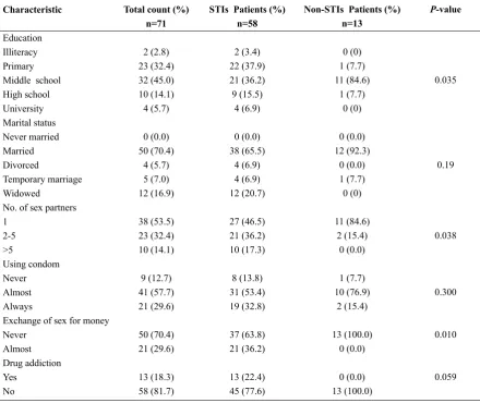
The descriptive data and the comparison of re-spective risk factors between the patients with and without STIs are presented in Table 1. The statistical analyses indicate significant differences in educa -tional status, number of sex partners, and number of patients who exchange sex for money between the STI and non-STI groups.
DISCUSSION
In the present study, four major STIs were investi
fig. 1. Distribution pattern of human papilloma virus (HPV), herpes simplex virus (HSV), Neisseria gonorrhoeae (NG) and Chlamydia tracomatis (CT) infections and respective co-infection among HIV-infected women in Shiraz, Iran (n=71).
fig. 3. Prevalence of human papilloma virus (HPV), herpes simplex virus (HSV), Neisseria gonorrhoeae (NG) and Chlamydia trachomatis (CT) infections in different age groups among HIV-infected women in Shiraz, Iran (n=71).
Table 1. Comparison of behavioral and demographic characteristics between HIV-infected women with or without STIs in Shiraz, Iran (n=71).
Characteristic
Education Illiteracy Primary Middle school High school University Marital status Never married Married Divorced
Temporary marriage Widowed
No. of sex partners 1
2-5 >5
Using condom Never Almost Always
Exchange of sex for money Never
Almost Drug addiction Yes
No
Total count (%) n=71
2 (2.8) 23 (32.4) 32 (45.0) 10 (14.1) 4 (5.7) 0 (0.0) 50 (70.4)
4 (5.7) 5 (7.0) 12 (16.9) 38 (53.5) 23 (32.4) 10 (14.1) 9 (12.7) 41 (57.7) 21 (29.6) 50 (70.4) 21 (29.6) 13 (18.3) 58 (81.7)
STIs Patients (%) n=58
2 (3.4) 22 (37.9) 21 (36.2) 9 (15.5)
4 (6.9) 0 (0.0) 38 (65.5)
4 (6.9) 4 (6.9) 12 (20.7) 27 (46.5) 21 (36.2) 10 (17.3) 8 (13.8) 31 (53.4) 19 (32.8) 37 (63.8) 21 (36.2) 13 (22.4) 45 (77.6)
Non-STIs Patients (%) n=13
0 (0) 1 (7.7) 11 (84.6)
1 (7.7) 0 (0) 0 (0.0) 12 (92.3)
0 (0.0) 1 (7.7) 0 (0) 11 (84.6)
2 (15.4) 0 (0.0) 1 (7.7) 10 (76.9)
2 (15.4) 13 (100.0)
0 (0.0) 0 (0.0) 13 (100.0)
P-value
0.035
0.19
0.038
0.300
0.010
in this area.
Based on our results, approximately half of the HIV-infected women carried the HPV genome in their cervical mucosa. In a study in China involving 95 women infected with HIV, a DNA hybridization assay identified close to 43% women with at least one type of high-risk HPV infection (16). Another study in Brazil conducted on 634 HIV-infected women re -vealed the prevalence of HPV infection to be 48%, of which 94% were infected with a high-risk HPV (17), whereas another report from Brazil showed a high -er prevalence of HPV (78.8%) among HIV-positive women (18). Previous reports suggest an increased prevalence of high-risk HPV types observed among HIV patients, compared to the HIV-negative patients (19). Given the high prevalence of HPV among the HIV patients in the present study, the existence of some cases with a co-infection with high-risk HPV genotypes is possible. As revealed in our study, HPV prevalence increased significantly by age, most prob -ably because of a greater exposure to the infectious agents as the women grew older and had increased number of sexual partners. Moreover, none of the studied patients had history of HPV vaccination. Thus, it may be concluded that performing intensive HPV vaccination programs among younger popula -tions in this region may help to decrease rate of in-fection in the future.
HPV infection, particularly of the high-risk HPV types, can be considered as a risk factor for develop -ing CT and NG infections (20, 21). Our data showed a high prevalence of CT infection among the patients (38%); although none was suffering from Lympho -granulomavenereum (LGV) when they initially par -ticipated in the study. Earlier studies indicate that the time between the first intercourse and STI onset is very short (22). The high prevalence of CT in our patients is indicative of the presence of untreated asymptomatic persistent infections or repeated epi-sodes of infections following inadequate treatment. In addition, no significant difference in CT frequen -cy was observed between patients of different age groups. However, previous studies have demonstrat -ed that the CT prevalence r-educ-ed with age, which could be due to repeated infections and immune re-sponse to the presence of the microorganism among older women. Franceschi et al. showed that CT prev -alence was significantly higher among 15-24 year-old women compared with women between 24-30 years old (20). The discrepancy between present findings
and other reports in terms of prevalence rates and correlations with age can be explained by the differ -ence in the study population, i.e. the entire popula-tion in our study were HIV positive.
In the present study, 5.6% of the patients were diagnosed with NG. Similar studies among HIV-in -fected and non-in-fected women have reported differ -ent NG prevalence worldwide. The prevalence of NG among symptomatic women from different national -ities in Dubai, UAE was 5.5% (23). Previous studies in China revealed the prevalence rate to be between 1.8-37.8% among female sex workers (24). Another study among women living with HIV in Denmark showed that none were infected with NG (25). How -ever, the study conducted in Papua New Guinea indicated a high prevalence of NG with the rate of 14.2% among the studied population (26). It has been observed that NG is becoming increasingly resistant to antibiotics worldwide, particularly among patients who receive repeated treatments for new infections (27, 28). Since the participants in the present study were among the high-risk groups with underlying diseases, i.e. HIV positive, it is very likely that they harbor emerging resistant strains and they may also serve as a reservoir for infecting others. Thus, devel-opment of comprehensive guidelines for screening and treatment of such STIs among the HIV-infected population seems to be vital.
In the present study, HSV DNA was detected in 10% of the HIV-infected women. In a similar study conducted on 369 HIV-positive women, more than 80% were observed to be HSV2 seropositive, and HSV DNA was detected in 7% of the respective cer -vico-vaginal samples (29). In a different study on 379 HIV-/HSV2- positive women, it was observed that 7% had viral secretion with a median load log 10 = 4.4 in their cervical os (30). HSV infection is com -mon a-mong HIV-positive women and is associated with an increased risk of HIV transmission. This observation might stem from the epithelial damage caused by HSV infection and consequently the in -creased shedding of HIV-infected cells and greater chance of HIV transmission to healthy individuals (31). Thus, with consideration of high prevalence of HSV infection (32), preventive strategies as well as appropriate HSV treatment (33) should be considered among such populations.
-gram in London and its outskirts, showed that close to 3% of CT cases were infected with NG as well, but here men accounted for 1/3 of the study population (34). A Korean study on 709 women, referred to the hospitals for a general check-up for 6 common STIs, revealed that the co-infection rate was 6.8%; howev -er, HPV and HSV were not addressed in this study (35). The relatively high rate of STI co-infection, similar to STI mono infection among the population in the present study, may be associated with their educational and socio-economic status. Educational status seems to play an important role in prevention, distribution and treatment of STIs (36, 37). Here, the majority of the studied patients had lower than high school education (80.2%) with a significant differ -ence between STI-harboring and non-STI patients. It has also been reported that the prevalence of STIs is higher in people having sexual contact with multiple sex partners, especially among high risk populations (38, 39). Our data also revealed that less than half of the studied patients had more than one sex partner (46.5%) and had been engaged in the practice of exchanging sex for money (29.6%). It was also shown that the prevalence of STIs was higher among illegal drug users; this may be due in part be-cause they use sex as a way to financially support their drug habits (40). Although in the present study, all individuals with drug addiction (18.3%) belonged to the STI group, no statistical difference was found between STI-harboring and non-STI patients. Thus, the high prevalence of STIs among this population may be related to their legal male partners.
It has been shown that the STIs can be transmitted to adults during unprotected sex with infected sex partners, and the use of condoms can notably reduce the rate of such transmission (25, 39). We did not find any significant difference in condom use rate between STIs and non-STI patients, and the major -ity of the studied population declare that they almost always use condoms (87.3%). However, this known preventive strategy should be promoted steadily in the society, especially among the youth. As for the patients in the present study, the existence of any STIs in addition to risky sexual behaviors such as having multiple sex partners may result in an in-crease in the incidence of STIs in the region.
In conclusion, the present study identified a high prevalence of STIs among HIV-infected women in the region of Fars Province, Iran. Moreover, it pro-vides useful information regarding STI-related risk
factors in this population, which can help policymak -ers and STI experts for future research and planning of health guidelines and policies. Significant atten -tion should focus on providing a comprehensive sex education to the potential patients, including recom-mending the use of condoms. In addition, a screen-ing program for CT and NG should be developed, and the patients who are positive for such bacterial infections must be fully treated. Finally, high preva-lence of HPV indicates the urgent need to develop a nationwide vaccination program aimed at females in the 10-12 years old age group.
ACKNOWLEDgEMENTS
Our thanks to Victor Leyva-Grado for critical re -view and Hassan Khajehei for copy editing of the manuscript. The study was supported by the Clinical Microbiology Research Center, Shiraz University of Medical Sciences, Shiraz, Iran.
REfERENCES
1. Aberg JA, Gallant JE, Anderson J, Oleske JM, Libman H, Currier JS, et al. Primary care guidelines for the management of persons infected with human immuno-deficiency virus: recommendations of the HIV medi-cine association of the Infectious Diseases Society of America. Clin Infect Dis 2004;39:609-629.
2. Moodley JR, Constant D, Hoffman M, Salimo A, Allan B, Rybicki E, et al. Human papillomavirus prevalence, viral load and pre-cancerous lesions of the cervix in women initiating highly active antiretroviral therapy in South Africa: a cross-sectional study. BMC Cancer 2009;9:275.
3. Tang Q, Qin D, Lv Z, Zhu X, Ma X, Yan Q, et al. Herpes simplex virus type 2 triggers reactivation of Kaposi's sarcoma-associated herpesvirus from latency and col-laborates with HIV-1 Tat. PLoS One 2012;7(2):e31652. 4. Qin D, Zeng Y, Qian C, Huang Z, Lv Z, Cheng L, et
al. Induction of lytic cycle replication of Kaposi's sar-coma-associated herpesvirus by herpes simplex virus type 1: involvement of IL-10 and IL-4. Cell Microbiol 2008;10:713-728.
Int J Infect Dis 2007;11:115-122.
6. Tanton C, Weiss HA, Le Goff J, Changalucha J, Rusizo-ka M, Baisley K, et al. Correlates of HIV-1 genital shed-ding in Tanzanian women. PLoS One 2011;6(3):e17480. 7. Buckner LR, Amedee AM, Albritton HL, Kozlowski
PA, Lacour N, McGowin CL, et al. Chlamydia tra-chomatis infection of endocervical epithelial cells enhances early HIV transmission events. PLoS One 2016;11(1):e0146663.
8. Fastring DR, Amedee A, Gatski M, Clark RA, Mena LA, Levison J, et al. Co-occurrence of Trichomonas vaginalis and bacterial vaginosis and vaginal shedding of HIV-1 RNA. Sex Transm Dis 2014;41:173-179. 9. Helleberg M, Kronborg G, Larsen C, Pedersen G,
Ped-ersen C, Gerstoft J, et al. Causes of death among Dan-ish HIV patients compared with population controls in the period 1995–2008. Infection 2012;40:627-634. 10. Galli L, Spagnuolo V, Salpietro S, Gianotti N,
Cos-sarini F, Lazzarin A, et al. Mortality of HIV-infected patients with or without cancer: comparison with the general population in Italy. Antivir Ther 2012;17:447-458.
11. Heard I. Prevention of cervical cancer in women with HIV. Curr Opin HIV AIDS 2009;4:68-73.
12. Aoki Y, Tosato G. Neoplastic conditions in the context of HIV-1 infection. Curr HIV Res 2004;2:343-349. 13. Yee C, Krishnan-Hewlett I, Baker C, Schlegel R,
How-ley P. Presence and expression of human papillomavi-rus sequences in human cervical carcinoma cell lines. Am J Pathol 1985;119:361-366.
14. Evander M, Edlund K, Boden E, Gustafsson A, Jons-son M, KarlsJons-son R, et al. CompariJons-son of a one-step and a two-step polymerase chain reaction with degenerate general primers in a population-based study of human papillomavirus infection in young Swedish women. J Clin Microbiol 1992;30:987-992.
15. Kessler HH, Mühlbauer G, Rinner B, Stelzl E, Berger A, Dörr H-W, et al. Detection of herpes simplex virus DNA by real-time PCR. J Clin Microbiol 2000;38:2638-2642.
16. Zhang H-Y, Tiggelaar SM, Sahasrabuddhe VV, Smith JS, Jiang C-Q, Mei R-B, et al. HPV prevalence and cervical intraepithelial neoplasia among HIV-infected women in Yunnan Province, China: a pilot study. Asian Pac J Cancer Prev 2012;13:91-96.
17. Grinsztejn B, Veloso VG, Levi JE, Velasque L, Luz PM, Friedman RK, et al. Factors associated with in-creased prevalence of human papillomavirus infection in a cohort of HIV-infected Brazilian women. Int J In-fect Dis 2009;13:72-80.
18. Corrêa CM, Teixeira NCP, de Araújo ACL, de Olivei-ra Carvalho N, Del Castillo DM, Campos RR, et al. Prevalence and multiplicity of HPV in HIV women in Minas Gerais, Brazil. Rev Assoc Med Bras (1992)
2011;57:425-430.
19. Sarkar K, Pal R, Bal B, Saha B, Bhattacharya S, Sen-gupta S, et al. Oncogenic HPV among HIV infected female population in West Bengal, India. BMC Infect Dis 2011;11:72.
20. Franceschi S, Smith JS, Van Den Brule A, Herrero R, Arslan A, Bosch FX, et al. Cervical infection with Chlamydia trachomatis and Neisseria gonorrhoeae in women from ten areas in four continents: A cross-sec-tional study. Sex Transm Dis 2007;34:563-569. 21. Samoff E, Koumans EH, Markowitz LE, Sternberg M,
Sawyer MK, Swan D, et al. Association of Chlamydia trachomatis with persistence of high-risk types of hu-man papillomavirus in a cohort of female adolescents. Am J Epidemiol 2005;162:668-675.
22. Tu W, Batteiger BE, Wiehe S, Ofner S, Van Der Pol B, Katz BP, et al. Time from first intercourse to first sexually transmitted infection diagnosis among adoles-cent women. Arch Pediatr Adolesc Med 2009;163:1106-1111.
23. Mehrabani D, Behzadi MA, Azizi S, Payombarnia H, Vahdani A, Namayandeh M, et al. Cervical infection with Herpes simplex virus, Chlamydia trachomatis, and Neisseria gonorrhoeae among symptomatic wom-en, Dubai, UAE: a molecular approach. Interdiscip Perspect Infect Dis 2014;2014:347602.
24. Luo L, Li X, Zhang L-l. Neisseria gonorrhoeae prev-alence, incidence and associated risk factors among female sex workers in a high HIV-prevalence area of China. Int J Infect Dis 2015;38:115-120.
25. Thorsteinsson K, Ladelund S, Storgaard M, Rønsholt FF, Johansen IS, Pedersen G, et al. Sexually transmit-ted infections and use of contraceptives in women liv-ing with HIV in Denmark–the SHADE cohort. BMC Infect Dis 2016;16:81.
26. Vallely LM, Toliman P, Ryan C, Rai G, Wapling J, Tomado C, et al. Prevalence and risk factors of Chla-mydia trachomatis, Neisseria gonorrhoeae, Tricho-monas vaginalis and other sexually transmissible in-fections among women attending antenatal clinics in three provinces in Papua New Guinea: a cross-section-al survey. Sex Health 2016;13:420-427.
27. Kubanova A, Frigo N, Kubanov A, Sidorenko S, Le-snaya I, Polevshikova S, et al. The Russian gonococ-cal antimicrobial susceptibility programme (RU-GASP)–national resistance prevalence in 2007 and 2008, and trends during 2005-2008. Euro Surveill 2010;15(14):19533.
28. Kirkcaldy RD. Neisseria gonorrhoeae antimicrobial susceptibility surveillance—the gonococcal isolate surveillance project, 27 sites, United States, 2014. MMWR Surveill Summ 2016;65:1-19.
cor-relates of cervicovaginal herpes simplex virus type 2 shedding among HIV-infected women in the Women's Interagency HIV Study. Int J STD AIDS 2011;22:273-277.
30. Aumakhan B, Hardick A, Quinn TC, Laeyendecker O, Gange SJ, Beyrer C, et al. Genital herpes evaluation by quantitative TaqMan PCR: correlating single detection and quantity of HSV-2 DNA in cervicovaginal lavage fluids with cross-sectional and longitudinal clinical data. Virol J 2010;7:328.
31. Horbul JE, Schmechel SC, Miller BR, Rice SA, South-ern PJ. Herpes simplex virus-induced epithelial dam-age and susceptibility to human immunodeficiency virus type 1 infection in human cervical organ culture. PLoS One 2011;6(7):e22638.
32. Ziyaeyan M, Japoni A, Roostaee MH, Salehi S, Solei-manjahi H. A serological survey of Herpes simplex vi-rus type 1 and 2 immunity in pregnant women at labor stage in Tehran, Iran. Pak J Biol Sci 2007;10:148-151. 33. Ziyaeyan M, Alborzi A, Japoni A, Kadivar M,
Davar-panah MA, Pourabbas B, et al. Frequency of acyclo-vir-resistant herpes simplex viruses isolated from the general immunocompetent population and patients with acquired immunodeficiency syndrome. Int J Der-matol 2007;46:1263-1266.
34. Skidmore S, Copley S, Cordwell D, Donaldson D, Ritchie D, Spraggon M. Positive nucleic acid amplifi-cation tests for Neisseria gonorrhoeae in young people tested as part of the national chlamydia screening
pro-gramme. Int J STD AIDS 2011;22:398-399.
35. Kim S-J, Lee DS, Lee S-J. The prevalence and clinical significance of urethritis and cervicitis in asymptom-atic people by use of multiplex polymerase chain reac-tion. Korean J Urol 2011;52:703-708.
36. Rajapure V, Tirwa R, Poudyal H, Thakur N. Preva-lence and risk factors associated with sexually trans-mitted diseases (STDs) in Sikkim. J Community Health 2013;38:156-162.
37. Ginindza TG, Stefan CD, Tsoka-Gwegweni JM, Dlamini X, Jolly PE, Weiderpass E, et al. Prevalence and risk factors associated with sexually transmitted infections (STIs) among women of reproductive age in Swaziland. Infect Agent Cancer 2017;12:29.
38. Street RA, Reddy T, Ramjee G. The generational ef-fect on age disparate partnerships and the risk for hu-man immunodeficiency virus and sexually transmitted infections acquisition. Int J STD AIDS 2016;27:746-752.
39. Kuete M, Huang Q, Rashid A, Ma XL, Yuan H, Es-calera Antezana JP, et al. Differences in knowledge, attitude, and behavior towards HIV/AIDS and sexu-ally transmitted infections between sexusexu-ally active foreign and Chinese medical students. Biomed Res Int 2016;2016:4524862.