CODEN [USA]: IAJPBB ISSN: 2349-7750
I
INNDDOO AAMMEERRIICCAANN JJOOUURRNNAALL OOFF
P
PH
HA
AR
R
MA
M
AC
CE
EU
U
TI
T
IC
CA
AL
L
S
SC
CI
IE
EN
NC
CE
ES
S
Available online at:
http://www.iajps.com Research Article
CONSEQUENCE OF FASCIOCUTANEOUS FLAPS IN 100
CASES WITH EXPOSED BONES OF LOWER LEGS
Dr. Abbas memon
1, Dr. Mehtab Ahmed Pirwani
2*, Dr. Faheem Ahmed Memon
3,
Dr. Irshad Ahmed Bhutto
41MBBS, FCPS, Assistant Professor, Department of Orthopaedic surgery of LUMHS 2MBBS, M.S, Associate Professor, Department of Orthopaedic surgery of LUMHS 3 MBBS, M.S, Assistant Professor, Department of Orthopaedic surgery of LUMHS
4 MBBS, M.S, Professor, Department of Orthopedic Surgery and Traumatology LUMHS, Jamshoro, Sindh-Pakistan.
Abstract:
Objective: To evaluate the outcome of fasciocutaneous flaps in 100 cases having exposed bones in lower leg. Methodology: This cross sectional study was conducted at the Department of Plastic Surgery & Orthopaedic Surgery, LUHMS Jamshoro with the 3 years duration august 2011 to September 2014. All the patients with all age groups either gender having uncomplicated or complex defects concerning legs due to any factor were selected for study. All the cases with chronic osteomyelitis including diabetic mellitus were excluded from the study. After taking complete medical history, physical examination, routine laboratory investigation and radiography all the patients were underwent reconstruction using different fasciocutaneous flaps. Flaps result was recorded as per partial or complete flap necrosis as well as valuable handling of the receiver’s defect and outcome was recorded in the Performa.
RESULTS: Majority of patients i.e 43(43%) belong to age group of 31-45. 91(91%) were male while 09(9%) were female. According to etiology, most common reason was RTA in 85(85%). Most common type of flap used was sural flap in 28(28%) patients followed by posterior calf flap in 13(13%) patients, lateral fasciocutaneous flap 14(14%), double fasciocutaneous flap 10(10%), medial fasciocutaneous flap 9(9%) , retrograde peroneal flap in 9(9%) , malleolar flap 07(7% . while primary closure was done in 02(2%) patients. According to the outcome partial flap necrosis was found in 6% patients followed by partial skin loss 4% patients and complete flap necrosis was only in 1 cases.
CONCLUSION: Fasciocutaneous flap can cover exposed bone at any site of lower legs; retrograde sural flap is perfect to cover exposed bones especially of foot and ankle joint.
Key words: Outcome, Fasciocutaneous Flapes, Lower Leg Defects
Correspondence author:
Dr. Mehtab Pirwani,
Associate Professor,
Department of Orthopaedic Surgery of LUMHS
Email: dr.sajidarain786@gmail.com 03132851728
Please cite this article in press as Mehtab pirwanin et al, To Evaluate the Outcome of Fasciocutaneous Flaps in Lower Leg Defects Authors, Indo Am. J. P. Sci, 2017; 4(07).
INTRODUCTION:
Administration of soft tissue around the lower regions of feet and legs have a substantial challenging stipulation for reconstructive surgery specialist due to the defects of composite tissue, poor circulation and insufficient as well as stiff local tissues.1 Tendon bones are often unprotected on account of the narrowness of subcutaneous tissues, turning skin implanting to have a poor alternative.2 A lasting flap having persistent vascularity, suitable skin texture, reliable rotational arc, dissection comfort as well as least donor region morbidity are the best preferred choices for handling such defects.3,4 Various local flaps concerning defects of hind foot including flap of dorsalis pedis artery, abductor muscle flaps of digiti minimi and hallucis, have insufficient tissue in addition to an inadequate rotational arc thus limiting their recurrent application. Flap of medial planter artery has been a best choice in cases of weight tolerating heel however its contribution in injury often prevents its application.5 Numerous current publications have demonstrated acceptance of flaps of lateral supramalleolar & superficial sural artery in order to handle defects regarding ankle & foot. These flaps may be upgraded effortlessly as well as replaced for the flaps of microsurgery of ankle & foot reconstruction and distal lower leg in certain conditions. There are benefits of these flaps to a certain extents of rotation, easy & quick raise, persistent vascularity and satisfactory donor-site morbidity. Though, a few complications are accounted, like partial flap & flap congestion failure due to insufficient venous return, whole flap failure due to rotation or compression of thick & wide adipofascial pedicle beneath subcutaneous tunnel, in addition to donor region morbidity.6,7 Baumeister et al8 established a general complication frequency of nearly 59% in an accurate analysis of seventy flaps, which comprise a wide patient group without & with comorbidities. The other reconstructive procedures for dorsal surface of the foot & ankle defects are flaps of without microsurgery. The transfer of microsurgical free flaps may escalate the supply of blood to united regions and offer handling of exposed crucial tissues at a stage.9-11 Free thoracodorsal artery perforator flaps are capable of resurfacing defects concerning any size as well as offer various forms of tissue to reconstruct composite defects. There are a few reports demonstrating the free thoracodorsal artery perforator flaps flap for reconstruction of dorsal surface of foot defects in literatures.12,13 Therefore this study has been conducted to evaluate the e the outcome of fasciocutaneous flaps in cases having exposed bones of lower legs at tertiary care hospital.
MATERIAL AND METHODS:
This cross sectional study was held at Plastic Surgery and Orthopaedic Surgery Department of LUHMS Jamshoro with the 3 years duration august 2011 to September 2014. All the patients with all age groups either gender having uncomplicated or complex defect concerning leg due to any reason were selected in this study. All the cases with chronic osteomyelitis, chronic liver disease, thrombocytopenia, altered PT, APTT and diabetes were excluded from the study. After taking complete medical history, physical examination, routine laboratory investigation and radiography all the patients were underwent reconstruction using different fasciocutaneous flaps. After surgery postoperative time the flap was observed for any pallor or venous congestion. Flaps result was recorded as per partial or complete flap necrosis, successful handling of receiver defect, easy or trouble in wearing shoes or walking, ambulatory condition of traumatic limb. All the information regarding age, gender, etiology, types of flaps, site of injury and outcome was recorded in the Performa and analyzed in SPSS version 16.0.
RESULTS:
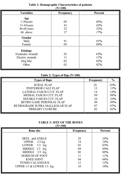
Majority of patients i.e 43(43%) belong to age group of 31-45 . while 17(17%) patients were above 60 years. 32(32%) patients belonged to age group of 40-60 years and 08 (8%) patients were in age group of 15-30 years. Regarding gender of participants, 91(91%) were male while 09(9%) were female. According to etiology, most common reason was road traffic accident in 85(85%) patients followed by electric wound in 11%, fight 2%, others 02(2%). TABLE:1.
Most common type of flap used was sural flap seen in 28(28%) patients followed by posterior calf flap was used in 13(13%) patients, lateral fasciocutaneous flap 14(14%), double fasciocutaneous flap 10(10%), medial fasciocutaneous flap 9(9%) , retrograde peroneal flap 9(9%) , malleolar flap 07(7%) . while primary closure was done in 02(2%) patients. TABLE:2.
Table 1: Demographic Characteristics of patients (N=100)
Variables Frequency Percent
Age 1-30years 31-45years 46-60 years 60 above
08 43 32 17
08% 43% 32% 17%
Gender Male Female
91 09
91% 09%
Etiology
Traumatic wounds Electric wounds
Dog bite Other
85 11 02 02
85% 11% 02% 02%
Table 2: Types of flap (N=100)
Types of flaps Frequency %
SURAL FLAP POSTERIOR CALF FLAP LATERAL FASCIO CUT: FLAP
MEDIAL FASCIO CUT: FLAP DOUBLE FASCIO CUT: FLAP
RETRO GADE PERONEAL FLAP RETROGRADE SUPRA MALLEOLAR FLAP
PRIMARY CLOSURE
28 13 14 09 10 09 07 02
28% 13% 14% 09% 10% 09% 07% 02%
TABLE 3: SITE OF THE BONES (N=100)
Bone site Frequency Percent
HEEL and ANKLE UPPER 1/3 leg LOWER 1/3 leg MIDDLE 1/3 leg MIDDLE 2/3 leg DORSUM OF FOOT
KNEE JOINT TENDO CACANEOUS UPPER 1/3 & LOWER 1/3 leg
28 14 03 09 08 11 04 12 10
Table 4: Outcome of flap (N=100)
Outcome Frequency Percent
COPLETE FLAP NECROSIS PARTIAL SKIN LOSS PARTIAL FLAP NECROSIS
01 04 06
01% 04% 06%
DISCUSSION:
Compound trauma of the lower libs needs to be covered by different flaps. This type of trauma most common affected were 2nd, 3rd and 4th age groups males those who moving around actively to bread win for the families.14 Though in this series majority of patients i.e 43(43%) belong to age group of 31-45 . while 17(17%) patients were above 60 years. 32(32%) patients belonged to age group of 40-60 years and 08 (8%) patients were in age group of 15-30 years. Regarding gender of participants, 91(91%) were male while 09(9%) were female. Similarly Ajmal S et al15 reported that 20(80%) cases were men whereas 5(20%) were women. Their age varied through 2-60 years, with 25 years of mean age. Franken JM et al16 reported that the male gender was most represented as; out of total cases 39 were male and 13 were female, mean age was 44.5 years with range of minimum 15 years and maximum 79 years. Suliman MT et al17 also found comparable results as 75% were male and 25% were female. We found most common reason was traumatic wound seen in 85(85%) patients followed by electric wound 11(11%), dog bite 02(2%), others 02(2%). Comparable results were found in study of Ajmal S et al15. In this study most common type of flap used was sural flap seen in 28(28%) patients followed by posterior calf flap seen in 13(13%) patients, lateral fasciocutaneous flap 14(14%), double fasciocutaneous flap 10(10%), medial fasciocutaneous flap 9(9%), retrograde peroneal flap 9(9%) , malleolar flap 07(7%). while primary closure was done in 02(2%) patients. Kumar Met al18 reported that the reconstructive method applied comprises the various faciocutaneous flaps applied comprises lateral fasciocutaneous flap, posterior calf flap, sural flap, double fasciocutaneous flap, medial fasciocutaneous flap, retrograde peroneal as well as retrograde supramalleolar. On other hand Suliman MT et al17 reported that peroneal artery perforator
flap used in 75% patients, superficial sural artery flap used in 17%, while posterior tibial artery perforator flap used only in 1 case.
In this series most common bone involved was heel and ankle seen in 28(28%) patients followed by upper 1/3rd of leg seen in 14(14%) patients, tendo
cacaneous in 12(12%) patients , dorsum of foot seen in 11(11%) patients ,upper 1/3rd and lower 1/3rd of leg was involved in 10(10%) patients, middle 1/3rd of leg seen in 09(9%) patients, middle 2/3rd of leg seen in 08(8%) patients. Knee joint was involved in 04(4%) patients. As well as Debbarma S et al19 stated that right leg was the predominant lower limb involved with incidence of 73.33% within group-I in addition to 60% within group II. Middle one third of the leg being the common site of the soft-tissue defect in both groups 66.7% and 60% in group I and group II respectively. Ajmal S et al15 reported that the defective site was distal 3rd of leg among 9 (36%) cases, lateral malleolus among 3 (12%), heel among 9 (36%), and 2 (8%) each for dorsum of foot as well as unprotected achilles tendon.
CONCLUSION:
We concluded that by fasciocutaneous flaps we can cover exposed bone at any site of lower legs. Retrograde flap is ideal to cover exposed heel, dorsum and the sole of foot and ankle joint. More studies are required to strong experience regarding flaps to cover exposed bones.
REFERENCES:
1. Fraccalvieri M, Boqetti P, Verna G, Carlucci S, Favi R, Bruschi S. Ditally based fasiocutaneous sural flap for foot reconstruction: a retrospective review of 10 years’ experience. Foot Ankle Int 2008;29:191–8. 2. Akhtar S, Hameed A. Versatility of the sural fasciocutaneous flap in the coverage of lower third leg and hind foot defects. J Plast Reconstr Aesthet Surg 2006;59:839–45.
3. Xu G, Jin LL. The coverage of skin defects over the foot and ankle using the distally based sural neurocutaneous flaps: Experience of 21 cases. J Plast Reconstr Aesthet Surg 2008;61:575–7.
4. Ahmed SK, Fung BK, Ip WY, Fok M, Chow SP. The versatile reverse flow sural artery neurocutaneous flap: A case series and review of literature. J Orthop Surg Res 2008;3(1):15–20. 5. Chen SL, Chen TM, Wang HJ. The distally based sural fasciomusculocutaneous flap for foot reconstruction. J Plast Reconstr Aesthet Surg 2006;59:846–55.
6. Touam C, Rostoucher P, Bhatia A, Oberlin C. Comparative study of two series of distally based fasciocutaneous faps for coverage of the lower one-fourth of the leg, the ankle, and the foot. Plast Reconstr Surg. 2001;107 (2):383-92.
7. Ulusal BG, Lin YT, Ulusal AE, Lin CH, Yen JT. Reconstruction of foot defects with free lateral arm fasciocutaneous faps: analysis of ffy patients. Microsurgery 2005;25(8):581- 8.
8. Baumeister SP, Spierer R, Erdmann D, Sweis R, Levin LS, Germann GK.A realistic complication analysis of 70 sural artery faps in a multimorbid patient group. Plast Reconstr Surg. 2003; 112 (1):129- 40.
9. Francel, T J., Kolk, C. A. V., Hoopes, J. E., Manson, P. N., and Yaremchuk, M. J. Microvascular sof-tissue transplantation for reconstruction of acute open tibial fractures: Timing of coverage and long-term functional results. Plast Reconstr Surg. 1992;89(3): 478-87.
10. Heller, L., and Levin, L. S. Lower extremity microsurgical reconstruction. Plast Reconstr Surg. 2001;108(4): 1029-41.
11. Kulahci Y, Sever C, Uygur F. Fasciocutaneous Flap Choices to Treat Dorsal Surface of Foot Defects. Journal of Clinical and Analytical Medicine. 2012 Jan 1;3(1):21-5.
12. Kim SE, Rhyou IH, Suh BG, Chung KC. Use of thoracodorsal artery perforator fap for sof tissue reconstruction in children. Ann Plast Surg. 2006; 56(4): 451- 4.
13. Lin CT, Huang JS, Yang KC, Hsu KC, Chen JS, Chen LW. Reliability of anatomical landmarks for skin perforators of the thoracodorsal artery perforator fap. Plast Reconstr Surg 2006;118(6):1376- 86. 14. Ramesh P, Kumar AS, Anita C, Krishnaveni A. A Comparative Study of Soft Tissue Cover in Compound Lower Limb Trauma at an Interval of a Decade. IJSR 2015;4;8;2094-2107
15. Ajmal S, Khan MA, Khan RA, Shadman M, Yousof K, Iqbal T. Distally based sural fasciocutaneous flap for soft tissue reconstruction of the distal leg, ankle and foot defects. J Ayub Med Coll Abbottabad. 2009;21(4):19-23.
16. Franken JM, Hupkens P, Spauwen PH. The treatment of soft-tissue defects of the lower leg after a traumatic open tibial fracture. European journal of plastic surgery. 2010 Jun 1;33(3):129-33.
17. Suliman MT. Distally based adipofascial flaps for dorsal foot and ankle soft tissue defects. The Journal of Foot and Ankle Surgery. 2007 Dec 31;46(6):464-9.
18. 12. Kumar M, Makhdoom A, M.S. Tahir, Zai ZY. Outcome of Fasciocutaneous Flaps in Exposed Bones of Leg. World Journal of Medical Sciences 11 (2): 268-272
19. Debbarma S, Singh NS, Singh PI, Singh S N, Singh A M, Meena RK. Fasciocutaneous flap as a method of soft tissue reconstruction in open tibial fractures. J Med Soc 2013;27:100-5
20. Follmar KE, Baccarani A, Steffen P, Baumeister L, Levin S, Erdmann D. The distally based sural flap. Plast Reconstr Surg 2007;119:138-48
21. Baumeister SP, Spierer R, Erdmann D et al. A realistic complication analysis of 70 sural artery flaps in a multimorbid patient group. Plast Reconstr Surg 2003;112:129–40.