Original Article
The Effect of Various Mixing Techniques on the Push-Out Bond Strength of
Calcium Enriched Mixture
Nooshin Sadat Shojaee 1, Alireza Adl 2, Fereshte Sobhnamayan 1, Fatima Vasei 3
1
Dept. of Endodontics, School of Dentistry, Shiraz University of Medical Sciences, Shiraz, Iran.
2 Dept. of Endodontics and Biomaterials Research Center, School of Dentistry, Shiraz University of Medical Sciences, Shiraz, Iran. 3
Undergraduate Student, School of Dentistry, Shiraz University of Medical Sciences, Shiraz, Iran.
KEY WORDS
Calcium Enriched Mixture;
Push-out Bond Strength;
Mixing Method;
Ultrasonic
Received March 2015;
Received in Revised form July 2015; Accepted October 2015;
ABSTRACT
Statement of the Problem: Correct proportioning and mixing are essential to ensure
cements attain their optimum physical properties.
Purpose: The aim of this experimental study was to evaluate the influence of various
mixing techniques including manual, mechanical mixing, and ultrasonic vibration on
push-out bond strength of calcium enriched mixture (CEM).
Materials and Method: Ninety 2-mm-thick dentin disks were prepared from
single-rooted human teeth and filled with CEM mixed with manual, trituration, or ultrasonic
methods. Push-out bond strength values of the specimens were measured by a
univer-sal testing machine after 3 and 21 days. The samples were then examined under a
stereomicroscope at 40× magnification to determine the nature of bond failure. Data
were analyzed by Kruskal-Wallis and Mann-Whitney test. (p< 0.05)
Results: The highest (7.59 MPa) and lowest (4.01 MPa) bond strength values were
recorded in conventional method (after 21 days) and trituration method (after 3 days),
respectively. There was no statistically significant difference between the three
tech-niques in 3 and 21 days.
Conclusion: According to the results, various mixing techniques had no effect on the
push-out bond strength of CEM cement.
Corresponding Author: Adl A., Dept. of Endodontics, Shiraz Dental School, Ghasrodasht Street, Shiraz.
P.O.Box: 7144833586 Email: [email protected] Tel: +98-71-36263193-4
Cite this article as: Sadat Shojaee N., Adl A., Sobhnamayan F., Vasei F. The Effect of Various Mixing Techniques on the Push-Out Bond Strength of Calcium Enriched Mixture. J Dent Shiraz Univ Med Sci., 2016 June; 17(2): 128-133.
Introduction
Many types of dental cements are available as powder
and liquid that should be mixed before application.
Cor-rect proportioning and mixing are essential to ensure
that the cements attain their optimum physical
proper-ties. [1] Encapsulating along with trituration in
compari-son to manual mixing has the potential to reduce air
spaces between adjacent particles. It results in a more
thorough wetting of the powder particles and improves
the unification of the resultant paste. [2]
Whilst trituration uses conventional mechanical
energy, there might be a potential for ultrasonic energy
to be more effective. Ultrasonic vibration has a
dispers-ing effect on the particles of materials, which frequently
cluster together. Ultrasonic treatment has been reported
to be effective in increasing the compressive strength,
[3-4] tensile bond strength, [5-6] and hardness [7] of
glass ionomer cements.
Little information is available on the effect of
var-ious mixing techniques on the physical properties of
mineral trioxide aggregate-like (MTA) materials.
Nek-oofar et al. compared ultrasonication, trituration, and
manual mixing and concluded that the application of
ultrasonic energy to MTA produced a significantly
higher surface microhardness value. [8] On the other
hand, Shahi et al. reported that different mixing
meth-ods had no significant effect on the push-out bond
showed that mechanical mixing of encapsulated MTA
resulted in higher compressive strength values than
those mixed manually. [10]
Calcium enriched mixture (CEM) cement was
in-troduced in 2008 with similar clinical application to
MTA but different chemical compositions. [11-12]
CEM is tooth-colored water-based cement which
con-sists of calcium oxide, calcium phosphate, calcium
car-bonate, calcium silicate, calcium sulphate, calcium
hy-droxide and calcium chloride. [13] This cement
exhibit-ed favourable results in regard to biocompatibility,
anti-bacterial effect, and sealing properties. [13-18]
There is no information about the effect of mixing
techniques on the push-out bond strength of CEM
ce-ment. Therefore, the purpose of this study was to
evalu-ate the effect of various mixing techniques including
ultrasonic vibration, trituration, and manual method on
push-out bond strength of CEM cement.
Materials and Method
Sixty freshly extracted human teeth including single
rooted mandibular premolars or maxillary incisors that
were either intact or contained only small carious lesion
were used in this study. Teeth with cracks or internal
resorption were excluded from the study. After
remov-ing the crowns by usremov-ing a diamond disk, the middle
thirds of the teeth were sectioned perpendicular to the
root long axis to obtain 90 dentin disks with the
thick-ness of 2±0.2 mm. A diamond saw microtome
(Mecatom T180; Presi SA, Angonnes, France) was used
to obtain root dentin slices. The internal disk canals
space was enlarged with Gates Glidden burs (Dentsply
Maillefer; Ballaigues, Switzerland) sizes 2 to 5 to
achieve a standard diameter of 1.3 mm. [9] The root
sections were immersed in 17% EDTA
(ethylenedia-minetetraacetic acid) (Asia Chemi Teb; Tehran, Iran),
and then in 2.5% sodium hypochlorite (Pakshooma;
Tehran, Iran) each for three minutes to remove the
smear layer. They were, then, washed with distilled
water and dried. [9] The root sections were randomly
divided into 6 groups (n=15), and the lumens were filled
with CEM cement (BioniqueDent; Tehran, Iran) as
fol-lowing.
In groups 1 and 4 the CEM cement was mixed
with conventional method; in groups 2 and 5, the CEM
cement was mixed with trituration in an amalgamator
(Farazmehr; Esfahan, Iran) at the speed of 4500
revolu-tions/min for 30 second (customized encapsulated
CEM); in groups 3 and 6, the CEM cement was mixed
with an ultrasonic tip (Ultradent Products; Inc., Logan,
UT, USA). The CEM cements in all instances were
mixed according to the manufacturer’s
recommenda-tions. The samples were wrapped in wet pieces of gauze
and kept in an incubator (Mart Microbiology B. V.;
Netherlands) at 37 ̊C and 95% humidity for 3 days
(groups 1, 2, and 3) or 21 days (groups 4, 5, and 6).
Push-out test
The push-out test was performed on the samples by
using a universal testing machine (Zwick/Roell, Z050;
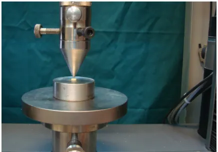
Zwick/Roell, Ulm, Germany) (Figure 1).
Figure 1: Universal testing machine
The cured specimens were placed on a metal slab
with a central hole and loaded with a 0.7-mm diameter
cylindrical stainless steel plunger at a speed of 1mm/
min. The maximum load applied to the CEM cement at
the time of dislodgement was registered in newton.
To express the bond strength in megapascals
(MPa), the recorded values were divided by the
adhe-sion surface area of CEM in mm2 calculated according
to the following formula:
2πr × h, where π is the constant 3.14, r is the root
canal radius, and h is the thickness of the root slice in
millimetres.
The modes of bond failure were evaluated under
the light microscope (Dino-light; Hsinchu, Taiwan) at
40× magnification. Each sample was categorized into
one of the three failure modes as adhesive failure at the
CEM and dentin interface, cohesive failure within the
CEM, and mixed failure mode. The data were analyzed
by using Kruskal-Wallis and Mann-Whitney test as
Results
The means and standard deviations (SD) of the push-out
bond strength of the groups are shown in Table 1.
Table 1: The Means and standard deviations of push-out bond strength of experimental groups
Group Mean(MPa)(SD)
Group1 (Conv, 3days) 4.860±1.413 Group 2 (Trit ,3 days) 4.016±1.322 Group 3 (Ultra ,3days) 4.848±2.122 Group 4 (Conv, 21days) 7.598±5.062 Group 5 (Trit ,21 days) 4.548±4.485 Group 6 (Ultra, 21days) 5.104±3.872
Abbreviations: Conv: Conventional method, Ultra: Ultrasonic meth-od, Trit: Trituration method
Although the groups that CEM was mixed with
trituration (group 2 and 5) showed the lowest value of
push-out bond strength, no statistically significant
dif-ference was found between the three techniques in 3 and
21 days (Figure 2).
Figure 2: The mean of push-out bond strength values of CEM by 3 mixing methods
Inspection of the samples revealed the bond
strength to be predominantly cohesive for conventional
technique but mixed for trituration and ultrasonic
tech-niques. (Table 2 and Figure 3) (p<0.05)
Discussion
In this study, push-out test was used to assess the bond
strength between CEM cement and dentinal walls.
Var-ious methods have been described to evaluate the
bond-ing quality of dental materials to dentin such as shear,
[19-20] compressive, [4] tensile, [4, 6] flexural [20] and
push-out bond strength. [9-10, 22-23] Among them,
push–out test has been shown to be efficient and
relia-ble. [23]
Table 2: Percent of each mode of failure among the experi-mental groups
Experimental Group Cohesive (%)
Adhesive (%)
Mixed (%)
Group1 (Conv, 3 days) 73.3 0 26.7 Group 2 (Amal, 3 days) 20 20 60 Group 3 (ultra, 3 days) 46.7 0 53.3 Group 4 (Conv, 21days) 40 13.3 46.7 Group 5 (Amal, 21 days) 0 20 80 Group 6 (Ultra, 21days) 20 33.3 46.7
Figure 3: Modes of failure a: adhesive failure; note the clean canal wall. b: cohesive failure within CEM. c: mixed failure; note the MTA residual inside the canal.
A scanning electron microscopy (SEM) and
ener-gy dispersive X-ray analysis (EDXA) study
demonstrat-ed that in the presence of normal saline as a storage
solution, hydroxyapatite crystals are formed and
pre-cipitated over the surface of CEM cement. The
compo-sition and structure of precipitated crystals were
compa-rable with that of standard hydroxyapatite. [24]
There-fore, in the present study, the root slices were wrapped
in pieces of gauze soaked in normal saline. [25]
To achieve optimal properties, the particles of
hy-draulic cements should be thoroughly mixed with water.
The mixing technique of cements is fundamental for
producing effective contact between the powder
parti-cles and liquid and a final set material with optimal
physical, chemical, and biological properties. [2] It has
been reported that mixing methods have a significant
effect on porosity and compressive strength of glass
ionomer cements. [1, 26] Regarding MTA, it has been
demonstrated that mechanical mixing enhanced the
compressive strength [10] and ultrasonic vibration
pro-duces a significantly higher surface microhardness. [8]
In addition to mixing techniques, root end preparation
techniques have been shown to influence the bond of
endodontic materials to dentinal walls. Shokouhinejad
et al. evaluated the push-out bond strength of two
Er,Cr:YSGG laser or ultrasonic technique and
conclud-ed that the bond strength of MTA and CEM to root-end
cavities were comparable and higher in ultrasonically
prepared cavities. [27]
In the present study, the influence of various
mix-ing techniques on push–out strength of CEM cement
was evaluated for the first time. The results showed that
trituration method compared with manual and ultrasonic
techniques results in lower push-out strength; however,
no statistically significant difference was found between
the three mixing techniques.
Another study on the effect of different mixing
techniques on the compressive strength showed that
mechanical mixing with amalgamator increased the
compressive strength of CEM cement. [28] The
con-flicting results of these two studies can be attributed to
the fact that push-out test and compressive strength have
different natures.
Interestingly, the studies on MTA showed similar
results. While Shahi et al. [9] showed that different
mix-ing methods had no significant effect on the push-out
bond strength of MTA; Basturk et al. [10] reported that
mechanical mixing of encapsulated MTA resulted in
higher compressive strength value than those mixed
manually.
Therefore, one may assume that different mixing
methods have impact on the compressive strength, not
on the push-out strength of hydraulic cements like MTA
and CEM.
The results of the current study showed that there
was no significant difference between the push out bond
strength of similar groups in 3 and 21 days. This finding
is in contrast with that of Rahimi et al. [29] who
report-ed an increase in the bond strength of CEM cement
from 24 hours to 7 days. The reason for the observed
disagreement may be related to the different
experi-mental set-up of the two studies. Gancedo-Caravia and
Garcia-Barbero [30] showed that curing conditions do
play an important role in the retention characteristics of
MTA. Therefore, different studies with different curing
condition should not be expected to have similar results.
In this study inspection of the samples revealed
the bond strength to be predominantly cohesive for
manual technique, but mixed for trituration and
ultra-sonic techniques. Therefore, it can be concluded that
mixing method can affect the pattern of bond failure.
Two separate studies with manual mixing technique
reported cohesive bond failure as the predominant bond
failure for CEM, which is in agreement with the results
of the current study. [31-32]
It should mention that the mechanical tests are
un-able to reflect the clinical situation; hence, future studies
are required to determine the effect of these techniques
on the bond strength of material in clinical applications.
Conclusion
Within the limitation of this in vitro study, it can be
concluded that different mixing techniques evaluated in
this study have no effect on the push-out bond strength
of the CEM cement
Acknowledgment
This manuscript was based on thesis by Dr. Vasei. The
authors thank the Vice Chancellery of Shiraz University
of Medical Sciences for supporting this research
(Grant#8069). We also thank Dr. Vossoughi from the
Center for Research Improvement of the School of
Den-tistry for the statistical analysis and Biomaterial
Re-search Center, School of Dentistry, Shiraz University of
Medical Sciences, Shiraz, Iran for technical support.
Conflict of Interest
The authors of this manuscript certify that they have no
conflict of interest.
References
[1] Hachmeister DR, Schindler WG, Walker WA 3rd, Thomas DD. The sealing ability and retention charac-teristics of mineral trioxide aggregate in a model of apexification. J Endod. 2002; 28: 386-390.
[2] Nomoto R, Komoriyama M, McCabe JF, Hirano S. Effect of mixing method on the porosity of encapsulat-ed glass ionomer cement. Dent Mater. 2004; 20: 972-978.
[3] Kleverlaan CJ, van Duinen RN, Feilzer AJ. Mechanical properties of glass ionomer cements affected by curing methods. Dent Mater. 2004; 20: 45-50.
Feilzer AJ. The influence of accelerating the setting rate by ultrasound or heat on the bond strength of glass ion-omers used as orthodontic bracket cements. Eur J Or-thod. 2005; 27: 472-476.
[6] Fagundes TC, Barata TJ, Bresciani E, Cefaly DF, Car-valho CA, Navarro MF. Influence of ultrasonic setting on tensile bond strength of glass-ionomer cements to dentin. J Adhes Dent. 2006; 8: 401-407.
[7] Towler MR, Bushby AJ, Billington RW, Hill RG. A preliminary comparison of the mechanical properties of chemically cured and ultrasonically cured glass ionomer cements, using nano-indentation techniques. Biomateri-als. 2001; 22: 1401-1406.
[8] Nekoofar MH, Aseeley Z, Dummer PM. The effect of various mixing techniques on the surface microhardness of mineral trioxide aggregate. Int Endod J. 2010; 43: 312-320.
[9] Shahi S, Rahimi S, Yavari HR, Samiei M, Janani M, Bahari M, et al. Effects of various mixing techniques on push-out bond strengths of white mineral trioxide aggregate. J Endod. 2012; 38: 501-504.
[10]Basturk FB, Nekoofar MH, Gunday M, Dummer PM. Effect of various mixing and placement techniques on the flexural strength and porosity of mineral trioxide aggregate. J Endod. 2014; 40: 441-445.
[11]Asgary S, Eghbal MJ, Parirokh M, Ghoddusi J, Kheirieh S, Brink F. Comparison of mineral trioxide aggregate's composition with Portland cements and a new endodontic cement. J Endod. 2009; 35: 243-250. [12]Zarrabi MH, Javidi M, Jafarian AH, Joushan B.
Histo-logic assessment of human pulp response to capping with mineral trioxide aggregate and a novel endodontic cement. J Endod. 2010; 36: 1778-1781.
[13]Asgary S, Shahabi S, Jafarzadeh T, Amini S, Kheirieh S. The properties of a new endodontic material. J En-dod. 2008; 34: 990-993.
[14]Mozayeni MA, Milani AS, Marvasti LA, Asgary S. Cytotoxicity of calcium enriched mixture cement com-pared with mineral trioxide aggregate and intermediate restorative material. Aust Endod J. 2012; 38: 70-75. [15]Asgary S, Akbari Kamrani F, Taheri S. Evaluation of
antimicrobial effect of MTA, calcium hydroxide, and CEM cement. Iran Endod J. 2007; 2: 105-109.
[16]Asgary S, Eghbal MJ, Parirokh M. Sealing ability of a novel endodontic cement as a root-end fillingmaterial. J Biomed Mater Res A. 2008; 87: 706-709.
[17]Mirhadi H, Moazzami F, Safarzade S. The Effect of Ac-
idic pH on Microleakage of Mineral Trioxide Aggre-gate and Calcium-Enriched Mixture Apical Plugs. Iran Endod J. 2014; 9: 257-260.
[18]Tabrizizade M, Asadi Y, Sooratgar A, Moradi S, Sooratgar H, Ayatollahi F. Sealing ability of mineral trioxide aggregate and calcium-enriched mixture ce-ment asapical barriers with different obturation tech-niques. Iran Endod J. 2014; 9: 261-265.
[19]Pakshir HR, Farbodan Z, Hedayati Z. Effect of Chang-ing Cross Head Speed on Shear Bond Strength of Or-thodontic Brackets and Mode of Adhesive Failure. J Dent Shiraz Univ Med Scien. 2008; 9: 127-136. [20]Davari AR, DaneshKazemi AR, Modaresi J,
Moham-madi Z, Akbarian L. The Effect of Light-Curing Time to Adhesive Layer on Shear Bond Strength of Compo-site to Dentin. J Dent Shiraz Univ Med Scien. 2007; 8: 10-18.
[21]Sharafeddin F, Alavi A, Talei Z. Flexural strength of glass and polyethylene fiber combined with three dif-ferent composites. J Dent Shiraz Univ Med Scien. 2013; 14: 13-19.
[22]Esfahanizadeh GR, Salari MH, Zahedi Rad S. Evalua-tion of the Influence of Resin Cement Thickness on the bond Strength of FRC Post. Shiraz Univ Dent J. 2011; 12: 321-326.
[23]Goracci C, Tavares AU, Fabianelli A, Monticelli F, Raffaelli O, Cardoso PC, et al. The adhesion between fiber posts and root canal walls: comparison between microtensile and push-out bond strength measurements. Eur J Oral Sci. 2004; 112: 353-361.
[24]Asgary S, Eghbal MJ, Parirokh M, Ghoddusi J. Effect of two storage solutions on surface topography of two root-end fillings. Aust Endod J. 2009; 35: 147-152. [25]Nekoofar MH, Stone DF, Dummer PM. The effect of
blood contamination on the compressive strength and surfacemicrostructure of mineral trioxide aggregate. Int Endod J. 2010; 43: 782-791.
[26]Nomoto R, McCabe JF. Effect of mixing methods on the compressive strength of glass ionomer cements. J Dent. 2001; 29: 205-210.
[27]Shokouhinejad N, Razmi H, Fekrazad R, Asgary S, Neshati A, Assadian H, et al. Push-out bond strength of two root-end filling materials in root-endcavities pre-pared by Er,Cr:YSGG la-ser or ultrasonic technique. Aust Endod J. 2012; 38: 113-117.
mixing and placement methods on the compressive strength of calcium-enriched mixture. Iran Endod J. 2015; 10: 104-106.
[29]Rahimi S, Ghasemi N, Shahi S, Lotfi M, Froughreyhani M, Milani AS, et al. Effect of blood contamination on the retention characteristics of two endodonticbio-materials in simulated furcation perforations. J Endod. 2013; 39: 697-700.
[30]Gancedo-Caravia L, Garcia-Barbero E. Influence of humidity and setting time on the push-out strength of mineral trioxideaggregate obturations. J Endod. 2006;
32: 894-896.
[31]Shokouhinejad N, Razmi H, Fekrazad R, Asgary S, Neshati A, Assadian H, et al. Push-out bond strength of two root-end filling materials in root-end cavities pre-pared by Er,Cr:YSGG laser or ultrasonic technique. Aust Endod J. 2012; 38: 113-117.