The effect of taurine on
dystrophic muscle tissue
function
Submitted by
Deanna Maree Horvath
BSc (Biomedical Science) (Hons)
School of Biomedical and Health Sciences
Faculty of Health, Engineering and Science, Victoria University
Submitted in fulfilment of the requirements for the degree of Doctor of Philosophy
i
ABSTRACT
Duchenne muscular dystrophy (DMD) is a lethal X-linked genetic disorder which results
in chronic degeneration of skeletal muscle, significantly impacting on the duration and
quality of life. Despite the genetic defect and the missing protein dystrophin having been
identified and characterised over 20 years ago, curative genetic therapies are still not
clinically applicable, and corticosteroids, which are the only significantly beneficial
treatment option currently available to DMD patients, are associated with several
side-effects. Thus, there is a need for additional therapeutic interventions that can improve
skeletal muscle function and delay the onset of severe pathology in dystrophy.
The amino acid taurine is essential for normal skeletal muscle function, and has been
shown to act on several factors thought to be key contributors to the development of
skeletal muscle pathology in dystrophy. Moreover, as dystrophic skeletal muscle
demonstrates a significant decrease in taurine content, it is possible that raising
intramuscular taurine stores may preserve muscle function in dystrophy, and thus have
potential therapeutic applications. Despite this, only two studies have ever examined the
effect of taurine supplementation on dystrophic muscle function.
The purpose of this thesis was to examine the effect of taurine on dystrophic skeletal
muscle function, which was performed in three studies using the dystrophic mdx mouse as a model for DMD. Study 1 (Chapter 4) investigated the effect of taurine
supplementation throughout the early mdx lifespan (time-points from day 28 to day 70), where skeletal muscle pathology is more homologous to the human condition, to
determine if taurine supplementation could significantly increase skeletal muscle taurine
content in the mdx, despite the significant muscle degeneration that occurs at this stage. The expression of excitation contraction (E-C) coupling, calcium (Ca2+) handling and contractile proteins were also investigated, to determine if the beneficial effects of
taurine supplementation on muscle function are associated with changes in protein
expression. Taurine supplementation successfully increased skeletal muscle taurine
ii
expression, and also increased the expression of the Ca2+ handling protein calsequestrin (CSQ) as well as the contractile proteins actin.
Study 2 (Chapter 5) examined the effect of long and short-term taurine supplementation
in the 6 month old mdx mouse on contractile function and the activity of several key metabolic enzymes. This study found that while taurine supplementation is unable to
improve peak tetanic or twitch force, that long-term taurine supplementation significantly
reduced fatigue in the mdx extensor digitorum longus (EDL) muscle. In addition, taurine supplementation altered the activity of phosphofructokinase (PFK) and beta-hydroxyacyl
CoA dehydrogenase (β-HAD) in the EDL, and creatine kinase (CK) and citrate synthase (CS) in the soleus (SOL).
The final study (Chapter 6) investigated the effect of altering skeletal muscle taurine
content in non-dystrophic and dystrophic fast and slow-twitch muscle on contractile
function, assessed using a protocol that utilised stimulation frequencies and a bath
temperature that is similar to what the muscles investigated would experience in vivo. Taurine effectively increased intramuscular stores of taurine while β-alanine significantly depleted them. Despite these alterations in taurine content, no significant differences in
peak tetanic or twitch force were observed in control or mdx mice, although taurine did significantly reduced susceptibility to fatigue in the EDL of the mdx mouse, while β -alanine significantly reduced fatigue in both the mdx and control mice.
In conclusion, this thesis has demonstrated that taurine supplementation is able to
significantly increase the intramuscular stores of taurine in dystrophic skeletal muscle,
while β-alanine treatment significantly depletes taurine stores. Taurine did not improve peak tetanic or twitch force in the mdx mouse, while taurine depletion with β-alanine did not reduce muscle function, an effect likely due to beneficial effects of β-alanine itself. Taurine did, however, significantly improve resistance to fatigue and recovery in
fast-twitch dystrophic skeletal muscle. In the young mdx mouse, taurine supplementation increased the expression of contractile proteins and CSQ, while in older (6 month) mdx
mice taurine supplementation has no significant effect on any of these measures. Thus,
it appears that the beneficial effects of taurine supplementation are highly dependent on
the age of the mice at experimentation, as well as the level of muscle damage at the
iii
VICTORIA UNIVERSITY
CANDIDATE DECLARATION
“I, Deanna Maree Horvath, declare that the PhD thesis entitled:
The effect of taurine on dystrophic muscle tissue function
Is no more than 100, 000 words in length including quotes and exclusive of tables,
figures, appendices, bibliography, references and footnotes. This thesis contains no
material that has been submitted previously, in whole or in part, for the award of any
other academic degree or diploma. Except where otherwise indicated, this thesis is my
own work”.
Full Name Deanna Maree Horvath
Signed ……….
iv
ACKNOWLEDGEMENTS
Completion of this PhD and thesis would not have been possible if not for the support
and guidance of many people.
Firstly, I would like to acknowledge and thank my supervisors, Dr Alan Hayes and Dr
Craig Goodman. To Alan, thank you for taking me on as a student and giving me the
opportunity to complete a PhD, for this, I am forever grateful. Thanks also for your
assistance in the lab when necessary, and your input into the final thesis. To Craig,
thank you for always showing interest in my work, offering practical and sound advice,
and your genuine enthusiasm and passion for research. Thank you also for involving me
in your research, and giving me my first publication in this field.
To Dr Robyn Murphy, thank you for welcoming me into your lab and teaching me the
techniques I needed to complete my PhD. I cannot begin to express how grateful I am
for all that you have done for me, and for your ongoing support. To Janelle Mollica, thank
you not only for your advice, support, and assistance in the lab, but also your friendship
over the last 6 years and your encouragement when I needed it the most. To the rest of
the current La Trobe University Zoolology lab members, you have been a pleasure to
work with, thanks for all your help.
To Jessica Ellis, you have been a constant support throughout my PhD and I know that I
would not be submitting this thesis if it were not for you. Your technical advice and
assistance was invaluable, as was (and is) your friendship.
A very special thank you to Dr Dawson Kidgell, you were always there to give me some
much needed perspective during this PhD, and your attitude in life is one that I have
always admired and respected. But above all, thank you for your loyal friendship and
v
Also to Phillip Seymour, I am so grateful for your unwavering support, willingness to
listen and sound advice that you have provided me with throughout my studies.
To Dr Amy Larsen, there is too much to thank you for, so I will just say how incredibly
grateful I am for the wonderful support and constant encouragement that you have
provided me with during this process. To the talented Dr Marissa Caldow, thank you for
all the work you put into formatting the final thesis, I am very lucky to have you as a
friend. To all my other friends left unmentioned directly, thank you.
To my family, Wanda, Frank and Wendy Horvath, thank you for all your love, support
and sacrifices that you have made over my many years of study. In particular, thank you
to my dad proof reading the final manuscript and to my mother for doing everything I had
no time to do while writing this thesis. To my sister, who is a science nerd at heart, thank
you for your encouragement and support.
Last, but certainly not least, I owe my deepest gratitude to Michael Azzopardi for his
love, dedication and persistent confidence in me. This would not have been possible
vi
LIST OF PUBLICATIONS AND AWARDS
PAPERS
Goodman, C. A., Horvath, D., Stathis, C., Mori, T., Croft, K., Murphy, R. M. & Hayes, A.
(2009). Taurine supplementation increases skeletal muscle force production and
protects muscle function during and after high-frequency in vitro stimulation. J Appl Physiol, 107, 144-54.
PRESENTATIONS
Horvath, D. M., Hayes, A., & Goodman, CA. (2010). The effect of taurine and β-alanine
supplementation on taurine content and contractile properties of skeletal muscle in the
vii
TABLE OF CONTENTS
ABSTRACT... i
CANDIDATE DECLARATION... iii
ACKNOWLEDGEMENTS... iv
LIST OF PUBLICATIONS AND AWARDS... vi
TABLE OF CONTENTS... vii
LIST OF FIGURES... xv
LIST OF TABLES... xviii
ABBREVIATIONS... xix
CHAPTER ONE: PERSPECTIVES 1.1 Thesis scope ... 1
CHAPTER TWO: REVIEW OF THE LITERATURE 2.1 Duchenne Muscular Dystrophy ... 4
2.1.1 Dystrophin structure and function ... 4
2.1.2 Mechanical damage and membrane weakness ... 6
2.1.3 Ion channel dysfunction ... 8
2.1.4 Impaired Ca2+ handling ... 11
2.1.5 Oxidative stress ... 12
2.1.6 Chronic inflammation ... 14
2.1.7 Impaired metabolism ... 16
2.1.8 Muscle function impairments ... 17
i) E-C coupling ... 17
ii) Whole muscle function ... 18
2.2 Mammalian models of dystrophy ... 21
2.2.1 Canine muscular dystrophy ... 21
viii
2.2.3 Mdx mouse ... 22
i) Muscular involvement ... 23
ii) Muscle function ... 24
iii) Force development ... 25
iv) Fatigue ... 26
v) Exercise tolerance ... 28
vi) Limitations of the mdx model ... 29
2.3 Current treatment approaches ... 31
2.3.1 Molecular therapies ... 31
2.3.2 Pharmacological therapies ... 32
2.3.3 Nutritional Interventions ... 33
2.4 Taurine ... 35
2.4.1 Properties of taurine ... 35
2.5 Taurine and the pathophysiology of DMD ... 37
2.5.1 Taurine and membrane stabilisation ... 37
2.5.2 Taurine and Ca2+ regulation ... 39
2.5.3 Taurine and oxidative stress ... 41
2.5.4 Taurine and inflammation ... 42
2.5.5 Taurine and metabolism ... 44
2.5.6 Taurine and contractile function ... 46
2.6 Taurine and the mdx mouse ... 49
2.6.1 Tissue taurine content in the mdx mouse ... 49
2.6.2 Taurine and muscle function in the mdx mouse ... 50
2.6.3 Taurine supplementation and the mdx mouse ... 51
2.7 Conclusions ... 53
2.8 Broad and specific aims of the thesis ... 53
2.8.1 Aims of study one ... 54
2.8.2 Aims of study two ... 55
ix CHAPTER THREE: GENERAL METHODS
3.1 Animals and supplementation... 57
3.2 Contractile protocol ... 58
3.2.1 Dissection procedures ... 58
3.2.2 Stimulation protocols ... 59
3.3 Taurine content ... 63
3.3.1 Muscle sample preparation ... 63
3.3.2 Taurine extraction ... 63
3.4 Enzyme activity ... 66
3.4.1 Homogenising procedure ... 66
3.4.2 CK (muscle and plasma) ... 66
3.4.3 PFK ... 67
3.4.4 β-HAD ... 68
3.4.5 CS ... 69
3.4.6 Protein determination ... 70
3.5 Western blotting ... 71
3.5.1 Tissue preparation ... 71
3.5.2 Gels and transfer conditions ... 71
3.5.3 Primary antibodies ... 71
3.5.4 Secondary antibodies ... 72
3.5.5 Imaging and analysis ... 72
CHAPTER FOUR: TAURINE SUPPLEMENTATION DURING ACUTE DYSTROPHIC PROGRESSION: IMPACT ON TISSUE TAURINE, TAURINE TRANSPORTER AND E-C COUPLING PROTEIN EXPRESSION IN KELETAL MUSCLE 4.1 Introduction ... 73
4.2 Aims and hypothesis ... 75
4.3 Methods ... 76
4.3.1 Experimental groups and treatment protocol ... 76
4.3.2 Sample collection ... 76
4.3.3 HPLC determination of taurine content ... 77
4.3.4 Western blot analysis of dystrophin expression ... 77
4.3.5 Western blot analysis of TauT expression ... 77
4.3.6 Western blot analysis of E-C coupling and Ca2+ handling proteins ... 78
x
4.4 Results ... 79
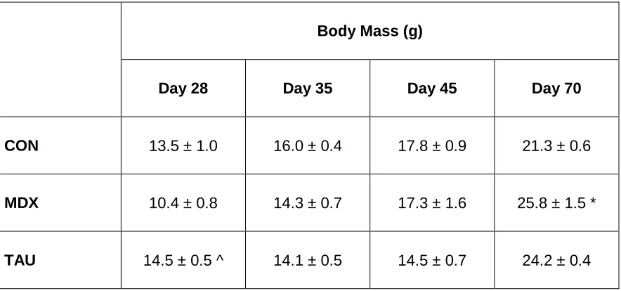
4.4.1 Body mass ... 79
4.4.2 Muscle mass and relative muscle mass ... 80
i) Day 28 ... 80
ii) Day 35 ... 80
iii) Day 45... 80
iv) Day 70 ... 81
4.4.3 Hind limb muscle % dw/ww ... 83
4.4.4 DIA muscle % dw/ww ... 85
4.4.5 Hind limb muscle taurine content ... 86
i) Day 28 ... 86
ii) Day 35 ... 86
iii) Day 45... 86
iv) Day 70 ... 87
4.4.6 DIA muscle taurine content ... 89
i) Day 28 ... 89
ii) Day 35 ... 89
iii) Day 45... 89
iv) Day 70 ... 89
4.4.7 Dystrophin expression ... 91
4.4.8 TauT protein expression ... 92
4.4.9 E-C coupling and Ca2+ handling protein expression ... 94
i) Contractile protein expression ... 94
ii) E-C coupling protein expression ... 97
iii) CSQ protein expression... 97
4.5 Discussion ... 102
4.5.1 Effect of taurine supplementation on body and muscle mass ... 102
4.5.2 Effect of taurine supplementation on skeletal muscle fluid content ... 103
4.5.3 Effect of taurine supplementation on skeletal muscle taurine content ... 105
4.5.4 Effect of taurine supplementation on TauT protein expression ... 107
4.5.5 Effect of taurine supplementation on E-C coupling and Ca2+ handling proteins ... 108
xi
CHAPTER 5: LONG AND SHORT-TERM TAURINE SUPPLEMENTATION ON
CONTRACTILE PROPERTIES AND ENZYME ACTIVITY IN MDX SKELETAL MUSCLE
5.1 Introduction ... 113
5.2 Aims and hypothesis ... 116
5.3 Methods ... 117
5.3.1 Animals ... 117
5.3.2 Contractile protocol ... 118
5.3.3 Plasma CK measurement ... 119
5.3.4 Enzyme activity analysis in muscle ... 119
5.3.5 Statistical Analysis ... 119
5.4 Results ... 120
5.4.1 Body mass ... 120
5.4.2 Muscle mass and relative muscle mass ... 121
i) EDL ... 121
ii) SOL ... 121
5.4.3 Plasma CK ... 122
5.4.4 EDL isometric contractile properties ... 123
i) Twitch characteristics, CSA and L0 ... 123
ii) Peak tetanic force ... 124
iii) Force-Frequency Relationship ... 126
iv) Fatigue ... 127
v) Percentage of original force post-fatigue ... 127
5.4.5 Enzyme activity for EDL ... 129
i) PFK ... 129
ii) CK ... 130
iii) β-HAD ... 131
iv) CS ... 132
5.4.6 SOL isometric contractile properties ... 133
i) Twitch characteristics, CSA and L0 ... 133
ii) Peak tetanic force ... 134
iii) Force-Frequency Relationship ... 136
iv) Fatigue ... 137
xii
5.4.7 Enzyme activity for SOL ... 139
i) PFK ... 139
ii) CK ... 140
iii) β-HAD ... 141
iv) CS ... 142
5.5 Discussion ... 143
5.5.1 Effect of taurine supplementation on body and muscle mass ... 144
5.5.2 Effect of taurine supplementation on plasma CK concentration ... 145
5.5.3 Effect of taurine supplementation on isometric contractile properties of EDL ... 147
5.5.4 Effect of taurine supplementation on isometric contractile properties of SOL ... 149
5.5.5 Effect of taurine supplementation on enzyme activity ... 150
5.6 Conclusions ... 154
CHAPTER 6: THE EFFET OF TAURINE AND β-ALANINE SUPPLEMENTATION ON CONTRACTILE PROPERTIES, TAURINE TRANSPORTER AND E-C COUPLING PROTEIN EXPRESSION IN C57BL/10 AND MDX MICE 6.1 Introduction ... 155
6.2 Specific aims and hypothesis ... 158
6.3 Methods ... 159
6.3.1 Animals ... 159
6.3.2 Dissection and contractile protocol ... 159
6.3.3 Measurement of skeletal muscle taurine content ... 161
6.3.4 Measurement of plasma CK ... 161
6.3.5 Western blot analysis of dystrophin expression ... 161
6.3.6 Western blot analysis of TauT protein expression ... 162
6.3.7 Western blot analysis of E-C coupling and Ca2+ handling protein expression ... 162
6.3.8 Statistical analysis ... 162
6.4 Results ... 163
6.4.1 Fluid consumption ... 163
i) CON ... 163
ii) Mdx ... 163
iii) Comparison of fluid consumption between CON and mdx mice ... 164
6.4.2 Body mass ... 166
6.4.3 Muscle mass and relative muscle mass ... 167
i)EDL ... 167
xiii
6.4.4 Freeze dried muscle % dw/ww ... 169
i)PLANT ... 169
ii)DIA ... 170
6.4.5 Muscle taurine content ... 171
i) PLANT ... 171
ii) DIA ... 171
6.4.6 Plasma CK concentration ... 173
6.4.7 The effect of treatments on the isometric contractile properties of EDL ... 174
i) Twitch characteristics, CSA and L0 ... 174
ii) Peak tetanic force ... 176
iii) Force-Frequency Relationship ... 178
iv) Fatigue ... 180
v) Recovery of EDL ... 181
a) CON ... 181
b) Mdx ... 181
6.4.8 The effect of treatments on the isometric contractile properties of SOL ... 183
i) Twitch characteristics ... 183
ii) Peak tetanic force ... 185
iii) Force-Frequency Relationship ... 187
iv) Fatigue ... 189
v) Recovery ... 190
6.4.9 Dystrophin expression ... 192
6.4.10 TauT protein expression ... 193
6.4.11 E-C coupling and Ca2+ handling expression ... 194
6.5 Discussion ... 198
6.5.1 The effect of taurine and β-alanine supplementation on fluid intake ... 199
6.5.2 The effect of taurine and β-alanine supplementation on body and muscle mass ... 200
6.5.3 The effect of taurine and β-alanine supplementation on skeletal muscle fluid and taurine content ... 200
6.5.4 The effect of taurine and β-alanine supplementation on plasma CK concentration ... 202
6.5.5 The effect of taurine and β-alanine supplementation on EDL contractile function ... 203
6.5.6 The effect of taurine and β-alanine supplementation on SOL contractile function ... 207
6.5.7 The effect of taurine and β-alanine supplementation on TauT protein expression ... 208
6.5.8 The effect of taurine and β-alanine supplementation on E-C coupling and Ca2+ handling protein expression ... 209
xiv
CHAPTER 7: CONCLUSIONS AND FUTURE DIRECTIONS
7.1 Summary of the major findings ... 212
7.2 The effect of taurine supplementation and depletion on skeletal muscle taurine content and TauT protein expression ... 213
7.3 The effect of taurine supplementation and depletion on contractile function ... 215
7.4 The effect of taurine supplementation on metabolic enzyme function, E-C coupling and Ca2+ handling proteins ... 217
7.5 Limitations ... 218
7.6 Future directions... 219
7.7 Conclusion ... 220
xv
LIST OF FIGURES
2.1 The link between dystrophin and the DAPC in skeletal muscle 5
2.2 EBD in non-dystrophic and mdx mouse muscle 7
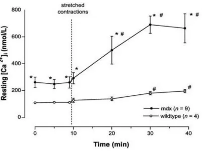
2.3 Resting Ca
2+ concentrations in control and mdx skeletal muscle after
exposure to ten stretch contractions 10
2.4 Schematic of the multiple pathways involved in DMD pathogenesis that
are activated by Ca2+ 11
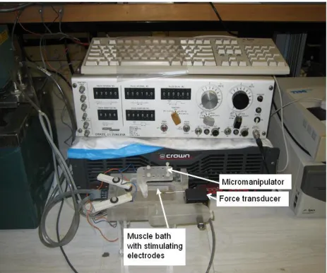
3.1 Contractile machine setup used for stimulation protocols 60
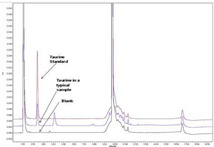
3.2 Typical HPLC trace for taurine content analysis 65
4.1 Taurine content in TA and GAST from day 28 to day 70 in CON, MDX
and TAU mice 88
4.2 Taurine content in DIA from day 28 to day 70 in CON, MDX and TAU
mice 90
4.3 Representative Western blot of dystrophin expression in day 28 and day
70 in CON, MDX and TAU mice 91
4.4 TauT expression in day 28 and day 70 in CON, MDX and TAU mice 93
4.5 Myosin expression in day 28 and day 70 in CON, MDX and TAU mice 95
4.6 Actin expression in day 28 and day 70 in CON, MDX and TAU mice 96
4.7 Day 28 and day 70 DHPR protein expression in CON, MDX and TAU
mice 98
4.8 Day 28 and day 70 RyR protein expression in CON, MDX and TAU mice 99
4.9 Day 28 and day 70 SERCA protein expression in CON, MDX and TAU
mice 100
4.10 Day 28 and day 70 CSQ protein expression in CON, MDX and TAU mice 101
5.1 Plasma CK concentration in CON MDX and taurine supplemented groups 122
5.2 Absolute and specific force production for the EDL muscles from CON
MDX and taurine treated mdx mice 125
5.3 Relationship between stimulation frequency and force production for the
xvi 5.4
Time taken for EDL force to fatigue to 70, 50 and 30% of original force and the percentage of original force attained at the end of the fatiguing stimulation
128
5.5 EDL PFK activity in CON MDX and taurine supplemented groups 129
5.6 EDL CK activity in CON MDX and taurine supplemented groups 130
5.7 EDL β-HAD activity in CON MDX and taurine supplemented groups 131
5.8 EDL CS activity in CON MDX and taurine supplemented groups 132
5.9
Absolute and specific force production for the SOL muscles from CON MDX mice as well as in association with long and short term taurine treatment
135
5.10
Relationship between stimulation frequency and relative force production for the SOL muscles between CON MDX mice as well as in association with taurine treatment
136
5.11
Time taken for SOL force to fatigue to 70, 50 and 30% of original force and the percentage of original force attained at the end of the fatiguing stimulation
138
5.12 SOL PFK activity in CON MDX and taurine supplemented groups 139
5.13 SOL CK activity in CON MDX and taurine supplemented groups 140
5.14 SOL β-HAD activity in CON MDX and taurine supplemented groups 141
5.15 SOL CS activity in CON MDX and taurine supplemented groups 142
6.1 Taurine content for PLANT and DIA for CON and mdx mice in
association with treatments 172
6.2 Plasma CK concentration for CON and mdx mice in association with
treatments 173
6.3 Absolute and specific force production for the EDL in CON and mdx mice
as well as in association with treatments 177
6.4
The relationship between relative peak force (% of maximum) and stimulation frequency for EDL in CON and mdx mice in association with treatments
179
6.5
Percentage of the original force at the end of 1 minute fatiguing stimulation at 70 Hz for 250 ms every 1 s in CON and mdx mice in association with treatments
180
6.6 Recovery of CON and mdx EDL muscles after a fatiguing stimulation
measured using a stimulation frequency of 100 Hz 182
6.7 Absolute and specific force for SOL in CON and mdx mice in association
xvii 6.8
The relationship between relative peak force (% of maximum) and stimulation frequency for SOL in CON and mdx mice in association with treatments
188
6.9
Percentage of the original force at the end of a 3 minute fatiguing stimulation ant 30 Hz for 500 ms every 1 s in CON and mdx mice in association with treatments
189
6.10 Recovery of force for CON and mdx SOL muscles after a fatiguing
stimulation measured using a stimulation frequency of 80 Hz 191
6.11 Dystrophin expression in CON and mdx mice in association with
treatment 192
6.12 TauT expression in CON and mdx EDL muscles in association with
treatment 193
6.13 Contractile protein expression in CON and mdx EDL muscles in
association with treatment 195
6.14 DHPR and RyR protein expression in EDL in CON and mdx EDL
muscles in association with treatment 196
6.15 SERCA and CSQ protein expression in CON and mdx EDL muscles in
xviii
LIST OF TABLES
3.1 Antibody supplier and dilution information for proteins investigated 72
4.1 Comparison of body mass between CON MDX and TAU groups 79
4.2 Comparison of muscle mass between CON MDX and TAU groups 81
4.3 Normalisation of muscle mass to body mass in CON, MDX and TAU mice 82
4.4 Percentage of dry weight to wet weight for TA across ages in CON MDX
and TAU mice 83
4.5 Percentage of dry weight to wet weight for GAST across ages in CON
MDX and TAU mice 84
4.6 Percentage of dry weight to wet weight for DIA across ages in CON MDX
and TAU mice 85
5.1 Body mass with taurine treatment 120
5.2 Muscle mass and relative muscle mass with taurine treatment 121
5.3 Isometric contractile properties of EDL with taurine treatment 123
5.4 Isometric contractile properties of SOL with taurine treatment 133
6.1 Fluid consumption with taurine and β-alanine treatment 165
6.2 Body mass with taurine and β-alanine treatment 166
6.3 EDL muscle mass and relative muscle mass with taurine and β-alanine
treatment 167
6.4 SOL muscle mass and relative muscle mass with taurine and β-alanine
treatment 168
6.5 PLANT % dw/ww 169
6.6 DIA % dw/ww 170
6.7 Isometric contractile properties of EDL with taurine and β-alanine
treatment 175
6.8 Isometric contractile properties of SOL with taurine and β-alanine
xix
ABBREVIATIONS
Abs/min Absorbance per minute
ACS Analytical consulting services
ADP Adenosine diphosphate
AMP Adenosine monophosphate
ANOVA Analysis of variance
ARC Animal resource centre
ATP Adenosine triphosphate
ATPase Adenosine triphosphatase
AUS Australia
β Beta
β-HAD Beta-hydroxyacyl CoA dehydrogenase
BAL Beta-alanine
BSA Bovine serum albumin
°C Degrees celsius
c centimetres
C57BL/10 C57 black ten mouse
Ca2+ Calcium
CAC Citric acid cycle
CaCl2 Calcium chloride
CAT Catalase
CCD Charge coupled device
CK Creatine kinase
Cl- Chloride
CNS Central nervous system
CO2 Carbon dioxide
CoA Coenzyme A
CON Control
Cr Creatine
CS Citrate synthase
xx
CSQ Calsequestrin
CXMDJ Canine X-linked muscular dystrophy (Japan)
DAPC Dystrophin associated protein complex
ddH20 Double distilled water
DHAP Dihydroxyacetone phosphate
DHPR Dihydropyridine receptor
DIA Diaphragm
DMD Duchenne muscular dystrophy
DSHB Developmental studies hybridoma bank
DTNB 5,5′-Dithiobis(2-nitrobenzoic acid)
dw/ww Dry weight per wet weight
EBD Evan’s blue dye
E-C Excitation-contraction
EDL Extensor digitorum longus
EDTA Ethylenediaminetetraacetic acid
EGTA Ethylene glycol-bis(2-aminoethylether)-N,N,N′,N′-tetraacetic acid
F-1,6-P2 Fructose 1,6- diphosphate
F-6-P Fructose-6-phosphate
FAD Flavin adenine dinucleotide
FMOC Fluorenylmethyloxycarbonyl
g Grams
g/cm3 Grams per centimetres cubed
G-3-P Glyceraldehyde- 3-phosphate
G-6-P Glucose-6-phosphate
G-6-PDH Glucose-6-phosphate dehydrogenase
GAST Gastrocnemius
GES Guanidinoethane sulfonate
GLUT1 Glucose transporter -1
GLUT4 Glucose transporter -4
GRMD Golden retriever muscular dystrophy
GSH Glutathione
H+ Hydrogen free radical
H2 Hydrogen
xxi
H2O2 Hydrogen peroxide
HCl Hydrogen chloride
HCO3- Bicarbonate
HK Hexokinase
HOCl Hypochlorous anion
HPLC High performance liquid chromatography
IGF-1 Insulin-like growth factor -1
IgG Immunoglobulin G
IL-1β Interleukin-1β
IL-6 Interleukin-6
IL-8 Interleukin-8
IP Intra-peritoneal
I-R Ischaemia reperfusion
K+ Potassium
KATP Potassium adenosine triphosphate
KCl Potassium chloride
kDa Kilodaltons
KH2PO4 Potassium dihydrogen phosphate
Lf Optimal muscle fibre length
Lo Optimal muscle length
L-Tau Long-term taurine supplemented
M Molar
m/min Metres per minute
MANOVA Multiple analysis of variance
mdx Mouse muscular dystrophy
MEP Mitochondrial encoded proteins
mg Milligram
mg/kg Milligram per kilogram
mg.kg-1 Milligram per kilogram per minute
Mg2+ Magnesium
MgCl2 Magnesium chloride
MgSO47H20 Magnesium sulfate heptahydrate
MHC Myosin heavy chain
xxii
µ/ml Microlitre/millilitre
µl Microlitre
µmol Micromole
µmol/g/ww Micromole per gram of wet weight
µmol/min/g Micromole per minute per gram
min Minutes
mM Millimolar
mN Millinewton
mN2 Millinewton squared
MPO Myeloperoxidase
msec Milliseconds
MT Mechanical threshold
Mt-CK Mitochondrial creatine kinase
MyoD Myogenic differentiation factor -1
n Number
N/cm2 Newton per centimetre squared
Na+ Sodium
Na2HPO4 Sodium phosphate dibasic
NaCl Sodium chloride
NAD+ β-Nicotinamide adenine dinucleotide
NADPH β-Nicotinamide adenine dinucleotide phosphate hydrate
NaF Sodium fluoride
NaHCO3 Sodium bicarbonate
NF-κB Nuclear factor kappa B
nM Nanomolar
NMR Nuclear magnetic resonance
nNOS nitric oxide synthase
NO Nitric oxide
O2 Oxygen
O2- Superoxide
Ox-Phos Oxidative phosphorylation
PBS Phosphate buffered saline
Pcr Phosphocreatine
xxiii
PFK Phosphofructokinase
PIC Protease inhibitor cocktail
PLANT Plantaris
Po Maximum tetanic force
Pt Peak twitch force
P-T Permeability-transition
½ RT Half relaxation time
ROS Reactive oxygen species
RPM Revolutions per minute
RyR Ryanodine receptor
SAC Stretch activated channels
SD Standard deviation
SEM Standard error of the mean
SERCA Sarco/endoplasmic reticulum Ca2+ ATPase pump
SOD Superoxide dismutase
SOL Soleus
sPo Maximum specific force
SR Sarcoplasmic reticulum
S-Tau Short-term taurine supplemented
TA Tibialis anterior
Tau Taurine
TauCl Taurine chloromine
TauT Taurine transporter
TBST Tris buffered saline
TNFα Tumour necrosis factor-alpha
tRNA Transfer RNA
TTP Time to peak
T-tubule Transverse-tubule
UNT Untreated
UTRN Utrophin
v Volume
V Volts
v/v Volume per volume
1
CHAPTER ONE
PERSPECTIVES
1.1 Thesis scope
Duchenne muscular dystrophy (DMD) is a lethal X-linked genetic disorder that results in
the absence of the cytoskeletal protein dystrophin and affects one in every 3,500 live
male births (Bushby et al., 2010b, Emery, 1995, Koenig et al., 1987). This makes DMD
the second most common single gene disorder in Western countries and the most
common of the muscular dystrophies (Bogdanovich et al., 2004). The lack of dystrophin
leads to chronic degeneration of skeletal muscle with progressive muscle weakness,
characteristic atrophy and replacement of myofibres with fat and endomysial fibrosis
(Desguerre et al., 2009, Turgeman et al., 2008). The eventual result of this damage for
affected boys is severe muscle wasting that necessitates permanent use of a wheelchair
by 10-12 years of age, and death from respiratory or cardiac failure by their early
twenties (Emery, 1995, Pellegrini et al., 2004).
Despite the fact that the genetic abnormality leading to the development of DMD was
identified and characterised more than 20 years ago, the disease remains incurable with
very few beneficial treatment options available to sufferers (Mendell et al., 2010).
Research continues into molecular therapeutic approaches such as gene replacement
therapy (Ohtsuka et al., 1998, Tremblay et al., 1993), stem cell transplantation (Ferrari et
al., 1998, Gussoni et al., 1999) and manipulation of myoregulatory growth and
development factors (Bogdanovich et al., 2002, Grounds and Torrisi, 2004, Lynch et al.,
2001a). However, the widespread use of these techniques as a treatment still seems
unlikely in the short term. Currently, the only significantly beneficial treatment available is
corticosteroids, commonly prednisolone and deflazacort (Bushby et al., 2010, Griggs et
al., 1993, Parreira et al., 2010). While corticosteroids unequivocally improve the quality
of life for DMD patients, the harmful side effects that are associated with their use,
2
For this reason alternative and adjunct treatment options have been sought, such as
surgery, physical therapy and nutritional interventions. To date there is some evidence
that nutritional compounds such as creatine (Escolar et al., 2005, Louis et al., 2004,
Passaquin et al., 2002, Pulido et al., 1998), green tea extract (Buetler et al., 2002,
Dorchies et al., 2009), conjugated linoleic acid (Davidson and Truby, 2009) and
glutamine (Granchelli et al., 2000, Mok et al., 2008) may be able to alleviate some of the
symptoms associated with DMD. However, the search for additional compounds that can
specifically target the cellular pathways involved in the development of skeletal muscle
pathology in DMD continues (De Luca et al., 2002).
It has recently been suggested that the sulphur-containing amino acid taurine may be a
potentially beneficial compound in DMD treatment (Conte Camerino et al., 2004, De
Luca et al., 2003, McIntosh et al., 1998a). Taurine, the most abundant free amino acid
found in skeletal muscle, has been shown to play a role in cytoprotection, membrane
and protein stabilisation, Ca2+ homeostasis, antioxidant defence, regulation of inflammation and modulation of E-C coupling (Bakker and Berg, 2002, Conte Camerino
et al., 2004, De Luca et al., 2001a, Huxtable, 1992, Schuller-Levis and Park, 2003,
Warskulat et al., 2004). Furthermore, significant depletion of taurine in skeletal muscle
(as evidenced by the taurine transporter knockout mouse) results in severely reduced
exercise capacity, muscle atrophy and cardiac abnormalities (Ito et al., 2010, Warskulat
et al., 2007).
Interestingly, many of taurine’s actions described above are key therapeutic targets of
DMD pathology, and depletion of taurine results in pathological changes within muscle
that are consistent with those observed in dystrophic conditions. As the ability to retain
taurine appears to be impaired in DMD patients and the mdx mouse (a commonly used model for human DMD) (De Luca et al., 2002, McIntosh et al., 1998a), and given the
importance of taurine in normal skeletal muscle function, supplementation with taurine
3
Despite the evidence suggesting that taurine may ameliorate DMD pathology there has
been very little research specifically investigating the effects of taurine in DMD, although
initial results from a few studies have shown promise (Conte Camerino et al., 2004,
Cozzoli et al., 2011b, De Luca et al., 1998, McIntosh et al., 1998a). The effect of in vitro
application of taurine on E-C coupling of the EDL muscle of the mdx mouse was examined by De Luca and colleagues in 1998 and again in 2001, with taurine shown to
have beneficial effects on membrane stabilisation through changes to mechanical
threshold (MT) (De Luca et al., 1998, De Luca et al., 2001a), with the MT shifting
towards more positive potentials (a profile associated with sedentary control animals),
and maintaining a high level of chloride conductance (De Luca et al., 2003, De Luca et
al., 2001a). Furthermore, in vivo supplementation with taurine was able to counteract exercise induced loss of forelimb strength (De Luca et al., 2003). Most recently, Cozzoli
et al. (2011) evaluated the potential of a combined treatment of taurine and prednisolone
on mdx muscle function to determine whether synergistic treatment was better than corticosteroid treatment alone. Combined treatment not only improved strength and
restored MT to control levels but was also able to decrease the over-activity of a subset
of calcium channels thought to be involved in the development of altered Ca2+ homeostasis in dystrophic muscle, a key factor in the progression of muscle wasting
(Cozzoli et al., 2011b). Given the promising results of the limited studies to date, further
investigation is warranted into the potential beneficial effects of taurine on dystrophic
skeletal muscle.
Thus, the aim of this thesis was to further investigate the effect of taurine on dystrophic
skeletal muscle function. This includes determining if taurine supplementation is able to
significantly increase skeletal muscle taurine content in the mdx mouse during peak damage, and in adult mice where a chronic but low level of muscle degeneration
persists. The protein expression of the taurine transporter (TauT) was also examined, as
it has been reported that while taurine content is low in dystrophic skeletal muscle, an
increase to 140% of control taurine content has been observed in the plasma of mdx
mice (De Luca et al., 2001a), suggesting that there may be a alterations in the TauT with
dystrophy. Finally, as improvements in muscle function are considered to be an
important outcome measure for pharmacological interventions in DMD, contractile
function including peak tetanic force, fatigue and recovery are all investigated, as well as
4
CHAPTER TWO
REVIEW OF THE LITERATURE
2.1 Duchenne Muscular Dystrophy
2.1.1 Dystrophin structure and function
Dystrophin has a large molecular mass of 427kDa and the gene responsible for
regulating its expression is the largest in the human genome (Zhou et al., 2006).
Structurally, dystrophin is a peripheral membrane protein which is present close to, but
not integrated into, the lipid bilayer of the cell, and is responsible for linking the myofibres
contractile machinery and associated cytoskeleton to the extracellular matrix (Ozawa et
al., 1999). Dystrophin has four distinct domains: 1) the actin domain attaching to the
muscle fibre cytoskeleton, 2) a cystein rich domain with two Ca2+ binding sites, 3) a rod domain and 4) a carboxy-terminal domain that binds directly to β-dystroglycan, α1-syntrophin, nitric oxide synthase (nNOS), calmodulin and α-dystrobrevin forming a link to
the sarcoglycans (Niebroj-Dobosz et al., 2001).
Together dystroglycans, sarcoglycans, syntrophin, and dystrobrevin form the dystrophin
associated protein complex (DAPC) that is associated with dystrophin expression and is
a crucial structural and signalling link across the sarcolemma (See Figure 2.1) (Zhou et
al., 2006). In DMD, the absence of dystrophin means this crucial link is not maintained
and the expression of the proteins in the DAPC are significantly reduced, with studies in
both DMD patients and the mdx mouse reporting an 85 % decrease in DAPC constituents and delocalisation of the proteins (Cullen et al., 1994, Ohlendieck and
5
Figure 2.1 The link between dystrophin and the DAPC in skeletal muscle
The figure demonstrates how dystrophin connects the myofibre cytoskeleton to the extracellular
matrix via the sarcolemmal DAPC, forming a key structural and signalling component of skeletal
6
The end result of this structural and signalling abnormality in DMD is a cascade of
events that increase the fragility of the muscle cell, predisposing the myofibres to
damage particularly during contraction (Dellorusso et al., 2001b, Rousseau et al., 2010).
This leads to increased membrane permeability and impaired Ca2+ homeostasis, which then results in further damage via protease activation and inflammation, impairing
contractile function (Deconinck and Dan, 2007). The excess cytosolic Ca2+ caused by this disruption also affects the mitochondria which act as sinks for excess Ca2+, causing impaired energy metabolism and an increase in oxidative stress within the muscle
(Brookes et al., 2004, Whitehead et al., 2006).
Initially, the muscle of DMD patients is able to repair itself, however, over time the
persistent cycles of degeneration-regeneration eventually exhausts this capacity
resulting in failure of regenerative processes, with myofibres becoming replaced with
adipose and fibrotic connective tissue (Abdel-Salam et al., 2009, Porter et al., 2002,
Taniguti et al., 2011). The muscles that are affected depend on the stage of progression
of the disease, with some muscles being more susceptible to damage than others. As
several mechanisms have been shown to be involved in the development of the DMD
pathology, it appears that the cause of the muscle damage is multifactorial and
cumulative (see Deconinck and Dan (2007) for a comprehensive review). Some of the
major factors thought to be involved in DMD pathogenesis are described briefly below;
2.1.2 Mechanical damage and membrane weakness
As one of the earliest findings in DMD patients was an elevation in muscle specific
enzymes (such as CK) in plasma, it was suggested that a lack of dystrophin, and
delocalisation of the DAPC, could compromise the integrity of the sarcolemma and thus
increase its susceptibility to damage, causing the leak of intracellular proteins (Emery,
2003). Under normal conditions, dystrophin and the DAPC distribute mechanical forces
evenly across the sarcolemma ensuring that sarcomere length is uniform across the
muscle fibre, thus minimising stress to the sarcolemma (Rousseau et al., 2010, Zhou et
al., 2006). In DMD the ability to sustain contraction, particularly eccentric (lengthening)
contractions, is significantly reduced (Dellorusso et al., 2001b, Head et al., 1994, Moens
et al., 1993) and it has been proposed that the stress imposed during this activity causes
7
from permeability studies using dyes such as Evans blue (EBD) and Procion orange
(Deconinck and Dan, 2007, Whitehead et al., 2006). Figure 2.2 demonstrates typical
results from this type of study, where EBD appears in dystrophic muscle but is absent
from control tissue, indicating increased permeability that is often attributed to tearing
(Rooney et al., 2009). Indeed, several studies using limb-immobilisation demonstrate
that reducing contractile activity results in significantly reduced signs of dystrophy in
young mdx mice (Mizuno, 1992, Mokhtarian et al., 1999), while upregulation of the dystrophin homologue utrophin can decrease susceptibility to damage, as evidenced by
a lower uptake of EBD, both at rest, and after eccentric contractions (Miura et al., 2009).
However, in direct contrast to this theory it has been suggested that while dystrophic
tissue does take up more membrane-impermeable dyes and is significantly more prone
to contractile-induced damage, this does not necessary support the theory of an
increase in membrane weakness and tearing (Allen and Whitehead, 2011, Whitehead et
al., 2006).
Figure 2.2 EBD in (A) non-dystrophic and (B) mdx mouse muscle
Displays the influx EBD into the dystrophic (B) cell (red portions) whereas in non-dystrophic
tissue (A), the dye is not seen as it is unable to permeate the membrane as sarcolemmal integrity
8
Some evidence exists that there is little or no difference in the strength of dystrophic
sarcolemma when this is measured directly via suction of membrane patches (Hutter et
al., 1991) and actual visualisation of membrane tearing post-contraction has never
successfully been observed. In addition, it has been shown that when holes are
artificially made in the mdx sarcolemma, the repair of this damage occurs in less than one minute, and that the capacity for repair is not different between mdx and control muscle (Bansal et al., 2003). These findings lend support to another key theory in
dystrophic muscle pathology which suggests that dystrophin may be involved in the
aggregation and normal functioning of ion channels in the sarcolemma, with its absence
resulting in alterations to normal function. Altered channel activity could lead to
increased Ca2+ entry into the cell, causing damage to the membrane and thus increased uptake of dyes, rather that dye uptake resulting from tearing in an inherently weak
sarcolemma. uptake of dyes in DMD.. Of particular interest are those channels that allow
Ca2+ entry into the muscle fibre, as altered Ca2+ homeostasis has been found in human, canine and murine models of DMD and is thought to be a key factor in the development
of necrosis (Bakker et al., 1993, Emery, 2003, Fong et al., 1990, Williams et al., 1990).
2.1.3 Ion channel dysfunction
Alteration in the expression and activity of two main Ca2+ channels in skeletal muscle have been investigated as possible mechanisms of the altered Ca2+ homeostasis observed in muscle from DMD patients, and various dystrophic animal models. The
Ca2+ leak channel normally opens in response to calcium depletion of the SR, allowing an increased influx of extracellular Ca2+ into the muscle to refill depleted SR stores(Alderton and Steinhardt, 2000, McCarter and Steinhardt, 2000). These
store-operated Ca2+ channels (SOC) play a key role in the maintenance of normal cytosolic Ca2+ concentrations.
As such, In both DMD boys and the mdx mouse, Ca2+ leak channels appear to be more active and have a greater open probability (McCarter and Steinhardt, 2000). Fong et al
(1990) has shown Ca2+ leak channels to be more active in resting dystrophic muscle cells compared to controls, while Turner et al (1993) found further increases in Ca2+ channel leak activity in association with contractile activity. A consequence of increased
9
leading to myofibre damage (Alderton and Steinhardt, 2000, McCarter and Steinhardt,
2000). Interestingly, Ca2+ activated proteolysis and leak channel activity appear to interact with each other in a positive feedback loop creating a self perpetuating cycle of
Ca2+ entry and protease driven damage, ultimately leading to muscle cell destruction (Alderton and Steinhardt, 2000, Turner et al., 1993, Whitehead et al., 2006). To date, the
exact mechanism that leads to this alteration in Ca2+ leak channel function in dystrophy remains unclear. However, recent investigations into two major components of SOC
channels, the stomal interaction molecules (STIM) and the Ori family of channels, are
providing further insight into SOC’s involvement in dystrophic muscle pathology
(Edwards et al., 2010, Launikonis et al., 2010).
Stretch-activated channels (SAC) are another type of Ca2+ channel under investigation as a possible source of excess Ca2+ entry into dystrophic muscle (Allen et al., 2010, Rolland et al., 2006, Suchyna et al., 2000, Yeung et al., 2005). In particular, two
members of the SAC family have been a key focus, including TRPC1 (transient receptor
potential channel 1) and TRPV2 (transient receptor potential V2) (Millay et al., 2009,
Rolland et al., 2006, Zanou et al., 2009), These mechanosensitive ion channels are not
only more abundant in dystrophic tissue, but have also been shown to have a greater
open probability in muscle fibres from mdx mice in response to membrane stretch (Vandebrouck et al., 2002, Whitehead et al., 2006). Furthermore, Whitehead et al (2006)
showed improved force production, an attenuation of the rise in cytosolic Ca2+ concentration and decreased membrane permeability in dystrophic muscle after exercise
when investigating two SAC blockers, streptomycin and GcMTx4.
It has also been shown that expression of mini-dystrophin in mdx muscles is able to reduce damage associated with stretch contractions, suggesting that dystrophin is
essential for normal channel function (Deconinck et al., 1996). These findings indicate
that Ca2+ entry through SAC leads to alterations in membrane permeability, and thus the initiator of the self-perpetuating cycle of Ca2+ entry and protease driven damage, rather than membrane damage being the primary cause of an increase in cytosolic Ca2+ (see Figure 2.4). This could also explain why dystrophic tissue uptakes membrane
10
Together, these observations provide strong evidence to suggest that both the Ca2+ leak and mechanosensitive SAC channels are a major contributor to the disturbed Ca2+ homeostasis observed in dystrophic muscle.
Figure 2.3 Resting Ca2+ concentration in control and mdx skeletal muscle after
exposure to 10 stretch contractions
A higher resting Ca2+ concentration is observed in mdx muscle which is then exacerbated
following stretch contractions. *,Significant difference at p < 0.05 between wild-type and mdx;
#, significantly different at p < 0.05 from control period before stretched contractions. (Allen et al.,
11
2.1.4 Impaired Ca2+ handling
Despite conflict in the literature regarding the source, it is well accepted that dystrophic
skeletal muscle has increased cytosolic concentration of Ca2+ and that this results in the activation of several downstream processes that result in myofibre damage and
eventually necrosis (Bakker et al., 1993, Emery, 2003, Fong et al., 1990, Williams et al.,
1990, Yeung et al., 2005). Elevated cytosolic Ca2+ causes a redistribution of Ca2+ into organelles, such as the SR and mitochondria, leading to further complications such as
impaired metabolism and an increase in oxidative stress (Ruegg et al., 2002). Figure
2.4 demonstrates some of the key complications associated with impaired Ca2+ handling in dystrophic myofibres.
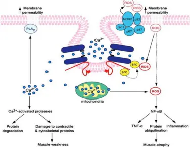
Figure 2.4 Schematic of the multiple pathways involved in DMD pathogenesis that
are activated by Ca2+
Increased cytosolic Ca2+ triggers multiple pathological events such as protein degradation,
inflammation, increased ROS production and impaired ATP synthesis. Figure sourced from (Allen
12
2.1.5 Oxidative stress
Oxidative stress was proposed as a mechanism for muscle injury in DMD as the
pathological changes observed are similar to what is seen under conditions of increased
oxidative stress, and many of the associated biochemical markers have been found in
dystrophic muscle (Disatnik et al., 1998, Hauser et al., 1995, Murphy and Kehrer, 1989).
In addition to this, dystrophic muscle cells seem inherently more susceptible to cellular
injury when exposed to oxidative stress (Rando et al., 1998), and increased free
radical-induced injury have been found in skeletal muscles of DMD patients and the mdx mouse (Hauser et al., 1995, Haycock et al., 1996, Murphy and Kehrer, 1989, Rando, 2002).
While often considered to be a downstream effect in response to other pathological
changes (such as impaired Ca2+ handling and necrosis) there is some suggestion that oxidative stress may be a primary rather than secondary cause of degeneration in DMD.
Disatnik et al (1998) found that in the pre-necrotic state, mdx muscle showed signs of lipid peroxidation and the induction of several antioxidant genes despite no active
cellular necrosis being present. This suggests that elevated oxidative stress precedes
the onset of muscle damage, and that the damage observed could be the direct result of
increased membrane lipid peroxidation (Disatnik et al., 1998). This theory is supported
by McArdle et al (1994) who demonstrated that the increased membrane permeability
that is characteristic of dystrophic tissue is not evident in young (14 day old) mdx mice, indicating that this pathology is secondary to the loss of dystrophin and not a primary
result of its absence.
Be it primary or secondary in nature, increased oxidative stress undoubtedly plays a key
role in muscle tissue pathogenesis in dystrophy and is perpetuated by other aspects of
DMD pathology, in particular, excessive Ca2+ entry. As seen in Figure 2.6, Ca2+ overload in the mitochondria can stimulate the generation of excessive ROS which have been
linked to the development of increased membrane permeability and damage (Brookes et
al., 2004, Whitehead et al., 2006). Interestingly, there appears to be a close and
self-perpetuating relationship between calcium entry and ROS production. ROS can not only
promote the release of Ca2+ from the SR through its actions on the RyR, but has also been shown to increase activity of SAC, establishing an intracellular positive feedback
13
Meissner, 2001).Oneof the primary sources of oxidative damage in dystrophy is NADPH
oxidase, with as much as a 2-fold increase in NADPH oxidase protein expression being
reported in pre-necrotic mdx muscles (Whitehead et al., 2010).(Cozzoli et al., 2011a, Whitehead et al., 2008).
Not surprising given the elevated levels of oxidative stress, antioxidant systems of
skeletal muscle are also altered in dystrophy, with significantly elevated antioxidant
enzymes and antioxidants being found in DMD patients and the mdx mouse (Austin et al., 1992, Dudley et al., 2006). Despite this adaptive change, it appears that this is
insufficient to compensate for chronic exposure to elevated oxidative stress that is
characteristic of dystrophy, as several studies show decreases in damage and improved
muscle function in association with antioxidant treatment (Dorchies et al., 2009, Dudley
et al., 2006, Selsby, 2011).
Forexample, Selsby (2011) recently demonstrated a 30-45% decrease in contraction
induced damage and a 25% reduction in the fatigability of mdx EDL muscle with catalase (enzyme involved in the breakdown of hydrogen peroxide radicals)
over-expression. Similarly, supplementation with green tea extract significantly improved
antioxidant potential, delayed necrosis and decreased fatigue in fast twitch mdx muscle (Dorchies et al., 2009). It appears that antioxidant therapy can also reduce stretch
induced increases in membrane permeability, preventing excess Ca2+ entry and therefore the cascade of events that then leads to increased muscle necrosis
(Whitehead et al., 2008).
Although significant functional improvements associated with antioxidant treatment are
seen in mouse models of dystrophy, findings from human trials attempting to attenuate
disease progression have been largely unsuccessful (Backman et al., 1988, Fenichel et
al., 1988). A possible explanation for this discrepancy is that human trials have utilised
patients already experiencing skeletal muscle degeneration and impaired function,
whereas successful trials in murine models have initiated treatment before significant
muscle damage has occurred (Selsby, 2011). Also, depending on the ROS that is being
produced, some of the antioxidants may be less effective in preventing oxidative
damage (Whitehead, 2006). Despite the conflicting results from animal and human trials,
14
increasing antioxidant availability may protect against disease progression in dystrophic
muscles. However, more research is required to further explore this potential therapeutic
target.
A final crucial point to make about ROS involvement in dystrophic pathology is the link
between ROS production and stimulation of the inflammatory response. ROS are known
to activate the ubiquitous nuclear factor kappa B (NF-κB) pathway (see Figure 2.6),
which is responsible for regulating the inflammatory response through the expression of
several pro-inflammatory cytokines (Acharyya et al., 2007). As chronic inflammation and
impaired repair mechanisms are considered to be a key feature of dystrophy,
inflammation is considered separately in the following section however, it should be
noted that there is a distinct link between excessive ROS production and chronic
inflammation in dystrophy.
2.1.6 Chronic inflammation
Skeletal muscle of DMD patients consistently exhibit inflammatory changes such as
upregulation of pro-inflammatory genes, increases in inflammatory mediators and
infiltration of inflammatory cells, all of which contribute to a state of chronic inflammation
and myofibre necrosis (Acharyya et al., 2007). The infiltration of inflammatory cells was
originally considered to be a non-specific response to muscle fibre damage; however it
appears that aberrant intracellular signalling cascades precede disease onset in
dystrophic skeletal muscle, and contribute substantially to pathology (Evans et al.,
2009). This may be due, in part, to the loss of the DAPC that has now been established
to have a key signalling role within muscle (Zhou et al., 2006). Interestingly,
corticosteroids, which are the most successful and commonly used drug in DMD
treatment, have potent anti-inflammatory effects (Barnes, 1998, Grounds and Torrisi,
2004) and several other immunosuppressive therapies have been shown to significantly
ameliorate muscle wasting when administered prior to disease onset (De Luca et al.,
2005, Hodgetts et al., 2006, Radley and Grounds, 2006).
While there has been several inflammatory response pathways investigated in
dystrophic tissue, two of the most commonly researched are the transcription factor
15
2007, Grounds et al., 2008a, Haslett et al., 2002, Hnia et al., 2008). NF-κB regulates the
expression of a plethora of genes involved in the inflammatory response such as
cytokines, chemokines, immunoreceptors and inflammatory enzymes (Hnia et al., 2008),
while TNF-α has been implicated in several muscle wasting disorders and is a potent
inducer of the inflammatory response (Evans et al., 2009, Figueras et al., 2005, Hnia et
al., 2008). Increased NF-κB activity has been found in the both hind limb and diaphragm
muscle of the mdx mouse, with defective signalling also evident before the onset of muscle degeneration (Acharyya et al., 2007, Kumar and Boriek, 2003). Interestingly,
inhibition of NF-κB activity has been shown to reduce macrophage infiltration by up to
80% in mdx mice coupled with a 77 % reduction in membrane lysis of muscle tissue (Acharyya et al., 2007), as well as improving the morphological appearance of muscle
cells by limiting necrosis (Messina et al., 2006b). It has been proposed that some of the
effects seen with antioxidant therapy are due to modulation of NF-κB activity, as
increased oxidative stress and reduced antioxidants are both key activators of this
pathway (Whitehead et al., 2006).
TNF-α is perhaps the most potent activator of NF-κB, both of which are increased in
DMD patients and the mdx mouse (Hodgetts et al., 2006, Kumar and Boriek, 2003, Messina et al., 2006a). TNF-α is primarily released from activated macrophages and
monocytes, although skeletal muscle is also capable of synthesising this cytokine
intrinsically (Ramos et al., 2004). TNF-α is known to reduce contractile function, activate
proteolytic pathways, increase ROS production and induce muscle wasting (Acharyya et
al., 2007, Evans et al., 2009). Not surprisingly, blockade of TNF-α has been successful
in attenuating the contraction-induced loss of muscle force in the mdx mouse (Piers et al., 2011) and can delay the onset of muscle pathology (Grounds and Torrisi, 2004).
The results from these and several other studies suggest that further investigation into
the role both TNF-α and NF-κB play in the initiation and progression of DMD pathology is
required, and that inflammation may be one of the key therapeutic targets to prevent
16
2.1.7 Impaired metabolism
It has been suggested that DMD results in an impairment of mitochondrial function
causing a compromised cellular energy status (Jongpiputvanich et al., 2005, Tseng et
al., 2002). As significant energy is required for muscle contraction, regeneration and
repair, as well as the activity of key ionic pumps within skeletal muscle, and given that
this demand is even greater under dystrophic conditions, it is likely that impairments to
mitochondrial function contribute significantly to dystrophic pathology (Ge et al., 2003,
Jongpiputvanich et al., 2005). The main source of this mitochondrial dysfunction is
thought be due to excess Ca2+ accumulation in dystrophic muscle. Despite Ca 2+ being a positive stimulator of oxidative metabolism under normal conditions, increased
mitochondrial Ca 2+ (reported as anywhere between two to six fold higher in dystrophy), when coupled with pathology can induce excessive ROS formation, the opening of the
permeability transition pore (PT pore) and apoptosis of muscle cells (Brookes, 2004).
Several studies have demonstrated abnormalities in mitochondrial oxidative metabolism
in muscular dystrophy (human and mouse) including decreases in the activity of
respiratory chain enzymes (Chinet et al., 1994, Even et al., 1994, Kuznetsov et al., 1998,
Onopiuk et al., 2009) and a lower rate of maximum oxygen consumption, although other
groups have reported no significant changes (Braun et al., 2001, Faist et al., 2001).
While research investigating changes in oxidative metabolism have yielded conflicting
results, it seems that alterations to creatine metabolism are widely accepted as a key
feature of dystrophic pathology. DMD patients have a higher urinary excretion of
creatinine coupled with significant reductions in total creatine within muscle tissue (Fitch
and Moody, 1969, Sharma et al., 2003). As PCr is a key energy source for SERCA and
the contractile filaments, it is likely that reduced PCr stores will impact muscle function
and as such creatine supplementation has been investigated as a potential therapy for
DMD. In healthy individuals, creatine has been shown to increase lean tissue mass,
muscle fibre area, strength and endurance capacity (Volek et al., 1999) and studies in
DMD have yielded similar results. Specifically, supplementation with creatine has been
shown to delay the progression of necrosis until day 34 in the mdx mouse, and improve mitochondrial respiration by 25% (Passaquin et al., 2002). In addition, creatine
17
studies have lead to several clinical trials using creatine supplementation in DMD
patients in an attempt to enhance muscle function. Interestingly, while creatine
demonstrates several beneficial effects in the short-term on muscle strength and
function, to date there is no evidence that long-term creatine treatment is beneficial, or
that treatment has any positive effect on patient lifespan (Banerjee et al., 2010).
Glycolytic metabolism has also been shown to be affected in DMD (Onopiuk et al., 2009,
Wehling-Henricks et al., 2009b). A likely candidate for this impairment is the rate-limiting
enzyme PFK, with activity reported to decrease by 45% in type II fibres of DMD patients
(Chi et al., 1987). Interest in this enzyme has recently been reignited due to the apparent
regulation of PFK by nitric oxide (NO), which is altered in DMD due to the lowered nNOS
that results from dislocation of the DAPC (Zhou et al., 2006). NO is known to be an
important signalling molecule in healthy skeletal muscle and is reported to play a role in
mitochondrial biogenesis, glucose transporter type 4 (GLUT-4) translocation and
expression (improving glucose uptake) and modulating neuromuscular transmission
(Wehling-Henricks et al., 2009b). Interestingly, nNOS itself has been found to directly
bind to PFK, suggesting that nNOS may be able to regulate enzyme activity
independently of NO (Wehling-Henricks et al., 2009b). Taken together, these results
suggest that the loss of nNOS and altered NO signalling may be the source of altered
glucose metabolism frequently reported in dystrophy, and a possible contributor to the
disease pathology (Wehling-Henricks et al., 2009b). Collectively, research into metabolic
function in DMD suggests that there is a link between the loss of dystrophin and
impairments in metabolism, which may contribute to disease pathology and impairments
to muscle function, albeit through indirect mechanisms.
2.1.8 Muscle function impairments
i) E-C coupling
While it is well accepted that dystrophic muscle displays significant impairments to whole
muscle function, the mechanisms responsible for the impairments observed in E-C
coupling and contractile function are currently poorly understood. Intact EDL muscle