Polycystic Ovary Syndrome, Obesity and Insulin
Resistance
by
Samantha Cassar
BSpSc, BAppSc(Hons)
This thesis is submitted in fulfilment of the requirements for the degree of
Doctor of Philosophy
College of Sport and Exercise Science
Institute of Sport, Exercise and Active Living
Victoria University
Melbourne Australia
Abstract
Polycystic ovary syndrome (PCOS) is a common and complex reproductive and metabolic
condition with major health consequences across the lifespan. Insulin resistance is thought to be a key underlying feature of PCOS, but its role in metabolic and reproductive
complications associated with the syndrome remains elusive. Therefore, the aim of the thesis was to comprehensively assess the role of IR in PCOS. I report that IR is definitively intrinsic to PCOS and exacerbated by BMI. But the effect of BMI on IR is more pronounced in PCOS
than controls. Diagnostic criteria and age seem to have little effect on IR in PCOS. Furthermore, IR in PCOS seems to be negatively correlated with testosterone and positively
correlated with sex hormone binding globulin. Gonadotropins seem to have little effect on IR. Various biomarkers associated with metabolic diseases appear more strongly associated with obesity rather than with PCOS status and Plasminogen activator inhibitor-1 may also be a
novel independent biomarker with the ability to predict IR in women with and without PCOS. This intrinsic IR in PCOS was not attributed to mitochondrial dysfunction and was not related
to the pathophysiology of reproductive dysfunction in PCOS as measured by Anti-Mullarian hormone (AMH). However, AMH was able to detect PCOS status and may also be useful in the diagnosis of PCOS. Collectively these chapters of related studies enhanced understanding
Doctor of Philosophy by Publication Student Declaration
I, Samantha Cassar, declare that the PhD thesis by publication entitled ‘Polycystic ovary syndrome, obesity and insulin resistance’ is no more than 100,000 words in length including
quotes and exclusive of tables, figures, appendices, bibliography, references and footnotes. This thesis contains no material that has been submitted previously, in whole or in part, for
the award of any other academic degree or diploma. Except where otherwise indicated, this thesis is my own work.
Thesis by Publication
All doctoral students at Victoria University are permitted to submit a thesis by publication. The thesis by publication is a thesis format that includes manuscripts that have been prepared
or accepted for publication. These manuscripts may have more than one author in which signatures from co-authors are required. The papers do not have to be rewritten for the thesis;
they can be inserted in their published format. The thesis must reflect a sustained and cohesive theme and framing or substantial linking text normally required in introducing the research and linking the chapter and manuscripts.
The work conducted in this thesis was part of a large National Health and Medical Research
Council (NHMRC) funded project investigating IR in PCOS. I joined the research team at Monash Centre for Health Research and Implementation, Monash University in 2009. My PhD supervisors, Associate Professor Nigel Stepto and Professor Helena Teede, initially instigated
this project, which focused on obese women with and without PCOS. I joined the team at the infancy stages of the second arm of study, at the beginning of my PhD, where my primary
role was to investigate IR in lean women with and without PCOS. I established a new clinical room on the premises and I was responsible for ethics amendments, recruitment and screening of all participants and was the primary contact for the study. I also organised and
accompanied each participant to their medical review and body composition appointments. I Organised and conducted fitness testing and assisted with euglycaemic-hyperinsunaemic
clamp appointments, including setting up all consumables, cannulation of some participants, blood sampling, analysing blood glucose levels, centrifuging blood samples, assisting with muscle biopsies and preparing muscle samples for analysis and ensuring the health and
correctly. I independently performed laboratory analysis and analysed and interpreted data collected.
My specific contributions are detailed at the beginning of each chapter. All of my work
Details of Included Papers: Thesis by Publication
Item/ Chapter
No.
Paper Title Publication Status Publication Title and Details
2
Insulin resistance in women with polycystic ovary syndrome: A systematic review and
meta-analysis of euglycaemic
Currently under review
Human Reproduction Update Impact factor: 8.6 Quartile ranking: Q1
3
Women with polycystic ovary syndrome have intrinsic insulin resistance on euglycaemic–
hyperinsulaemic clamp.
Published
Human Reproduction, Vol.28, No.3 pp. 777– 784, 2013
Accepted 18 December 2012 Impact factor: 4.6 Quartile ranking: Q1 Scopus citation number: 45
4a
Biomarkers and insulin sensitivity in women with PCOS: Characteristics and predictive
capacity.
Published
Clinical Endocrinology, DOI:10.1111/cen.12619 Accepted 22 September 2014
4b Response to ‘insulin sensitivity and leptin in
women with PCOS’ Published
Clinical Endocrinology, DOI:10.1111/cen.12678 Letter to the Editor
Accepted: 17 November 2014 Impact Factor: 3.35
Quartile ranking: Q1
5
Mitochondrial dysfunction as a potential mechanism underlying intrinsic insulin resistance in Polycystic Ovary Syndrome
Not Submitted
6
Polycystic ovary syndrome and anti-Müllerian hormone: role of insulin resistance, androgens,
obesity and gonadotrophins
Published
Clinical Endocrinology, Vol.81, Issue 6, pp. 899-906
Accepted 10 July 2014 Impact Factor: 3.35 Quartile ranking: Q1
Declaration by: Signature: Date:
Publications
Publications arising directly from this thesis:
*NK Stepto, *S Cassar, AE Joham, SK Hutchison, CL Harrison, RF Goldstein, HJ Teede, Women with polycystic ovary syndrome have intrinsic insulin resistance on euglycaemic–
hyperinsulaemic clamp, Human Reproduction, Vol.28, No.3 pp. 777–784, 2013 (*Joint first authors)
S Cassar, HJ Teede, LJ Moran, AE Joham, CL Harrison, BJ Strauss, NK Stepto, Polycystic ovary syndrome and anti-Mullerian hormone: role of insulin resistance, androgens, obesity
and gonadotrophins, Clinical Endocrinology, 2014 doi: 10.1111/cen.12557
S Cassar, HJ Teede, CL Harrison, AE Joham, LJ Moran, NK Stepto, Biomarkers and insulin
sensitivity in women with PCOS: Characteristics and predictive capacity, Clinical Endocrinology, 2014 DOI 10.1111/CEN12619
S Cassar, HJ Teede, CL Harrison, AE Joham, LJ Moran, NK Stepto. Response to ‘insulin sensitivity and leptin in women with PCOS, Clinical Endocrinology, 2014 DOI
Conference Presentations
Poster Presentations
S Cassar, NK Stepto, SK Hutchison, CL Harrison, HJ Teede, Exercise training enhances but does not restore insulin sensitivity and signalling in obese women with PCOS, 17th European College of Sport Science Congress 4-7 July 2012 Bruges, Belgium
S Cassar, NK Stepto, SK Hutchison, CL Harrison, HJ Teede, Mitochondrial function and
insulin resistance in women with polycystic ovary syndrome, 15th International Biochemistry of Exercise Conference (IBEC) Congress 17-21 June 2012
Oral Presentations
S Cassar, NK Stepto, LJ Moran, SK Hutchison, CL Harrison, HJ Teede, Markers of
Metabolic Disease Risk in Polycystic Ovary Syndrome, The Annual Scientific Meeting of the Endocrine Society of Australia and the Society for Reproductive Biology 2013
S Cassar, HJ Teede, M Misso, WG Hopking, NK Stepto, Insulin sensitivity in women with a polycystic ovary syndrome: A systematic review and meta-analysis, The Annual Scientific
Additional Conference Presentations Arising from this Thesis
Anju Elizabeth Johan, Samantha Cassar, Nigel Stepto, Cheryce L Harrison, Samantha Kate
Acknowledgements
The work presented in this thesis would not have been possible without the support and encouragement of many colleagues, family and friends.
I would like to express my sincere gratitude to my supervisory team; Associate Professor Nigel Stepto, for his continual encouragement, guidance, feedback and financial support
throughout my candidature. Professor Helena Teede for her enthusiasm, prompt and insightful feedback with manuscript submissions and the thesis, positive attitude, invaluable
advice, expertise in the area of women’s health and financial support; and Dr Christopher Shaw for his support, feedback and advice.
Dr Rebecca Goldstein and Dr Anju Joham for conducting the medical reviews, muscle biopsies and eulycaemic hyperinsulinaemic clamps. Your expertise and professionalism are
second to none.
Melanie Gibson-Helm and Nicole Ng for their tireless efforts, amazing problem-solving
skills, ‘mental health’ support, making me laugh when I wanted to cry, and friendship. I couldn’t have done it without both of you.
Dr Lisa Moran for your time, vital advice, profound knowledge, patience and rapid reply to e-mails. There are no words to express my sincere gratitude and appreciation.
Dr Marie Misso and Professor Will Hopkins for their imperative assistance with the
Samantha Hutchison and Cheryce Harrison for their tireless work with the obese arm of this study.
Professor Boyd Strauss for generously providing the DEXA and CT scans and interpretation
of results.
Southern Health Pathology and Dr Michael Daskalakis from the Department of Biochemistry
and Molecular Biology, Monash University for performing biochemical analysis including HbA1c, cholesterol, and triglycerides, testosterone, luteinising hormone, follicle stimulating
hormone and sex hormone globulin binding.
Past and present postgraduate students, who helped me acquire new laboratory skills and
were always available for assistance, advice or a friendly chat.
The participants for their time, patience, and dedication to research. Without you, none of this would be possible.
Lastly, I must thank my family and friends for their continual support and encouragement, especially my mum who made sure I never went hungry and always had clean clothes and
Table of Contents
Abstract ... ii
Doctor of Philosophy by Publication Student Declaration ... iii
Thesis by Publication ... iv
Details of Included Papers: Thesis by Publication ... vi
Publications ... viii
Conference Presentations ... ix
Poster Presentations ... ix
Oral Presentations ... ix
Additional Conference Presentations Arising from this Thesis ... x
Acknowledgements ... xi
List of Tables ... xix
List of Figures ... xx
Abbreviations ... xxi
Chapter 1 Review of Literature ... 1
1.0 Introduction and definition of polycystic ovary syndrome ... 1
1.1 Diagnostic Criteria of PCOS ... 2
1.3 Significance and Economic Burden of PCOS ... 6
1.4 PCOS Aetiology... 7
1.4.1 Genetics... 7
1.4.2 Prenatal Androgen Excess ... 8
1.4.3 Hypothalamic-Pituitary Axis ... 9
1.4.4 Obesity ... 10
1.5 Health Complications of PCOS ... 12
1.5.1 Reproductive Consequences ... 12
1.5.2 Pregnancy Risks ... 13
1.5.3 Metabolic Complications ... 14
1.5.4 Cardiovascular Complications ... 14
1.5.5 Carcinogenic Complications ... 15
1.5.6 Psychosocial Complications ... 16
1.6 Management Strategies in PCOS ... 16
1.7 Insulin Resistance in PCOS ... 17
1.7.1 Definition of Insulin Resistance... 17
1.7.2 Importance of Insulin Resistance in PCOS ... 18
1.7.4 Effect of Diagnostic Criteria on Prevalence of Insulin Resistance ... 20
1.7.5 Insulin Resistance - Aetiology in PCOS ... 21
1.7.6 Possible Defects in Skeletal Muscle of Women with PCOS ... 22
1.7.7 Insulin Resistance and Mitochondrial Dysfunction in Skeletal Muscle ... 24
1.8 Summary and Research Gaps ... 26
1.9 Aims of the Thesis ... 27
1.10 Organisation of the Thesis ... 27
Chapter 2 Insulin Resistance in Women with Polycystic Ovary Syndrome: A Systematic Review and Meta-Analysis of Euglycaemic-Hyperinsulinaemic Clamp Studies ... 28
Declaration of Co-Authorship and Co-contribution: Papers incorporated in thesis by publication... 29
2.0 Introduction ... 32
2.1 The Euglycemic-Hyperinsulinemic Clamp... 32
2.2 Fasting Insulin ... 34
2.3 Surrogate Indices for Insulin Resistance ... 35
2.4 Oral Glucose Tolerance Test (OGTT) ... 36
2.5 Assessment of Insulin Resistance ... 37
2.6.1 Testosterone ... 38
2.6.2 Sex-Hormone Binding Globulin and Insulin Resistance ... 39
2.6.3 Gonadotropins ... 40
2.7 Assessment of Literature... 41
2.7 Summary ... 42
2.8 My Role ... 42
Chapter 3 Women with Polycystic Ovary Syndrome have Intrinsic Insulin Resistance on Euglycaemic-Hyperinsulaemic Clamp. ... 167
Declaration of Co-Authorship and Co-Contribution: Paper Incorporated in Thesis by Publication ... 168
3.0 Introduction ... 171
3.1 Insulin Resistance in PCOS ... 171
3.2 My Role ... 173
Chapter 4a Biomarkers and insulin sensitivity in women with PCOS: Characteristics and predictive capacity. ... 181
Declaration of Co-Authorship and Co-Contribution: Paper Incorporated in Thesis by Publication ... 182
4.0 Introduction ... 185
4.2 Bio-Plex Immunoassay ... 187
4.3 Summary ... 188
4.4 My Role ... 188
Chapter 4b Response to ‘Insulin Sensitivity and Leptin in Women with PCOS.’ ... 197
Declaration of Co-Authorship and Co-Contribution: Paper Incorporated in Thesis by Publication ... 198
4.5 Introduction ... 200
4.6 My Role ... 200
Chapter 5 Mitochondrial Dysfunction as a Potential Mechanism Underlying Intrinsic Insulin Resistance in Polycystic Ovary Syndrome ... 204
5.0 Introduction ... 205
5.1 Role of mitochondria in insulin resistance ... 205
5.3 Techniques to measure Mitochondrial Function ... 207
5.4 Summary ... 208
5.5 My Role ... 208
Chapter 6 Polycystic ovary syndrome and Anti-Müllerian hormone: Role of insulin resistance, androgens, obesity and gonadotropins. ... 227
6.0 Introduction ... 231
6.1 AMH in Polycystic Ovary Syndrome ... 231
6.2 AMH and Hyperandrogenism ... 232
6.3 AMH and Insulin Resistance ... 232
6.4 AMH and Obesity ... 233
6.5 AMH and Gonadotropins ... 233
6.6 Summary ... 234
6.7 My Role ... 234
Chapter 7 Discussion and Conclusion ... 242
7.0 Discussion and Conclusions ... 242
7.1 Limitations ... 245
7.2 Key Future research goals ... 246
7.3 Overall Conclusion ... 247
References ... 248
Appendix 1 Copyright agreement for Chapter 3 ... 285
Appendix 2 Copyright agreement for Chapter 4a ... 288
Appendix 3 Copyright agreement for Chapter 4b ... 294
List of Tables
Chapter 2
Table 1 Studies included in meta-analysis ……..……… Page 60
Table 2 Studies included in meta-analysis investigating moderating ………. Page 65 effects of key hormones
Table 3 Meta-analysed overall mean effect of PCOS and insulin sensitivity .. Page 68 Table 4 Associations of differences in hormone concentrations with …….…. Page 73
differences in insulin sensitivity between women with and without PCOS
Table S1 MOOSE Checklist ……… Page 94
Table S2 Search strategy for systematic review ……….. Page 97
Table S3 Quality appraisal of included studies ……….... Page 98
Chapter 5
List of Figures
Chapter 2
Figure 1 Flow diagram of the systematic review study selection process …. Page 59
Figure 2 The effect of BMI on the difference in insulin sensitivity ……...… Page 70 between PCOS and control groups
Figure S1 Scatter plot of t-statistic associated with each study ……… Page 166
Chapter 5
Figure 1 Oxidative phosphorylation antibody linearity gel ………. Page 217 Figure 2 Mitochondrial respiration as measured by high resolution ………… Page 220
respirometry in lean women with and without PCOS.
Figure 3 Mitochondrial respiration changes as a result of the addition of …… Page 220 ADP.
Figure 4A Mitochondrial protein expression in women with and without ……... Page 221 PCOS.
Figure 4B Representative blot of mitochondrial protein expression for ……….. Page 221 complexes I-V.
Figure 5 Citrate synthase activity as an indicator of mitochondrial ………… Page 222
Abbreviations
AES Androgen Excess Society AMH anti-mullerian Hormone
ASRM American Society of Reproductive Medicine ATP adenosine triphosphate
AU arbitrary units BMI body mass index CI confidence interval
CVD cardiovascular disease CYP-17 cytochrome P450c17
DHEA dehydroepiandrosterone
DHEAS dehydroepiandrosterone sulphate DNA deoxyribonucleic acid
ECT electron transport chain
ESHRE European Society of Human Reproduction and Embryology
FAI free androgen index
FCCP carbonyl cyanide-4-(trifluoromethoxy) phenylhydrazone FFA free fatty acids
FSH follicle stimulating hormone GDM gestational diabetes mellitus
GIP glucose-dependant insulinotropic polypeptide GIR glucose infusion rate
G/I ratio fasting glucose to insulin ratio
GnRH gonadotropin releasing hormone HbA1c glycated haemoglobin
HOMA homeostatic model of assessment HPA hypothalamic-pituitary axis
HRP horseradish peroxidase HRR high resolution respirometry IGT impaired glucose tolerance
IKK-β inhibitor of nuclear factor kappa-B kinase subunit beta INSR insulin receptor
INSR-1 insulin receptor substrate-1 INSR-1 insulin receptor substrate-2 IR insulin resistance
IVGTT intravenous glucose tolerance test JNK C-Jun N-terminal kinase
LH luteinising hormone
MOOSE Meta-analysis of Observational Studies in Epidemiology mRNA messenger ribonucleic acid
mtDNA mitochondrial deoxyribonucleic acid NADH nicotinamide adenine dinucleotide hydride
NAFLD non-alcoholic fatty liver disease NIH National Institute of Health
NHMRC National Health and Research Medical Council
OR odds ratio
OXPHOS Oxidative phosphorylation
PCO polycystic ovary
PCOM polycystic ovary morphology
PCOS polycystic ovary syndrome
PGC-1α proliferator-activated receptor γ coactivator α
PKB protein kinase B
PI3K phosphoinositide 3-kinase
PRISMA Preferred Reporting Items for Systematic reviews and Meta-analyses
QUICKI quantitative insulin sensitivity check index QUOROM Quality of Reporting of Meta-analyses
ROS reactive oxygen species ROX residual oxygen consumption SD standard deviation
SHBG sex hormone binding globulin T2DM type 2 diabetes mellitus
TGFβ transforming growth factor β
VO2max Maximal oxygen consumption
Chapter 1 Review of Literature
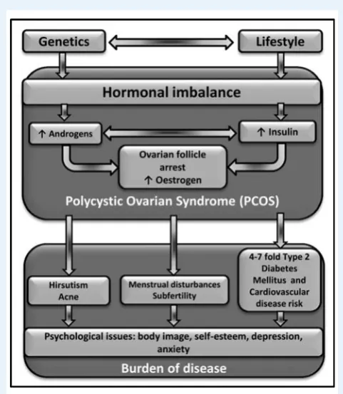
1.0 Introduction and definition of polycystic ovary syndrome
Polycystic ovary syndrome (PCOS) is a common metabolic and endocrine disorder affecting
6 to 21% of reproductive aged women depending on population, mean body mass index (BMI) and diagnostic criteria used (March et al., 2010, Boyle et al., 2012). High prevalence are rates seen in women whom are overweight or have an Indigenous or Asian background
(Boyle et al., 2012, March et al., 2010). The features of PCOS, including menstrual dysfunction, infertility and hirsutism have been described in medical records for more than
2,000 years (Azziz et al., 2011). The syndrome was officially recognised in the 1930’s by Stein and Leventhal who associated polycystic ovaries (PCO) to the clinical features of menstrual dysfunction, infertility, hirsutism and obesity (Stein and Leventhal, 1935). Since
the 1980’s, researchers expanded on these observations to report an association between hyperinsulinaemia and hyperandrogenism bringing to light possible aetiologies and a
complicated metabolic and reproductive condition with psychosocial and economic consequences across the lifespan (Burghen et al., 1980, Dunaif, 1989, Shoupe et al., 1983, Teede et al., 2010). These ground-breaking studies also caused great debate as to whether
insulin resistance (IR) is a unique feature of PCOS contributing to clinical features and health consequences.
During the past two decades skeletal muscle dysfunction has been associated with the pathogenesis of IR, including dysfunctional insulin signalling and mitochondria, and the
release of cytokines. However, there is still little consensus regarding the underlying mechanisms of IR in PCOS. Therefore further investigation into the aetiology of PCOS and
The present chapter provides an overview of the background of this thesis by describing the literature regarding PCOS and proposed aetiology, which is followed by an explanation of IR
and the current understanding of its role in the development of PCOS and associate reproductive consequences. It also concludes by highlighting gaps in our current knowledge.
1.1 Diagnostic Criteria of PCOS
The diagnostic criteria for the syndrome continue to evolve with advances in both the
understanding of the condition and the precision of medical equipment and technology. Hyperandrogenism, menstrual irregularity, and PCO have been proposed as the diagnostic features of PCOS (Rotterdam, 2004). Hyperandrogenism is assessed by clinical and
biochemical measurements. Clinical features of hyperandrogenism include hirsutism, acne, female-pattern alopecia (hair loss) and acanothosis nigricans, which occur in 73.9%, 53.3%,
34.8% and 5.3% of women with PCOS respectively (Ozdemir et al., 2010). These differ across ethnicities, are often subjective and altered by cosmetic treatment such as laser (McGill et al., 2007). This makes clinical features difficult to interpret when diagnosing
PCOS. Biochemical hyperandrogenism is determined by free testosterone and/or free androgen index (FAI) and are composite measures that include assessments of circulating
levels of serum testosterone and sex hormone binding globulin (SHBG) (Norman et al., 2007). However, it is unclear which androgens are best measured, what constitutes normal reference ranges in women and which analytical technique should be used, with recent
publications questioning the specificity and accuracy of commonly used immunoassays (Barth et al., 2007, Handelsman and Wartofsky, 2013, O'Reilly et al., 2014)
anovulation; the absence of menstruation for more than three months without pregnancy (Norman et al., 2007). Approximately 90% of women with oligoovulation or anovulation are
diagnosed with PCOS and up to 95% of women with PCOS present with these symptoms (Rotterdam, 2004). During adolescence, perimenopause and postmenopause, interpreting the
cause of irregular menstrual cycles is challenging and makes diagnosis more difficult.
Polycystic ovarian morphology (PCOM) is most commonly measured using transvaginal
ultrasound and is defined as the presence of 12 or more follicles in one or both ovaries, measuring 2-9 mm in diameter, and/or having an ovarian volume greater than 10 mL
(Rotterdam, 2004). Over 90% of women with PCOS have PCOM and women with PCO can have up to six times more early developing antral follicles compared to the ovaries of healthy women. PCO remains non-specific for PCOS and prevalence rates of PCO are increasing
because of more advanced imaging equipment (Leonhardt et al., 2014). Specificity of PCOM in PCOS diagnosis remains controversial. Anti-mullerian hormone (AMH) concentrations are
reflective of the number of pre-antral and small antral follicles in the ovary and raised concentrations are evident in women with PCO and PCOS. This makes AMH concentrations a potential tool to aid in the diagnosis of PCOS (Dewailly et al., 2014, Homburg et al., 2013).
Currently, IR is not included as a diagnostic criterion for PCOS, even though it is acknowledged to be a central feature of the condition (Teede et al., 2011). This is largely that
IR cannot be simply and accurately measured in clinical practise, hence routine testing is not recommended in PCOS.
To date, there are three definitions or diagnostic criteria for PCOS, which can make the diagnosis experience confusing and lengthy for both the clinician and woman involved
National Institutes of Health (NIH) consensus group and were based on opinion rather than clinical evidence (Azziz et al., 2006). PCOS was defined as having clinical and/or
biochemical signs of hyperandrogenism and oligoanovulation. In 2003, the second iteration of diagnostic criteria for PCOS was established by the European Society for Human
Reproduction and Embryology and the American Society of Reproductive Medicine (ESHRE/ASRM) consensus group to include any two of the three criteria; clinical and/or biochemical signs of hyperandrogenism, oligo-anovulation and PCO on ultrasound. This set
of criteria is referred to as the Rotterdam criteria and introduces different PCOS phenotypes; classic (hyperandrogenism and oligoanovulation), ovulatory (hyperandrogenism and PCO)
and normoandrogenic (oligoanovulation and PCO) (Rotterdam, 2004). In 2006, the Androgen Excess Society (AES) published a position statement, based on an evidence based review that suggested hyperandrogenism should be the key component to diagnose PCOS together with
oligoanovulation or PCO or both (Azziz et al., 2006, Azziz et al., 2009). All three definitions require the exclusion of other conditions that cause clinical features of PCOS. These
conditions include congenital adrenal hyperplasia, Cushing’s disease, thyroid dysfunction and hyperprolactinemia (Azziz et al., 2006, Azziz et al., 2009, Rotterdam, 2004).
The NIH and AES criteria tend to diagnose women with the more severe spectrum of the disease. However, the Rotterdam criteria are now internationally accepted by the NIH,
Australian guidelines and European societies. Given that an estimated 70% of PCOS cases are undiagnosed in Australia (March et al., 2010), reducing confusion and promoting awareness of the endorsed diagnostic criteria is important (Rotterdam, 2004, Teede et al.,
1.2 Epidemiology
The lack of prior consensus on the definition and diagnosis of PCOS has undermined
investigations attempting to accurately determine population-based prevalence rates for the condition (Hart et al., 2004, March et al., 2010). Despite PCOS being the most common endocrine disorders in reproductive aged women, prevalence estimates are highly variable
ranging from 2-21% depending on the diagnostic criteria used, recruitment strategy, population studied (selected and unselected populations) and ethnicity (Asuncion et al., 2000,
Boyle et al., 2012, Chen et al., 2008b, Diamanti-Kandarakis et al., 1999, Farah et al., 1999, Knochenhauer et al., 1998, Kumarapeli et al., 2008, March et al., 2010). The introduction of the Rotterdam criteria broadened the previous NIH definition by including PCO morphology
and therefore two additional phenotypes and as a consequence prevalence rates rose from an estimated 6-8% to 12-20% (Boyle et al., 2012, March et al., 2010, Yildiz et al., 2012).
The only community based study in Australia to assess prevalence rates of PCOS using current international diagnostic criteria (Rotterdam) reported a prevalence of 11.9% in 728
women between 27-34 years of age (March et al., 2010). The prevalence increased to 17.8% using the Rotterdam criteria when imputed data was included for participants not consenting
to ultrasounds (March et al., 2010). Furthermore, the prevalence of PCOS is higher in Australian Indigenous communities where it affects up to 21% of women (Davis et al., 2002, Boyle et al., 2012). A community based Iranian study investigated prevalence rates in 1126
women aged 18-45 years and found similar rates to that of the Australian study, 7.1%, using the NIH definition and 14.6% using the Rotterdam criteria (Tehrani et al., 2011). Prevalence
Lankan population (Rotterdam, n = 2,915) (Kumarapeli et al., 2008), 6.5% in women (NIH, n = 154) living in Spain volunteering to donate blood (Asuncion et al., 2000), 6.8% (n = 192) in
a Greek population (Diamanti-Kandarakis et al., 1999) and 26% in the United Kingdom (n = 230) (Michelmore et al., 1999).
Additionally, the prevalence of PCOS increases by 9.2% for every single unit increment in body mass index (BMI) (Teede, 2013) and PCOS is five times higher in obese populations
compared to women within a healthy weight range (Alvarez-Blasco et al., 2006). In contrast, reported prevalence rates of PCOS in underweight, normal-weight, overweight, obese and
moderate-obese women to be 8.2%, 9.8%, 9.9%, 9.0% and 12.4% respectively, leaving authors to concluded that PCOS is likely due to intrinsic or inherited factors (Yildiz et al., 2008). Therefore, obesity may have a small effect on prevalence rates of PCOS, but it has
profound effects on the presentation of clinical features and degree of IR in PCOS potentially due to the endocrine function of adipose tissue (Yildiz et al., 2008).
1.3 Significance and Economic Burden of PCOS
The short and long term health consequences associated with PCOS cause a large economic
burden. Calculations of the health related economic burden are based on the estimates of prevalence rates, co-morbidities and the expense of diagnosing and treating the condition (Azziz et al., 2005). In 2005, the estimated economic burden of diagnosing and providing
care for women with PCOS was $US4 billion annually in the United States (Azziz et al., 2005), equating to an estimated AU$800 million in Australia (Teede et al., 2011). PCOS
infertility care (Azziz et al., 2005). Furthermore, PCOS is the most common cause of anovulatory infertility in women, with high costs in infertile obese Australian women (Clark
et al., 1998). In the estimates detailed above, the NIH criteria were used to derive prevalence rates and the economic burden of PCOS. This may have underestimated the financial burden,
as prevalence rates are two to three fold higher when Rotterdam criteria is used. Surprisingly, only 2.3% of the economic burden was attributed to diagnosis and evaluation of the condition (Azziz et al., 2005). It is now recognised that there is a need to increase awareness of PCOS
and investment to aid in the early diagnosis and treatment preventing the onset of serious sequelae including infertility, IR, type 2 diabetes mellitus (T2DM) and cardiovascular disease
(CVD) (Azziz et al., 2005).
1.4 PCOS Aetiology
The aetiology of PCOS is complex and remains elusive. A combination of both genetic and environmental factors including ovarian dysfunction, hormonal disturbances, underlying hyperandroginism, IR, obesity and abnormalities in gonadotropin secretion have been
implicated in the aetiology of PCOS.
1.4.1 Genetics
A family history of PCOS and genetics has been implicated in the aetiology of PCOS and variations in phenotypical expression. PCOS has high heritability with monozygotic twin
sisters (tetrachoric correlation 0.71) being twice as likely to have PCOS compared to dizygotic twin sisters and other female siblings (tetrachoric correlation 0.38) (Vink et al.,
reported to cluster in families with PCOS. Mothers, sisters and brothers of women with PCOS are reported to have a defect in steriodogenesis leading to androgen excess compared
to controls (Legro et al., 2002b, Yildiz et al., 2012). First-degree relatives of women with PCOS also have IGT or IR (Yildiz et al., 2012). Genome-wide association studies in Han
Chinese women have reported 11 genetic loci linked to PCOS and these loci are found on regions that house genes responsible for gonadotropins, insulin signalling, reproductive hormones and T2DM (Chen et al., 2011, Shi et al., 2012). A replication study confirmed that
some of these variations in loci were also evident in European women and may be important in the aetiology of PCOS independent of ethnicity (Goodarzi et al., 2012).
1.4.2 Prenatal Androgen Excess
Although beyond the scope of this thesis, the prenatal environment and in particular androgen
excess intrauterine, is thought to play a pathophysiologic role in PCOS by contributing to reproductive and metabolic dysfunction in offspring (Abbott et al., 2005). Metabolic and
reproductive dysfunctions including hyperandrogenism, PCO, elevated luteinising hormone (LH) concentrations, IR, hyperlipidemia, glucose intolerance, and increased risk of T2DM have been reported in a variety of pre-androgenised animal models (Abbott et al., 1998, Birch
et al., 2003, Dumesic et al., 1997, Eisner et al., 2002, Manikkam et al., 2004, Recabarren et al., 2005). In rodent models the degree of reproductive or metabolic dysfunction was dose
dependent upon testosterone exposure (Foecking et al., 2005, Wu et al., 2010). Women with PCOS are reported to have elevated androgen levels during gestation and have higher concentrations of enzymes that convert unconjugated steroids into androstenedione and
subsequently testosterone in the placenta (Maliqueo et al., 2013, Sir-Petermann et al., 2002). Furthermore, testosterone levels are reported to be elevated to male levels in the umbilical
finding has not been replicated and is still a topic of debate (Anderson et al., 2010). Overall, the prenatal environment and excess androgen exposure intrauterine may play a role in the
development of PCOS.
1.4.3 Hypothalamic-Pituitary Axis
The Hypothalamic-pituitary axis (HPA) is a complex feedback loop comprising of the hypothalamus (containing gonadotropin releasing hormone (GnRH) neurons), pituitary gland
(responsible for the secretion of LH and follicle stimulating hormone [FSH]) and the ovary, which responds to changes in gonadotropin concentrations by follicular maturation and
ovulation (Roland and Moenter, 2014). It is proposed that abnormalities in the HPA exist in women with PCOS. These abnormalities result in an increased GnRH pulse frequency and disruption in the release of LH and FSH leading to an increase in immature follicles on the
ovary and menstrual dysfunction. Various studies have reported an increase in LH concentrations, LH/FSH ratio, and GnRH and LH pulse frequency and amplitude in women
with PCOS (Taylor et al., 1997, Waldstreicher et al., 1988). Whether defects in gonadotropin release are inherent to PCOS or secondary to developing the condition is under debate. Insulin and hyperandrogenism are factors proposed to play a role in gonadotropin regulation.
Hyperinsulinaemia causes an increase in LH receptor expression and premature release of the follicle, which combine to cause follicular arrest and subfertility or infertility
(Diamanti-Kandarakis, 2008). Evidence for a regulatory role of androgens in altered gonadotropin secretion is supported by the finding that prenatally androgenised rhesus monkeys and sheep produce female offspring with LH hypersecretion, hypothesised to occur due to altered
increase AMH through augmenting follicular growth and these high AMH concentrations may also negatively feedback on FSH production.
1.4.4 Obesity
Obesity is defined as excessive fat accumulation, which is commonly assessed using BMI. A BMI of 18.5 to <25 km/m2 is considered to be healthy, whereas a BMI of 25 to 29 km/m2 is classified as overweight and >30 km/m2 as obese. Obesity may have a bidirectional
relationship with PCOS; women with PCOS are predisposed to weight gain and excess weight gain appears to increase the prevalence of PCOS (Shorakae et al., 2014, Teede, 2013).
Adipose tissue acts as an endocrine organ secreting proteins known as adipokines (including resistin, visfatin, and Plasminogen activator inhibitor-1 [PAI-1]), which play a role in energy metabolism through their interactions with the liver, skeletal muscle, brain, pancreas and
reproductive system (Scherer, 2006). Obesity is strongly related to PCOS, with up to 61% of women with the condition being overweight or obese, although this varies with ethnicity and
the cause and association are still being investigated (Lim et al., 2012). Obesity has been reported to precede oligoovulation and hyperandrogenism and has deleterious effects on metabolic features of PCOS, in particular insulin sensitivity (Gambineri et al., 2002).
Reproductive disturbances of oligoovulation and anovulatory infertility are more common in
obese women compared to lean women irrespective of PCOS diagnosis (Teede, 2013). The risk of these reproductive disturbances worsens with increasing BMI (Teede, 2013). Obesity may also increase the risk of miscarriages and impair the outcome of assisted reproductive
technologies (Pasquali et al., 2014). Furthermore, obese women with PCOS tend to have higher testosterone levels compared to lean PCOS women. Excess adipose tissue may
consequently increasing the bioavailability of sex steroids including testosterone (Holte, 1996).
Metabolically, IR is compromised by obesity in both the general population and in women
with PCOS. The risk and degree of IR rises with increasing BMI; 27 kg/m2 is the critical point where a marked decrease in insulin sensitivity is observed (Garcia-Estevez et al., 2004). Furthermore, obese women with PCOS display higher fasting and glucose-stimulated insulin
concentrations and lower whole body insulin sensitivity compared to their non-PCOS counterparts (Dunaif et al., 1989). Although the degree of obesity is important in the
development of IR, the distribution of this adipose tissue also seems to have an effect. Individuals with a high intra-abdominal fat distribution tend to be less insulin sensitive compared to individuals with a more subcutaneous fat distribution (Carey et al., 1996, Cnop
et al., 2002, Fujimoto et al., 2000, Kahn, 2003).
It appears that obesity worsens the features of PCOS. Specifically, women diagnosed by the NIH criteria (menstrual dysfunction and hyperandrogenemia) were more obese compared to their sisters with the less severe phenotype of regular menstrual cycles and
hyperandrogenemia. Healthy sisters unaffected by PCOS were reported to have a lower body mass compared to both PCOS groups (Kahn et al., 2006, Legro et al., 2002a). While obesity
modifies the phenotype and/or severity of the features of PCOS, it is unclear whether it is a key player in the aetiology of the syndrome. To better understand the interaction between obesity and PCOS, this thesis will explore the impact of obesity on reproductive and
1.5 Health Complications of PCOS
PCOS is a lifelong condition associated with numerous clinical sequelae including
reproductive, metabolic, cardiovascular, carcinogenic and psychosocial comorbidities (Teede et al., 2010). These comorbidities vary in severity across the lifespan with features of hyperandrogenism most prominent among adolescents, whereas fertility and reproductive
issues are prominent among women in their 20’s and 30’s. Metabolic features, weight gain, and psychosocial challenges affect all ages, with metabolic features occurring earlier in
individuals who are overweight (Teede et al., 2011).
1.5.1 Reproductive Consequences
Women with PCOS may be subfertile with ovulatory dysfunction exacerbated by hyperandrogenism and obesity associated with the syndrome (Group, 2012, Rotterdam,
2004). Subfertility refers to reduced fertility in couples unsuccessfully trying to conceive (Gnoth et al., 2005) and is most commonly a result of anovulation, which affects 55-91% of women with PCOS (Loumaye et al., 2003). In a large epidemiological study comprising
4,535 women from North Finland, women with self-reported PCOS symptoms suffered more frequently from subfertility (26% versus 17% in healthy controls) and took longer to
conceive for the first time (Koivunen et al., 2008). Interestingly, the number of women conceiving and their family size (number of children) were similar between women with and without PCOS symptoms (Koivunen et al., 2008). It is feasible that women with PCOS took
longer to conceive because of fewer ovulatory cycles (Liang et al., 2011). With time the overall fertility of women with symptoms of PCOS may not be greatly impaired.
IR and hyperandrogenism are two mechanisms proposed to contribute to PCOS
consequences. Hyperinsulinaemia increases the availability of testosterone and other steroids by suppressing the production of SHBG. IR also augments LH and ovarian androgen
production, leading to hyperandrogenism. This accelerates pre-antral and antral follicular growth in the ovary and elevated LH results in premature luteinisation causing follicular arrest and decreased oocyte quality (Piouka et al., 2009, Diamanti-Kandarakis and Dunaif,
2012). Obesity has an additional inhibitory effect on gonadotropin release due to an increase in aromatization of androgens in adipose tissue resulting in the suppression of LH and the
consequent inhibition of the dominant follicle (Grossman et al., 2008). Treatment with insulin lowering medication induces regular menstrual cycles and improves pregnancy rates (Brettenthaler et al., 2004). The role of IR, androgens and gonadotropins in ovarian
dysfunction will be further discussed in Chapter 7.
1.5.2 Pregnancy Risks
Along with subfertility, women with PCOS may also experience complications during pregnancy (Roos et al., 2011); increased risk of miscarriage during the first trimester
(30-50% versus 10-15% in control women) (Jakubowicz et al., 2002) and higher risk of gestational diabetes mellitus (GDM; OR 2.9, 95% CI 1.7-5.1), pregnancy-induced
hypertension (OR 3.7, 95% CI 1.9-6.8), pre-eclampsia (OR 3.47, 95% CI 1.9-6.2) and preterm birth (OR 1.75, 95% CI 1.2-2.6) (Boomsma et al., 2006). In utero, the embryo may be exposed to excess androgens and/or insulin that may have both short and long-term health
effects on the child. Long-term epigenetic programming may be disrupted particularly in genes responsible for reproduction and metabolism (Hickey et al., 2006, Li and Huang,
increased risk of admission to a neonatal intensive care unit (OR 2.3, 95% CI 1.3-4.3) and have a higher perinatal mortality (OR 3.07, 95% CI 1.0-9.2) (Boomsma et al., 2006). This
may be compounded by the impact of obesity, which is independently associated with spontaneous miscarriage, pre-eclampsia and GDM (Wax, 2009). The effect of PCOS and
obesity alone or in combination can be detrimental to pregnancy outcomes.
1.5.3 Metabolic Complications
Women with PCOS are at an increased risk of developing impaired glucose tolerance (IGT; OR 2.5, 95% CI 1.6-3.8) and T2DM (OR 4.43, 95% CI 4.1-4.8) (Moran et al., 2010).
Furthermore, women with PCOS are proposed to have a more rapid progression from IGT to T2DM prompting the International Diabetes Federation to identify PCOS as a significant non-modifiable risk factor for T2DM (Alberti et al., 2007, Norman et al., 2001). The
increased risk of IGT and T2DM in PCOS may be attributed to a variety of factors including, adipose tissue distribution, IR, abnormal beta cell function, androgen excess, GDM and a
family history of T2DM (Ciampelli et al., 1997, Dunaif et al., 1996, Dunaif et al., 1989, Legro et al., 1999, Holte et al., 1995, Vrbikova et al., 2004).In light of the increased risk of T2DM, evidence based guidelines for PCOS and the AES recommend annual to biannual 75g
oral glucose tolerance tests for women with PCOS to enable early detection and treatment for IGT and T2DM (Azziz et al., 2009).
See section 1.7 for a more detailed discussion on IR.
1.5.4 Cardiovascular Complications
Cardiovascular disease (CVD) is one of the leading causes of death in Australian women
atherosclerosis (endothelial dysfunction, impaired pulse wave velocity, increased carotid intima media wall thickness, carotid plaque and coronary artery calcification) but also
increased prevalence of many cardiometabolic risk factors including hyperinsulinaemia, dyslipidaemia, hypertension, IR and diabetes, which are all made worse by the presence of
obesity (Legro et al., 1999, Meyer et al., 2005a, Meyer et al., 2005b, Wild et al., 2011). Hyperandrogenism and low levels of SHBG have also been linked to increased CVD risk in both premenopausal and postmenopausal women (Sutton-Tyrrell et al., 2005). Although a
large body of evidence reports an increased prevalence of the clustering of cardiovascular risk factors in PCOS, evidence that PCOS is associated with increased CVD is scarce and
hampered by the lack of use of accepted diagnostic criteria, retrospective diagnosis of PCOS, and small sample size (Dokras, 2013).
1.5.5 Carcinogenic Complications
An association between PCOS and cancer was first reported almost 80 years ago. There are
numerous potential risk factors that may mediate the development of cancer including chronic anovulation and hyperandrogenism with unopposed oestrogen action (Genazzani et al., 2001, Key and Pike, 1988), nulliparity, obesity and T2DM (Carmina and Lobo, 1999,
ESHRE/ASRM, 2003, Chittenden et al., 2009). Women with PCOS are 3 times (OR 2.7, 95% CI 1.0-7.3) more likely to develop endometrial cancer and 2.5 times (OR 2.52, 95% CI
1.1-5.9) more likely to develop ovarian cancer compared to women without PCOS (Chittenden et al., 2009, Haoula et al., 2012, Schildkraut et al., 1996). Women with PCOS do not appear to be more likely to develop breast cancer (OR 0.9, 95% CI 0.4-1.8) and there are a lack of
studies investigating a link between POCS and other cancers including cervical cancer. While an awareness of association between PCOS and various cancers is recommended, routine
present .
1.5.6 Psychosocial Complications
Women with PCOS are also at increased risk of mental health disturbances including
depression, anxiety, reduced quality of life, eating disorders, psychosexual dysfunction (Barnard et al., 2007, Barry et al., 2011, Deeks et al., 2011, Moran et al., 2012, Banting et al., 2014, Legro et al., 2013) and bipolar disorders (Klipstein and Goldberg, 2006, Rassi et al.,
2010). It is difficult to identify the main cause of concern as PCOS involves several potentially distressing symptoms that vary in severity. Symptoms and co-morbidities
associated with PCOS include coping with the condition itself, subfertility/infertility, loss of femininity and sexuality, body image dissatisfaction (weight, hirsutism and acne) and lower self-worth (Deeks et al., 2011, Kitzinger and Willmott, 2002). It has been suggested that
younger women with PCOS are more likely to be affected by their appearance compared to older women with the syndrome (Farrell and Antoni, 2010). These negative feelings can
interfere with emotional development and lead to social fears that limit interactions with friends, family and the community (Benson et al., 2009). Psychosocial consequences are highly relevant in clinical care as they can adversely affect lifestyle management, often
considered to be first line treatment in PCOS.
1.6 Management Strategies in PCOS
Given that PCOS is a chronic and complex condition, a patient focused, self-management approach is encouraged, with emphasis on the short and long-term reproductive, metabolic and psychological features (Teede et al., 2011). Ongoing management is important and
line of defense for PCOS.
A growing body of research has demonstrated that weight loss, achieved through lifestyle
management, decreases abdominal fat, hyperandrogenism and IR, and improves lipid profiles, menstrual cyclicity, fertility, risk factors for T2DM and CVD and psychological
health in women with PCOS who are overweight (Huber-Buchholz et al., 1999, van Hooff et al., 2000). There is currently insufficient data investigating the effects of lifestyle modification and weight loss in PCOS.
1.7 Insulin Resistance in PCOS
IR is a common metabolic disorder that underpins the pathophysiology of diabetes, metabolic
syndrome, obesity and PCOS and their various health complications (Peppa et al., 2010). The mechanisms of IR are not fully elucidated and most commonly involve complex interactions
between multiple intrinsic and extrinsic factors.
1.7.1 Definition of Insulin Resistance
Insulin acts to stimulate glucose uptake in peripheral tissues (primarily skeletal muscle) as well as to supress hepatic glucose production in order to maintain blood glucose homeostasis
(Bergman, 2007, DeFronzo and Tripathy, 2009). A reduced ability of insulin to exert its physiological effects is termed IR. This can occur through impaired insulin-stimulated glucose uptake and glycogen synthesis at in skeletal muscle, adipose tissue and liver. Lower
suppression of hepatic glucose output, increased adipose tissue lipolysis; and impaired mitogenic processes (alterations in growth, differentiation, DNA synthesis, regulation of gene
with IR (Bergman et al., 1985, Kahn, 1985). The World Health Organisation (WHO) specifically defines IR as a glucose uptake (i.e. insulin sensitivity) below the lowest quartile
for background population under hyperinsulinemic-euglycemic conditions (Grundy 2004).
1.7.2 Importance of Insulin Resistance in PCOS
IR is a common feature in women with PCOS and a compensatory increase in circulating insulin concentrations are required to maintain glucose homeostasis (Munir et al., 2004). It is
hypothesized that this increase in insulin may contribute to hyperandrogenism, dysfunctional ovulatory cycles and altered follicular development in PCOS (Romualdi et al., 2011).
Evidence in support of this hypothesis arises from studies investigating the role of insulin sensitizing medication. When women with PCOS are treated with metformin or troglitazone, an improvement in peripheral insulin sensitivity is reported as well as reductions in androgen
concentrations and restoration of ovulatory cycles (Dunaif et al., 1996, Hasegawa et al., 1999, la Marca et al., 2000).
The mechanisms by which insulin mediates the production of androgens in the ovary are not completely understood. Alterations in LH receptor, insulin receptor (INSR), and cytochrome
P450c17 (CYP-17) as a result of hyperinsulinaemia contribute to excess production of progesterone, 17α-hydroxyprogesterone and testosterone as compared to normal theca cells
(Diamanti-Kandarakis et al., 2008, Diamanti-Kandarakis and Papavassiliou, 2006). The role of insulin in ovarian function becomes evident from the observations of severe ovarian hyperandrogenemia in women with syndromes of extreme IR (Poretsky et al., 1999).
Furthermore, INSR are found in ovarian theca, granulosa and stomal cells identifying the ovary as a target for insulin activity (Dunaif et al., 2001). In theca cells, the binding of insulin
activity of 17α-hydroxylase, which is a key mediator of androgen production (Munir et al.,
2004). Furthermore, decreased phosphorylation of MEK1/2 and MAPK-ERK1/2 in cultured
theca cells was associated with increased P450c17 expression, which plays a key role in androgen synthesis (Nelson-Degrave et al., 2005). Therefore, insulin plays a role in the
clinical features of PCOS and this concept will be explored in Chapter 6.
1.7.3 Prevalence of Insulin Resistance in PCOS
The prevalence of IR in PCOS is reported to be up to 70% (Moghetti et al., 2013). However, limited studies are available and prevalence rates are dependent on the population studied and
methods used for assessment. Furthermore, many studies have not used weight, age or ethnicity-matched controls, factors that have been shown to affect IR and even fewer have assessed the prevalence of IR in PCOS using the gold standard
euglycaemic-hyperinsulinaemic clamp. The different diagnostic criteria and phenotypic expressions also complicate research in this area.
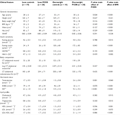
IR prevalence was first identified in a small seminal study using weight-matched controls and the euglycaemic-hyperinsulinaemic clamp, where insulin stimulated glucose utilisation was
below the lower range of weight-matched controls in 26% of obese and 60% of lean PCOS women (Dunaif et al., 1989). In another study of 40 obese PCOS women, 53% were insulin
resistant when the frequently sampled intravenous glucose tolerance test (IVGTT) was used and IR was defined using the 10th percentile of the normal distribution in the age, weight and ethnicity matched controls as a cut-off value (Legro et al., 1998). In a larger study using
controls that were not weight–matched, a higher prevalence of IR was reported in women with PCOS (Carmina and Lobo, 2004). IR was identified in 65% of PCOS women using
(HOMA) and qualitative insulin sensitivity check index (QUICKI) (Carmina and Lobo, 2004). Other studies using HOMA and QUICKI have reported lower prevalence rates of 18%
(Fulghesu et al., 2006) and up to 51% (de Paula Martins et al., 2007) of IR in PCOS. In a study with unmatched controls but adjusting for age, ethnicity and BMI, IR was present in
64% of women with PCOS using HOMA with the upper 95th percentile of adjusted values for controls to establish the upper normal limit for HOMA-IR (DeUgarte et al., 2005). These published studies demonstrate high variability, making it difficult to gage the prevalence of
IR in PCOS, due to different definitions, cut-off values, and measurement techniques of IR. Furthermore, confounding factors such as age and BMI are often overlooked. Given the
important aetiological role of IR in PCOS, there exists a need for quality assessment of prevalence rates of IR using gold standard techniques and for evidence synthesis of studies using these methods in PCOS. This is addressed in a systematic review in Chapters 2 and in
original research in Chapter 3.
1.7.4 Effect of Diagnostic Criteria on Prevalence of Insulin Resistance
Prevalence rates of IR in PCOS are also confounded by the transition of diagnostic criteria from the original NIH to Rotterdam. Women diagnosed with the classic phenotype of PCOS
(hyperandrogenism and anovulation) tend to have higher prevalence rates of IR compared to those diagnosed with the ovulatory (Carmina et al., 2005a, Moghetti et al., 2013) and
normoandrogenetic phenotypes (Broekmans et al., 2006, Goverde et al., 2009, Mehrabian et al., 2011). However, this is not always reported (Chae et al., 2008, Panidis et al., 2012, Shroff et al., 2007, Wang et al., 2010). In comparison to healthy women without PCOS acting as
controls, the NIH phenotype was generally insulin resistant (Carmina et al., 2005a, Chae et al., 2008, Wang et al., 2010, Dewailly et al., 2006) but results were not clear in the ovulatory
2008, Dewailly et al., 2006, Panidis et al., 2012). There are a number of limitations with the current studies making the interpretation of prevalence rates difficult. All, except one study
(Moghetti et al., 2013), have used surrogate measures of IR, which may not be sensitive enough do detect IR in PCOS, with a need for more studies using the gold standard
euglycaemic hyperinsulanaemic clamp (Buchanan et al., 2010). Furthermore, many studies did not take confounding factors, such as body composition, into account when assessing IR in different phenotypes. Therefore, to improve our knowledge in the area, Chapter 3 will
investigate the prevalence of IR using gold standard clamps across in the NIH and Rotterdam diagnostic criteria for PCOS.
1.7.5 Insulin Resistance - Aetiology in PCOS
A decrease in insulin sensitivity has been reported in women with PCOS (Dunaif et al., 1989,
Ciampelli et al., 1997, Diamanti-Kandarakis et al., 1998, Glintborg et al., 2006, Morin-Papunen et al., 2000). However, several studies have not supported this finding, especially in
lean women with PCOS, when confounding factors are taken into account including BMI, ethnicity, fat distribution, family history and diagnostic criteria, making relationships less clear (Holte et al., 1994, Ovesen et al., 1993, Vrbikova et al., 2004). The prevalence of IR in
PCOS is estimated to be up to 65% and occurs independently of obesity, but the effect of obesity on IR is additive to that of PCOS (Carmina and Lobo, 2004, Teede et al., 2011). The
presence of IR is a precursor to the development of other metabolic complications including T2DM. Large population studies using weight, age and ethnicity-matched controls are required.
International PCOS research agendas highlight aetiology and therapies as key priority areas
of PCOS (Azziz et al., 2006) but its aetiology remains unclear in PCOS as well as in other insulin resistant states. Current theories suggest intrinsic IR (genetic, inherent and unique to
PCOS) and extrinsic factors (obesity/physical inactivity and adipokines/cytokines) work synergistically to promote an insulin resistant state (Corbould et al., 2005, Dunaif, 1997).
Women with PCOS have hyperinsulinemia and decreased glucose-stimulated insulin secretion. Impaired muscle glucose uptake and whole body IR has been attributed to impaired insulin responsiveness of skeletal muscle and adipose tissue and specific abnormalities in
insulin metabolism including basal hyperinsulinemia, reductions in glucose-stimulated insulin secretion (Dunaif, 1997, Dunaif et al., 1996), reduced hepatic glucose uptake,
impaired suppression of hepatic gluconeogenesis (Dunaif et al., 1989) and abnormalities in insulin signalling in skeletal muscle (Dunaif, 1997, Corbould, 2008b, Corbould et al., 2005).
1.7.6 Possible Defects in Skeletal Muscle of Women with PCOS
Skeletal muscle accounts for up to 85% of whole body insulin-stimulated glucose uptake
(DeFronzo and Tripathy, 2009). Insulin stimulates glucose uptake in skeletal muscle by increasing the translocation of glucose transporter 4 (GLUT4) from intracellular vesicles to the cell surface, mainly mediated through the activation of the PI3-K and AKT or protein
kinase B (AKT/protein kinase B [PKB]) signalling pathways (Diamanti-Kandarakis and Papavassiliou, 2006). Women with PCOS have IR that is independent of obesity and this may
be attributed to INSR and/or post-binding defect in the insulin signalling pathways (Dunaif, 1997, Dunaif et al., 1992, Dunaif et al., 2001). Muscle biopsies taken from women with PCOS during basal conditions have normal insulin receptor substrate 1 (IRS-1) and PI3K
activity, however IRS-1-associated PI3-K activity was significantly reduced compared to age, weight and ethnicity matched control women during a euglycaemic hyperinsulinaemic clamp
IRS-1, or the p85 regulatory subunit of PI3-K in women with PCOS, but there was an increased abundance of insulin receptor substrate 2 (IRS-2), suggesting a compensatory
adjustment to help to maintain insulin mediated glucose uptake (Dunaif et al., 2001). Signalling abnormalities are also reported in the phosphorylation of AKT at Serine473 and
Threonine308 sites and AKT’s downstream target for GLUT4 translocation AS160, independently of obesity (Glintborg et al., 2008). Treatment with insulin sensitising medication (pioglitazone) improved insulin-stimulated glucose uptake, but not to normal
control levels (Hojlund et al., 2008). IRS-1 phosphorylation on Serine312, a key site for inhibiting insulin-mediated IRS-1 tyrosine phosphorylation and activation, was also reported
to be increased in PCOS (Corbould et al., 2005).
In contrast, others have failed to find changes in IRS-1-associated PI3-K activity and
AKT/PKB activation in skeletal muscle biopsies in women with PCOS following insulin infusion, despite impaired rates on insulin mediated glucose disposal (Ciaraldi et al., 2009,
Hojlund et al., 2008). The discrepancies between studies could be attributed to differences in the time course of muscle biopsies taken following insulin infusion (15 and 30 minutes post insulin infusion versus 3 hours) and the concentration of insulin achieved (physiological
levels or supraphysiological levels) (Dunaif et al., 2001, Hojlund et al., 2008).
Insulin sensitivity in skeletal muscle may also be reduced due to dysfunction in mitogenic insulin signalling pathways. An attenuation in the insulin stimulated ERK mitogenic pathway and an increase in basal phosphorylation of ERK 1/2 were reported in a small group of
women with PCOS (n=9) (Rajkhowa et al., 2009). The IRS/PKB pathway was similar in PCOS and controls. Together, this evidence highlights that impairments in the insulin
However, further work is required to define the defects responsible for impairments in insulin-mediated glucose disposal present in PCOS.
Other possible mechanisms for the development of IR in skeletal muscle are adipokines and
inflammatory markers. These markers have the ability to disrupt insulin signalling pathways either directly by inhibiting serine phosphorylation of the IRS or indirectly through inflammatory pathways including the c-Jun N-terminal kinase (JNK) and I-kappa B kinase β (IKKβ)/NFκB pathways (Tilg and Moschen, 2008). Some adipokines that may play a role in
modulating IR include leptin, resistin, visfatin, and (PAI-1 (Makki et al., 2013). Little
attention has been given to the interaction of these markers in the development of IR in PCOS; rather the majority of the research has focused on measuring these markers in isolation. Therefore, potential markers of IR in PCOS are further explored in Chapter 4.
1.7.7 Insulin Resistance and Mitochondrial Dysfunction in Skeletal Muscle
Another theory on the aetiology of IR became popular in the 1990’s and implicates abnormalities in mitochondrial function in the development of IR (Morino et al., 2006). Mitochondria are surrounded by an outer and an inner bilipid membrane. The inner
membrane consists of many folds that form cristae where the five oxidative phosphorylation enzymes are located. Nicotinamide adenine dinucleotide hydride (NADH) dehydrogenase
(complex I) is the first enzyme in the electron transport chain (ETC) and catalyses the transfer of electrons from NADH molecules to coenzyme Q. Complex II (succinate dehydrogenase) transmits electrons from succinate to coenzyme Q and directly connects the
citric acid cycle to the respiratory chain. From coenzyme Q, electrons can be transferred to complex III (cytochrome c reductase). Cytochrome c mediates electron transfer from
transferred to an oxygen molecule, which is reduced to water. ATP synthase, which is the prominent enzyme in complex V, is responsible for this proton gradient that drives ATP
synthesis from ADP and phosphate (Dudkina et al., 2010).
The function of mitochondria is to produce energy, mainly ATP, by oxidative phosphorylation (OXPHOS) (Brand and Nicholls, 2011). Other functions include fatty acid oxidation, cell signalling, reactive oxygen species (ROS) production, mediating oxidative
stress, regulation of apoptosis and cellular aging (Goodpaster, 2013, Holloszy, 1967, van Gurp et al., 2003). Metabolic homeostasis is tightly controlled by the mitochondria through
the oxidisation of both carbohydrates and lipids and by transitioning between these substrates in response to insulin, substrate concentrations and the contractile status of the muscle (rest versus contraction) (Kelley et al., 1993). Abnormality in any of these processes can be termed
mitochondrial dysfunction (Brand and Nicholls, 2011).
A role for mitochondria in IR emerged when researchers began to report defects in skeletal muscle mitochondria function in a range of different insulin-resistant populations; obese, T2DM and PCOS and in non-diabetic individuals with a family history of T2DM (Bullon et
al., 2014, Kelley et al., 2002). Defects in mitochondria include a reduction in mitochondrial size, content (Kelley et al., 2002, Morino et al., 2005, Ritov et al., 2005) and oxidative
enzyme activity (Heilbronn et al., 2007a, Ritov et al., 2005), decreased fat oxidation in skeletal muscle (Kelley et al., 1999, Kelley et al., 2002, Kim et al., 2000, Simoneau et al., 1999), down-regulation of genes involved in mitochondrial biogenesis and OXPHOS
(Mootha et al., 2003, Patti et al., 2003), decreased messenger ribonucleic acid (mRNA) and/or protein expression of mitochondrial genes/proteins (Heilbronn et al., 2007a, Heilbronn
(mtDNA) levels (Ritov et al., 2005) and decreases in mitochondrial oxidative capacity (Befroy et al., 2007, Mogensen et al., 2007, Petersen et al., 2004). Collectively, these studies
support the role of mitochondrial dysfunction in the aetiology of IR. However, much debate exists in this area, as a large body of research also report no association between IR and
mitochondrial function (De Feyter et al., 2008, Fisher-Wellman et al., 2014, Lefort et al., 2010, Rabol et al., 2011, Trenell et al., 2008) The mechanisms and associations of mitochondrial dysfunction and in IR in PCOS will be further explored in Chapter 5 of this
thesis.
1.8 Summary and Research Gaps
PCOS is a very common condition with a significant reproductive, metabolic, psychological and economic burden. Given the role of IR in the pathophysiology and clinical consequences
of PCOS, the syndrome appears to be an insulin resistant state and exploration of the aetiology of IR in PCOS is needed to better understand the condition and improve treatment options. Key research gaps remain in this area and include:
1. Evidence for the presence of intrinsic and extrinsic (obesity related) IR in PCOS using gold standard assessments of insulin sensitivity;
2. The need for accurate prevalence rates of IR in PCOS given the confounding factors such as age, BMI, diagnostic criteria, ethnicity and various methods used in measuring insulin sensitivity;
3. An understanding of factors that modulate IR in PCOS including sex steroids, inflammatory markers and adipokines;
4. Novel and optimal methods to measure IR in PCOS;
1.9 Aims of the Thesis
Given the gaps in our knowledge, the body of work presented in this thesis aims to:
i) Systematically review and meta-analyse the literature to determine the impact of PCOS status on IR, age, BMI and PCOS diagnostic criteria and to investigate factors that potentially mediate IR in PCOS using a systematic approach (Chapter 2);
ii) Investigate prevalence of IR in PCOS in a cross-sectional study using gold standard euglycaemic hyperinsulinaemic clamp studies (Chapter 3);
iii) Explore other potential markers of IR in PCOS (Chapter 4);
iv) Explore the role of mitochondrial function in the underlying aetiology of intrinsic IR in PCOS (Chapter 5);
v) Explore role of IR in reproductive consequences, specifically ovarian dysfunction (Chapter 6).
1.10 Organisation of the Thesis
The following chapters explore IR in PCOS. An overall introduction in Chapter 1 is followed
by Chapter 2, which is original research presented in the form of a systematic review and meta-analysis aimed at determining whether IR is intrinsic to PCOS and hormonal changes that may be associated with IR. Chapter 3 extends on the previous chapter by further
exploring the intrinsic nature of IR in PCOS through a comprehensive cross-sectional study. In this chapter I also described the prevalence of IR in PCOS based on different diagnostic
criteria. Chapter 4 details techniques to measure IR and proposes biomarkers, which may be useful in detecting IR in PCOS. Chapter 5 explores the role of mitochondrial dysfunction in IR and PCOS and finally Chapter 6 explores the relationship between of IR, ovarian function
Chapter 2
Insulin Resistance in Women with Polycystic
Ovary Syndrome: A Systematic Review and Meta-Analysis of
Declaration of Co-Authorship and Co-contribution: Papers incorporated in
thesis by publication
This declaration is to be completed for each conjointly authored publication and placed at the beginning of the thesis chapter in which the publication appears.
Declaration by: Signature: Date
Samantha Cassar 27/11/2014
Paper Title:
Insulin resistance in women with polycystic ovary syndrome: A systematic review and meta-analysis of euglycaemic hyperinsulinaemic clamp studies.
In the case of the above publication, the following authors contributed to the work as follows:
Name Contribution % Nature of Contribution
Samantha Cassar 70
Designed the research, performed the literature search, independently reviewed the articles and assessed risk of bias, contributed to data analysis and interpretation, wrote the manuscript, constructed tables and figures, provided critical review of the manuscript and
approved the final version for publication.
Marie L. Misso 5
Designed the research, performed the literature search, assisted with reviewing
articles, undertook critical revision for important intellectual content and approved
the final version for publication.
William G. Hopkins 5
Designed the meta-analysis models, performed statistical analysis and interpretation, wrote the manuscript, provided
critical revision for important intellectual content and approved the final version for