Prepare and Characterization of Polymer Coat Gold Nanoparticles and Their
Application in Targeted Drug Delivery for Breast Cancer
Hassan Lafta Atiyah M.Sc., Biotechnology
Department of Genetics & Biotechnology University College of Science, Osmania University
Abstract:
It has been very nearly 4 decades since the "war on cancer" was proclaimed. It is currently by
and large trusted that customized medication is the future for cancer persistent administration.
Having exceptional potential for early recognition, exact finding, and customized treatment of
cancer, nanoparticles have been broadly contemplated in the course of the most recent decade. In
this audit, we will condense the present best in class of gold nanoparticles in biomedical
applications focusing on cancer. Gold nanospheres, nanorods, nanoshells, nanocages, and surface
upgraded Raman dispersing nanoparticles will be examined in insight in regards to their
utilizations in vitro measures, ex vivo and in vivo imaging, cancer treatment, and medication
conveyance. Multifunctionality is the key component of nanoparticle-based operators. Focusing
on ligands, imaging marks, restorative medications, and different functionalities can all be
coordinated to take into account focused on atomic imaging and sub-atomic treatment of cancer.
Enormous strides have been made and numerous verification of-rule studies have been
effectively performed. The future looks brighter than at any other time yet numerous obstacles
stay to be won. A multifunctional stage in view of gold nanoparticles, with numerous receptor
focusing on, multimodality imaging, and various remedial elements, holds the guarantee for an
"enchantment gold projectile" against cancer. This audit is engaged basically on the combination
and uses of gold nanoparticles in the field of solution and focused on medication conveyance.
Nanotechnology has gotten to be a standout amongst the most intriguing and propelled territories
circumstances in this field because of their one of a kind properties, little size and high surface
zone to-volume proportion. These particles have been generally utilized as a part of different
biomedical applications and medication conveyance frameworks because of their latent nature,
security, high dispersity, non-cytotoxicity and biocompatibility.
INTRODUCTION:
Cancer is the third driving reason for death
(after coronary illness and stroke) in created
nations and the second driving reason for
death (after coronary illness) in the United
States. Ponders have demonstrated that there
were 10 million new cases, 6 million
passings, and 22 million individuals living
with cancer worldwide in the year 2000.
These numbers speak to an expansion of
around 22% in occurrence and mortality
from that of the year 1990. It is anticipated
that the quantity of new instances of all
cancers worldwide will be 12.3 and 15.4
million in the year 2010 and 2020,
individually. In 2008, a sum of 1,437,180
new cancer cases and 565,650 cancer
passings were evaluated to happen in the
United States alone. Nanotechnology, an
interdisciplinary research field including
science, building, science, and solution, has
extraordinary potential for early location,
precise determination, and customized
treatment of cancer. Nanoparticles are
commonly littler than a few hundred
nanometers in size, practically identical to
extensive organic atoms, for example,
compounds, receptors, and antibodies. With
the measure of around one hundred to ten
thousand times littler than human cells,
these nanoparticles can offer exceptional
collaborations with biomolecules both on
the surface of and inside the cells, which
may reform cancer finding and treatment.
The most all around concentrated on
nanoparticles incorporate quantum spots,
carbon nanotubes, paramagnetic
nanoparticles, liposomes, gold nanoparticles,
and numerous others.
In the course of the most recent decade,
there have been numerous nanotechnology
focuses set up around the world. In the
United States alone, more than six billion
dollars have been put resources into
nanotechnology research and more than
sixty focuses, systems, and offices, financed
by different offices, are in operation or soon
to open. In the wake of setting up an
interdisciplinary nanotechnology workforce,
it is normal that nanotechnology will
develop into a clinically helpful field sooner
One of the significant uses of
nanotechnology is in biomedicine.
Nanoparticles can be designed as
nanoplatforms for viable and focused on
conveyance of medications and imaging
marks by overcoming the numerous natural,
biophysical, and biomedical boundaries. For
in vitro and ex vivo applications, the upsides
of best in class nanodevices (eg, nanochips
and nanosensors) over conventional test
strategies are self-evident. Nonetheless, a
few hindrances exist for in vivo applications
in preclinical and conceivably clinical
utilization of nanotechnology, among which
are the biocompatibility, in vivo energy,
tumor focusing on viability, intense and
unending harmfulness, capacity to get away
from the reticuloendothelial framework
(RES), and cost-adequacy. In this audit, we
will abridge the present best in class of gold
nanoparticles in biomedical applications.
Synthesis of gold nanoparticles
There are numerous subtypes of gold
nanoparticles in light of the size, shape, and
physical properties. The most punctual
contemplated gold nanoparticles are gold
nanospheres (in spite of the fact that not
precisely circular in a strict sense). Thusly,
nanorods, nanoshells, and nanocages have
all been accounted for. Another kind of
gold-based nanoparticles, with incredible
surface-improved Raman scrambling
properties (named "SERS nanoparticles"),
will likewise be talked about in this survey.
In the accompanying content, the expression
"gold nanoparticle(s)" will allude to a
gathering of all these subtypes and the
subtype of gold nanoparticles utilized as a
part of every study will be indicated at
whatever point conceivable. With proceeded
with improvement in the union procedures
in the course of the most recent two decades,
a large portion of these gold nanoparticles
can now be created with all around
controlled size circulation, in some cases
with staggering accuracy (eg, nanocages).
Gold nanospheres
Gold nanospheres (otherwise called gold
colloids) of 2 nm to more than 100 nm in
measurement can be incorporated by
controlled decrease of a fluid HAuCl4
arrangement utilizing diverse diminishing
specialists under fluctuating conditions. The
most normally utilized lessening specialist is
citrate, which can create almost
monodisperse gold nanospheres. The span of
the nanospheres can be controlled by
changing the citrate/gold proportion. For the
most part, littler measure of citrate will yield
constraints of this strategy are the low yield
and the limitation of utilizing water as the
dissolvable. A two-stage strategy, roused by
the two-stage framework utilized by Faraday
as a part of 1857, equipped for delivering
thermally and air stable gold nanospheres of
decreased dispersity and all around
controlled size (as a rule <10 nm in
measurement) was accounted for in 1993.
This strategy was later enhanced using a
stage exchange reagent, for example,
tetraoctylammonium bromide. Once more,
the thiol/gold molar proportions can
influence the normal size of the
nanospheres. Bigger thiol/gold proportions
and quicker expansion of the reductant in
cooled arrangements will yield littler and
more monodispersed gold nanospheres. A
few different strategies have been
researched for gold nanosphere union, for
example, the utilization of different
reductants or ligands. There are various
writing gives an account of the utilization of
dendrimers as layouts or stabilizers for gold
nanosphere arrangement. Biocompatible
piece copolymers have been utilized for the
union of sterically settled gold nanospheres
in watery arrangement. The size and state of
the gold nanospheres could be promptly
controlled by tuning the combination
parameters, for example, the square piece,
and the relative/supreme groupings of the
square copolymer and HAuCl4.
Development of gold nanospheres in human
cells has additionally been accounted for.
Normally, gold nanospheres show a solitary
retention top in the noticeable range between
510 nm and 550 nm. With expanding
molecule estimate, the retention top
movements to a more drawn out wavelength
and the width of the assimilation spectra is
identified with the size conveyance extend.
Numerous different sorts of gold
nanoparticles with various size/shape, for
example, nanorods, nanoshells, and
nanocages, have been investigated to get
optical properties appropriate for biomedical
applications.
Gold nanorods
The synthesis of gold nanorods has been
accounted for utilizing a wide assortment of
techniques. Gold nanorods are normally
orchestrated utilizing the format technique,
in light of the electrochemical testimony of
gold inside the pores of nanoporous
polycarbonate or alumina layout layers. The
breadth of the gold nanorod is dictated by
the pore measurement of the format film,
while the length of the nanorod can be
controlled through the measure of gold
principal burden of this strategy is the low
yield since one and only monolayer of
nanorods is readied. Arrangement of gold
nanorods through electrochemical synthesis
has additionally been accounted for. In this
approach, numerous exploratory parameters
can decide the length of the nanorod hence
influencing its viewpoint proportion
(characterized as the length isolated by the
width).
Seed-mediated synthesis, maybe the most
entrenched strategy for gold nanorod
arrangement, can give higher angle
proportions than those arranged by different
strategies. Normally gold seeds are made by
concoction lessening of a gold salt with a
solid decreasing operator, for example,
NaBH4. These seeds, serving as the
nucleation locales for nanorods, are then
added to a development arrangement of gold
salt with a frail decreasing specialist, for
example, ascorbic corrosive and
hexadecyltrimethylammonium bromide. The
perspective proportions of the gold nanorods
can be controlled by differing the measure
of gold seeds as for the gold antecedent. In
addition, gold nanorods can be delivered in
quantitative yield with the expansion of
AgNO3. Other than the techniques said
over, a few different methodologies have
additionally been explored for the creation
of gold nanorods, including bio-lessening,
development on mica surface, and
photochemical synthesis.
Gold nanoshells
Optical imaging, incorporate those that
utilizations gold nanoparticles as the
differentiation specialists, has exceptionally
restricted applications in human studies.
Notwithstanding, in the close infrared locale
(NIR; 700–900 nm), the absorbance of all
biomolecules achieves least which gives a
moderately clear window to optical imaging.
By changing the piece and measurements of
the layers, gold nanoshells can be outlined
and created with surface plasmon
reverberation (SPR) tops running from the
unmistakable to the NIR area. For a given
sythesis of gold nanoshell, the SPR pinnacle
can be tuned by changing the proportion of
the center size to its shell thickness. Gold
nanoshells with SPR crests in the NIR area
can be set up by covering silica or polymer
dabs with gold shells of variable thickness.
Silica centers are developed utilizing the
Stöber procedure, the essential lessening of
tetraethyl orthosilicate in ethanol. To coat
the silica nanoparticles with gold in a fluid
situation, a seeded development method is
(2–4 nm in breadth) can be joined to the
silica center utilizing an amine-ended silane
as a liner atom, permitting extra gold to be
diminished until the seed particles mixed
into an entire shell. The breadth of the gold
nanoshell is to a great extent dictated by the
measurement of the silica center, and the
shell thickness can be controlled through the
measure of gold stored on the surface of the
center. Gold nanoshells have likewise been
combined through in situ gold nanoparticle
development utilizing thermosensitive
center shell particles as the layout. The
utilization of microgel as the center offers
fundamentally diminished molecule
conglomeration, and also thickness control
of the gold nanoshells utilizing electroless
gold plating. In one study, an infection
platform has been utilized to amass gold
nanoshells. This approach may possibly
furnish centers with a smaller size
appropriation and littler distances across
(<80 nm) than those of silica.
Gold nanocages
Gold nanocages with controllable pores at
first glance have been incorporated by
means of galvanic substitution response
between truncated silver nanocubes and
watery HAuCl4. Silver nanostructures with
controlled morphologies can be created
through polyol lessening, where AgNO3 is
decreased by ethylene glycol to produce
silver iotas and after that nanocrystals or
seeds. Ensuing expansion of silver particles
to the seeds delivers the fancied
nanostructures through controlling the silver
seed crystalline structures within the sight of
poly(vinylpyrrolidone), a polymer that is
prepared to do specifically official to the
(100) surface. The silver nanostructures,
utilized as a conciliatory layout, can then be
changed into gold nanostructures with
empty insides through the galvanic
substitution. The measurement and divider
thickness of the resultant gold nanocages
could be promptly controlled, to high
accuracy, by changing the molar proportion
of silver to HAuCl4.
SERS nanoparticles
SERS is an optical method that offers
numerous points of interest over customary
advancements, for example, fluorescence
and chemiluminescence, including better
affectability, large amounts of multiplexing,
heartiness, and unrivaled execution in blood
and other natural networks. In a
spearheading report, gold nanospheres (~13
nm in measurement) changed with
Cy3-named, alkylthiol-topped oligonucleotide
nearness of particular target DNA strands.
The Cy3 gathering was picked as the Raman
mark in light of its expansive Raman
cross-section.Subsequently, a few different reports
have additionally utilized SERS
nanoparticles. In one study, gold
nanospheres (60 nm in breadth) were
encoded with a Raman correspondent and
settled with a layer of thiolated polyethylene
glycol (PEG). Another sort of SERS
nanoparticle is made out of a gold center, a
Raman-dynamic sub-atomic layer, and a
silica covering. The silica covering can
guarantee physical heartiness, idleness to
different natural conditions, and
straightforward surface adjustment by means
of silica science. The thiol bunches that were
along these lines presented onto the silica
shell can be conjugated with
maleimide-initiated PEG chains for enhanced
biocompatibility.
Applications of nanoparticles
Nanotechnology has been a to a great degree
interesting issue in the course of the most
recent decade. A straightforward inquiry of
"Nano" in PubMed returned more than 6000
productions. Two noteworthy zones of
nanoparticle applications are material
science and biomedicine. Enormous strides
have been made in the material science field.
The way that gadgets are getting speedier,
better, and littler every month is a
reasonable and solid confirmation for such
accomplishment. Be that as it may,
utilizations of nanoparticles in the
biomedical field have not satisfied the
desires. Not very many nanoparticle-based
specialists are in clinical testing or
popularized for cancer conclusion or
treatment, and the vast majority of them
depend on liposomes which were produced
quite a few years prior. There is still far to
go before nanotechnology can really reform
quiet care the same number of have trusted it
would. Next, we will compress the advance
to date with respect to the utilization of gold
nanoparticles for biomedical applications.
Biomedical applications of gold
nanoparticles
Cancer nanotechnology is an
interdisciplinary region with expansive
potential applications in battling cancer,
including atomic imaging, sub-atomic
conclusion, focused on treatment, and
bioinformatics. The proceeded with
improvement of cancer nanotechnology
holds the guarantee for customized oncology
in which hereditary and protein biomarkers
can be utilized to analyze and treat cancer in
individual patient. Gold nanoparticles have
been examined in assorted territories, for
example, in vitro measures, in vitro and in
vivo imaging, cancer treatment, and
medication conveyance.
In vitro assays
Oligonucleotide-topped gold nanoparticles
have been accounted for polynucleotide or
protein, (for example, p53, a tumor silencer
quality) identification utilizing different
recognition/portrayal strategies, for
example, nuclear compel microscopy
(AFM), gel electrophoresis, scanometric
measure, chronocoulometry, opened up
voltammetric location, SPR imaging, and
Raman spectroscopy. In a few reports,
picomolar even femtomolar convergences of
DNA targets have been identified.
Bifunctional DNA-based adsorbate particles
have been assessed as atomic rulers, in light
of the SERS flags that fluctuate freely in
force as a component of the separation from
the gold nanoshell surface. Gold
nanoparticles have additionally been utilized
for some different applications, for example,
immunoassay, protein examine,
time-of-flight auxiliary particle mass spectrometry,
slender electrophoresis, and location of
cancer cells. In one report, dynamic light
dissipating (DLS) empowered quantitative
estimation of the grouping of intravenously
infused gold nanoshells in mouse blood.
This strategy may likewise be relevant
towards evaluating the course life time of
other strong nanoparticles. Gold nanoshells
functionalized with a pH-delicate SERS
journalist atom, 4-mercaptopyridine, were
appeared to be receptive to the pH of the
encompassing media inside the scope of 3 to
7. Another study has assessed the utilization
of gold nanoshells as optical biosensors for
constant discovery of streptavidin-biotin
collaborations in weakened human blood.
Notwithstanding, both the affectability (~3
µg/mL) and the dynamic range (3–50
µg/mL) were extremely poor. As a rule,
writing reports have blended discoveries. It
will be perfect if distinctive measures can be
analyzed next to each other utilizing a
similar model framework, which can
altogether help in choosing which examines
are the best possibility for potential clinical
testing. The National Cancer Institute (NCI)
cooperation for nanotechnology in cancer
has required each of its financed focuses to
test their recently created nanosensors
utilizing a similar standard examples, which
is relied upon to promptly distinguish which
new sensors really emerge from the huge
pool of new atomic sensors. Extending a
extensive scope of research labs the nation
over would be profoundly gainful to cancer
patients. Picking the right hopeful at an early
stage spares valuable time, as well as can
drastically decrease the cost for new
measure advancement.
Cancer therapy
Traditional methodologies for cancer
mediation incorporate surgery,
chemotherapy, and radiation treatment.
Exploiting their one of a kind properties,
most investigations of gold
nanoparticle-based cancer treatment have utilized
photothermal treatment for the devastation
of cancer cells or tumor tissue, which might
be conceivably helpful in the clinical setting.
At the point when illuminated with centered
laser heartbeats of appropriate wavelength,
focused on gold nanospheres, nanorods,
nanoshells, and nanocages can eliminate
microscopic organisms and cancer. It was
assessed that 70–80 °C was accomplished
through light ingestion by the gold
nanoparticles and up to 150 antibodies can
be conjugated to a nanoshell through a
bifunctional PEG linker. One charming
perception is that the greater part of these
studies focused on either EGFR or human
epidermal development consider receptor 2
(HER2), clearly because of the prepared
accessibility of monoclonal antibodies
(effectively affirmed by the Food and Drug
Administration [FDA] for cancer treatment)
that perceive these two proteins.
Since the absorbance wavelength (in
the unmistakable range) of little gold
nanospheres is not ideal for in vivo
applications, the gathering of gold
nanoclusters on the phone layer was
researched. It was found that the
arrangement of nanoclusters prompted
expanded neighborhood retention and
red-moving, contrasted with cells that did not
have nanoclusters. Noteworthy improvement
in laser-prompted cancer cell slaughtering
was watched utilizing a NIR laser. Gold
nanoshells are adequately huge (around
100–300 nm in distance across) to have SPR
tops in the NIR locale. In one spearheading
study, human bosom carcinoma cells
brooded with gold nanoshells were found to
experience photothermally prompted
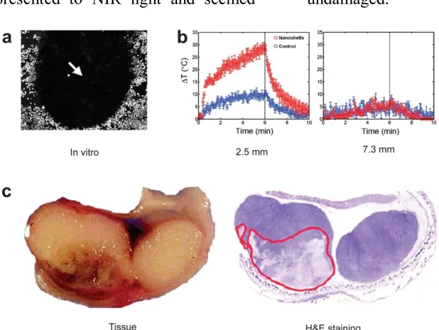
bleakness upon presentation to NIR light. In
vivo testing uncovered that introduction to
low measurement NIR light in strong tumors
treated with gold nanoshells brought about
huge normal temperature increment, fit for
inciting irreversible tissue harm, while the
controls (not treated with nanoshells)
when presented to NIR light and seemed undamaged.
Figure 1: Gold nanoshells can pulverize
cancer cells both in vitro and in vivo. a.
Cells brooded with gold nanoshells can be
executed by NIR light (dull range). b.
Fleeting plots of most extreme temperature
change of NIR-lighted tumors with and
without nanoshells at profundities of 2.5 mm
and 7.3 mm underneath the tissue surface. c.
Net pathology after in vivo treatment with
nanoshells and NIR laser uncovered
draining and loss of tissue birefringence
underneath the apical tissue surface.
Hematoxylin/eosin (H&E) recoloring inside
a similar plane affirms tissue harm inside the
range that contains nanoshells. In a late
report, it was proposed that 5000 gold
nanoshells per prostate cancer cell was
expected to accomplish cell murder.
PEG-covered nanoshells with pinnacle retention
in the NIR district were intravenously
infused into tumor-bearing naked mice. In
one concentrate, all tumors treated with the
NIR laser were removed and the mice
showed up tumor free for a while tumors in
control creatures (NIR laser treatment
without nanoshell infusion) kept on
developing. In another study, 93% of tumor
corruption and relapse was seen in a high
dosage nanoshell (8.5 µL/g) treated
gathering. Shockingly, a somewhat bring
down nanoshell dosage (7.0 µL/g) just
brought about tumor development capture at
21 days yet not tumor removal. The
motivation behind why such an unobtrusive
distinction in nanoshell measurement could
adequacy merits cautious examination. It is
significant that all these in vivo cancer
treatment concentrates just include
uninvolved tumor focusing on however not
particular sub-atomic focusing on. Detached
tumor focusing on is expected to the
non-particular collection of the nanoshells in the
tumor, named "the upgraded penetrability
and maintenance (EPR) impact", since the
tumor vasculature is typically more flawed
than ordinary veins and there is no
lymphatic waste in the tumor. The
enrollment of monocytes into hypoxic
districts inside tumors has been misused for
photograph actuated cell slaughtering with
gold nanoshells. Other than photothermal
treatment, gold nanoparticles have
additionally been examined in other
restorative studies. Phthalocyanine (a
photosensitizer) balanced out gold
nanospheres (2–4 nm in measurement) have
been accounted for photodynamic treatment
of refined tumor cells. Gold nanoparticles
have been appeared to upgrade the
antiproliferation and apoptosis of human
hepatoma cells prompted by Paclitaxel, a
chemotherapeutic medication. A late study
has demonstrated that improvement of
radiosensitivity can be accomplished
because of the expanded assimilation of
ionizing radiation by the gold nanoparticles,
which thus created softens up single-and
twofold stranded DNA. In spite of the fact
that it was recommended that focusing on
the DNA of cancer cells with gold
nanoparticles may offer a novel approach
that is by and large material to outer pillar
radiotherapy medicines, accomplishing
DNA focusing in vivo is to a great degree
troublesome.
Drug delivery
A few studies have reported the utilization
of gold nanoparticle as medication
conveyance vehicles. Tumor putrefaction
calculate alpha (TNF-α), a cytokine with
fantastic anticancer viability, is systemically
poisonous which extremely restricted its
remedial applications. A nanoparticle
conveyance framework, comprising of PEG
covered gold nanoparticle stacked with
TNF-α, was developed to amplify the tumor
harm and minimize the systemic
harmfulness of TNF-α. Blend of nearby
warming and nanoparticle-based
conveyance of TNF-α brought about
improved remedial viability than either
treatment alone. Thermally-instigated tumor
development deferral was upgraded by
pretreatment with the nanoparticle, when
given intravenously at the correct
stream concealment, and in addition tumor
perfusion abandons, recommended vascular
harm intervened tumor cell murdering.
Shockingly, taking after intravenous
organization, almost no aggregation in the
RES (eg, liver and spleen) or other solid
organs of the creatures was watched. Along
these lines, this nanoparticle conjugate has
likewise been utilized to annihilate the
tumor inside an iceball, again without
noteworthy systemic harmfulness. Stage I
clinical trials of this conjugate, along these
lines named "CYT-6091", are as of now
progressing to assess its security,
pharmacokinetics, and clinical viability.
Methotrexate (MTX), an inhibitor of
dihydrofolate reductase, is a
chemotherapeutic specialist for treating an
assortment of cancers sorts. MTX-gold
nanoparticle conjugate was readied and the
cytotoxic/antitumor impact was analyzed in
vitro and in vivo. Organization of the
conjugate smothered tumor development in
a mouse model of Lewis lung carcinoma,
though an equivalent measurements of free
MTX had no antitumor impact. Nanoshells
have been tried for medication conveyance.
In one early study, composites of hydrogels
and gold nanoshells were created for
photothermally-balanced medication
conveyance. Light at 1064 nm was
consumed by the nanoshells and changed
over to warmth, which prompted the fall of
the hydrogel accordingly essentially
upgrading the medication discharge. In this
way, tweaked sedate conveyance of
methylene blue, insulin, and lysozyme was
accomplished by illumination of the
medication stacked nanoshell-hydrogel
composites, with the medication discharge
rate subordinate upon the atomic weight of
the remedial particle. Empty gold nanoshells
can likewise embody chemicals, for
example, horseradish peroxidase (HRP),
which stayed dynamic inside the nanoshells
for little, yet not expansive, substrate atoms.
As anyone might expect, HRP did not
demonstrate any movement when caught
inside strong gold nanoparticles.
Tranquilize conveyance utilizing gold
nanoparticles, in blend with their
characteristic capacity for photothermal
treatment, ought to be investigated later on.
At present, which kind of gold nanoparticle
is the most reasonable for medication
conveyance applications is still easily
proven wrong. It was found that the
intracellular take-up of various measured
and molded gold nanoparticles are
measurements. The retention/diffusing
proficiency and optical reverberation
wavelengths have been figured for three
generally utilized classes of gold
nanoparticles: nanospheres, nanoshells, and
nanorods. The restricted range in the SPR
pinnacles of nanospheres (~520–550 nm)
brought about extremely constrained use for
in vivo applications. The SPR pinnacles of
gold nanoshells lie positively in the NIR
locale. The aggregate elimination of
nanoshells has a direct reliance on the
general size, however free of the center/shell
span proportion. The relative dissipating
commitment to the termination can be
quickly expanded by expanding the
nanoshell estimate or diminishing the
proportion of the center/shell sweep. Gold
nanorods were found to have tantamount
optical properties at much littler successful
size, with assimilation and scrambling
coefficients a request of size higher than
those for nanoshells and nanospheres. While
nanorod with a higher perspective
proportion and a littler successful span is a
superior photoabsorbing nanoparticle
reasonable for remedial applications, that
with a bigger compelling range is more ideal
for imaging purposes.
Examines have demonstrated that
femtosecond beat excitation (at 400 nm
wavelength) of DNA-changed nanoparticles
can prompt desorption of the thiolated DNA
strands from the nanoparticle surface by
breaking the gold-sulfur security. This
property could be abused later on for
controlled medication discharge. The
dependability of gold nanoparticle
bioconjugates in high ionic quality media
has been described as an element of the
nanoparticle estimate, PEG length, and the
monolayer piece. It was found that
nanoparticle security expanded with
expanding PEG length, diminishing
nanoparticle measurement, and expanding
PEG mole portion. Imperatively, gold
nanoparticles changed with PEG chains of
atomic weight (MW) 5000 were disguised as
productively as practically equivalent to
conjugates with PEG chains of MW 900. In
light of this discovering, gold nanoparticles
functionalized with ideal estimated PEG
chains (at any rate of MW 5000 to
productively sidestep the RES), with course
half-existence of no less than a couple of
hours, might be the most adequate for cancer
treatment.
Keeping in mind the end goal to make gold
conveyance and other biomedical
applications (imaging and treatment), they
should be successfully, particularly, and
dependably coordinated to a particular organ
or malady site without modification.
Particular focusing in vivo has not been
accomplished for gold nanoparticle-base
medication conveyance, because of the
moderately extensive general size of the
conjugate (normally more than 50 nm in
width) which forbids proficient
extravasation. In spite of the fact that use of
latent focusing on just has been appeared to
be viable in certain xenograft subcutaneous
tumor models, they may not really mirror
the clinical circumstance. Transgenic and
orthotopic tumor models are all the more
clinically pertinent and these tumors
ordinarily have a great deal less flawed
vasculature than subcutaneous ones, which
will make uninvolved focusing on
inadmissible for either cancer imaging or
treatment. Sub-atomic cancer markers
over-communicated on the tumor vasculature
might be the objectives of decision.
Methods of preparation
Top–down and bottom–up are the two
strategies used to create miniaturized scale
and nanoparticle sedate bearers. In the last
mentioned, the particulate framework is set
up from a condition of sub-atomic scattering
sort and is permitted to connect with
resulting development of strong particles.
Bottom–up methods, in this way, look to
orchestrate littler segments into gatherings
of complex structure, While the previous
begins with substantial size materials and
separates these into littler particles.
Traditional nanoparticle synthesis more
often than not relies on upon bottom–up
strategies.
Diverse techniques have been used
in the planning of chitosan small scale and
nanoparticles. The molecule measure,
soundness of the dynamic constituent and
the last item, leftover poisonous quality
present in the last item, and the active of the
medication discharge profile are components
that ought to be considered amid choice of
the strategy. Amid the planning of chitosan
particulate frameworks, the measure of the
readied particles is significantly reliant on
chitosan sub-atomic weight, chitosan
compound structure, especially the level of
deacetylation, and on the strategy for
arrangement. When in doubt, higher
sub-atomic weight chitosan produces bigger size
particles. Distinctive strategies are
accessible to get ready chitosan small
is for the most part bound to chitosan by
hydrogen holding, electrostatic connection,
or hydrophobic linkage. For the most part,
stacking the remedial specialist into chitosan
smaller scale/nanoparticles might be
accomplished either amid the readiness
procedure or after the particles have been
framed. In the previous, the restorative
operator is joined and inserted in the
chitosan framework, though in the last the
remedial specialist is adsorbed on the
molecule surface. More often than not, the
point is to accomplish high capture
productivity, which could be refined by
consolidation into the lattice, however the
remedial operator could be influenced by the
planning technique, added substances, and
so on. By and large, determination of the
technique is enormously reliant on the way
of helpful specialist and the kind of gadget
used in the conveyance. A rundown of
techniques utilized as a part of the readiness
of these particles. Every one of these
strategies include the bottom–up creation
prepare, in which get together of the broke
up atoms is accomplished to frame a clear
miniaturized scale or nanoparticulate
structure.
The procedures utilized as a part of the
arrangement of chitosan smaller
scale/nanoparticles stacked with
thermosensitive or less steady substances,
for example, proteins, peptides, hormones,
antibodies, plasmid DNA, and antigens
might be comprehensively ordered into
cross-connecting systems and drying
methods. Cross-connecting could be
accomplished synthetically or physically.
The strength of these thermosensitive or less
steady substances are firmly influenced by
the natural dissolvable and the
cross-connecting specialist utilized, with the result
of denaturation or concoction alteration. In
this way, physical cross-connecting and
drying procedures, for example, splash
drying, are favored and generally utilized for
these substances. As of late, turn around
micellar strategy has been presented. These
previously mentioned systems –
cross-connecting, drying, and turn around micellar
– notwithstanding sieving and dissolvable
vanishing were utilized as a part of the
planning of different medications of various
pharmacotherapeutic bunches. A knowledge
on these techniques is portrayed in the
accompanying areas.
CROSS-LINKING TECHNIQUES
The ionic cross-connecting strategy is the
most widely recognized among physical
cross-connecting methods since the
arrangement system is straightforward, does
not include utilization of natural dissolvable
or high temperature, and no synthetic
communication is included. These focal
points make this technique effective and ok
for creation of thermosensitive remedial
operators, for example, proteins, peptides,
hormones, and antibodies stacked into
chitosan particulate frameworks. Get
together and development of the particles is
accomplished by ionic cross-connecting
between chitosan or one of its subordinates,
being cationic in nature, and either
contrarily charged macromolecules or
anionic cross-connecting operators. Acidic
arrangement of chitosan is readied, and the
ionic cross-linker is included dropwise
alongside mixing and sonication. On the off
chance that cross-connecting is
accomplished by anionic cross-linkers, for
example, sodium sulfate or tripolyphosphate
(TPP), the procedure is called ionic gelation,
though adversely charged polyelectrolyte
macromolecules, for example, cyclodextrin
subsidiaries, dextran sulfate, and
poly-γ-glutamic corrosive deliver electrostatic
polyelectrolyte buildings (PEC) of ionic
cross-connecting sort.
Detailing of felodipine-stacked chitosan
microparticles has been accomplished by
ionic gelation. The chitosan sub-atomic
weight and fixation, centralization of the
cross-connecting operator (TPP), and TPP
pH have been accounted for to assume a
critical part in the medication discharge
design. Slower felodipine discharge was
acquired from TPP arrangement of low pH
and higher TPP fixation, and higher chitosan
sub-atomic weight and focus. Triclosan and
furosemide, two hydrophobic medications,
were stacked into chitosan nanoparticles by
ionic cross-connecting of chitosan with TPP,
and the discharge profile of both
medications from the arranged nanoparticles
was described by quick starting discharge
took after by a controlled-discharge
organize. TPP is broadly used to get ready
chitosan nanoparticles that have been
effectively utilized as a bearer for proteins
and antigens, for example, insulin, lockjaw
toxoid, egg whites, and flu subunit antigen.
Chitosan-stacked interleukin-2 (IL-2)
microparticles were readied utilizing sodium
sulfate as the anionic cross-linker.
Complex coacervation is a procedure of
liquid–liquid stage detachment that happens
oppositely charged particles are blended,
bringing about the development of an ionic
complex. Plasmid DNA was effectively
stacked into chitosan nanoparticles of size
range 450–820 nm by this strategy, and the
embodiment was more than 90% for
chitosan of high level of deacetylation and
the discharge was reached out for 24 hours.
As chitosan is a cationic polymer that is
dissolvable in acidic arrangement, it is
likewise conceivable to hasten chitosan from
its watery arrangement by expansion of
soluble base, which is the hypothetical
standard of the precipitation/coacervation
strategy. The system includes planning of
chitosan watery arrangement that is brought
through a spout into fluid or hydroalcoholic
arrangement of sodium hydroxide or
ethanediamine by blowing or dropping. The
encouraged chitosan microparticles are
isolated by filtration/centrifugation lastly
washed with hot and icy water. A
cross-connecting operator might be added to build
the hardness of the got particles, which can
amplify the medication discharge.
Prednisolone-stacked chitosan microspheres
were set up by the precipitation technique
and could upgrade the vehicle of
prednisolone over the epithelial boundary.
Complex coacervation, as officially
portrayed, has been utilized for the readiness
of ketorolac tromethamine chitosan
microspheres that controlled the medication
discharge, while possibly enhancing quiet
consistence because of the reduction in
dosing recurrence.
PEC chitosan nanoparticles created
through ionic collaboration with the
contrarily charged dextran sulfate and
stacked with insulin or vascular endothelial
development calculate (VEGF) was
accounted for. In another concentrate,
poly-γ-glutamic corrosive has been utilized as a
part of the planning of PEC chitosan
nanoparticles, which lessened the
transepithelial electrical resistance of Caco-2
cell monolayers. As of late, self-gathered,
electrostatic PEC chitosan particles have
been created for conveyance of a few
proteins, for example, insulin, heparin,
hyaluronan, and VEGF. Self-get together is
accomplished through electrostatic
connection between the cationic amino
gathering of chitosan and the artificially
changed N-anionic chitosan amines, for
Thermal cross-linking is another
method for physical cross-linking in which
citric corrosive, an ordinarily utilized
cross-linking specialist as a part of this technique,
is added to a fluid chitosan acidic
arrangement in a steady molar proportion
between citric corrosive and chitosan. The
blend is cooled to 0°C and added while
mixing to a sleek stage, for example, corn
oil or sesame oil, beforehand cooled to 0°C.
The emulsion is thermally cross-connected
at 120°C, and the got microspheres are
sifted, washed, lastly dried.
Indomethacin-stacked chitosan microspheres were set up
by this strategy.
Chemical cross-linking
In this strategy, chitosan small
scale/nanoparticles are framed through a
synthetic communication between a
cross-linking specialist and the essential amino
gatherings of chitosan. Regular cross-linkers
are glutaraldehyde, p-phthaldehyde,
ascorbyl palmitate, and dehydroascorbyl
palmitate. Chitosan microparticles arranged
utilizing ascorbyl and dehydroascorbyl
palmitate have bring down poisonous
quality and high insulin-stacking
effectiveness, and discharge insulin in a
controlled-discharge way, for around 80
hours, when contrasted with that delivered
utilizing di-aldehydes, glutaraldehyde, and
p-phthaldehyde as compound cross-linkers.
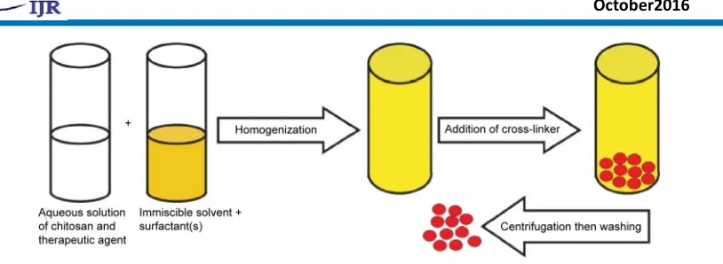
Concoction cross-linking may happen either
through maybe a couple steps. The
technique includes development of a
water/oil (w/o) emulsion in which chitosan
and the remedial specialist are in the fluid
stage that is emulsified into outer immiscible
dissolvable. The cross-linking specialist is
bit by bit included and, at long last, the
readied particles are isolated and washed
with fitting dissolvable to yield the coveted
molecule. Ascorbyl palmitate and
dehydroascorbyl palmitate were utilized as a
concoction cross-linking operator amid the
arrangement of chitosan-stacked insulin
microparticles in an outside mineral oil
stage. Development of these particles in the
inner watery period of w/o emulsion
enhances the capture of the remedial
specialist as the outer oil stage keeps the
Figure 2: Schematic representation of production of chitosan micro/nanoparticles by chemical
cross-linking.
A few added substances might be added to
upgrade solidness and exemplification
productivity of the restorative operator or
abatement its spillage. Gelatin has been
added to the watery stage to improve insulin
solidness and exemplification. Readiness of
chitosan microparticles containing cow-like
serum egg whites (BSA) has been accounted
for by this technique. Medications of various
pharmacotherapeutic gatherings, for
example, nonsteroidal mitigating,
antineoplastic, antifungal, antiseizure,
opioid, methyl xanthenes,
angiotensin-changing over protein inhibitor, and
bone-related medications have been stacked into
chitosan microparticles. Glutraldehyde,
glutraldehyde separated in toluene, and
ascorbyl palmitate are normally utilized
cross-linkers, while fluid paraffin or a blend
of mineral oil/petroleum ether is utilized as
outer oil stage. Basic cases of medications in
each pharmacotherapeutic bunch. As of late,
Ahmed and El-Say have created rabeprazole
chitosan nanoparticles inside a w/o
nanoemulsion by emulsifying the fluid stage
into paraffin oil containing a surfactant
blend of ranges and tweens. The streamlined
nanoparticles demonstrated a nanosize run,
120±32 nm, and were circular fit as a fiddle
as showed by the filtering electron
micrograph
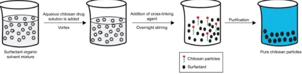
Reverse micellar method
Reverse micelles are water droplets in the
nanometer size (1–10 nm) scattered in
natural dissolvable in view of the impact of
surfactants. The watery center of these
nanosized droplets can be utilized as a
reactor to plan nanoparticles. The planning
strategy incorporates readiness of fluid
drug–chitosan arrangement that will be
added under mixing to a blend of natural
cross-linking operator is included, and the blend is
left on the stirrer overnight for finish
cross-linking. The natural dissolvable is then
vanished to get a dried mass. To evacuate
the surfactant, the got dried mass is scattered
in water and an appropriate salt is added to
accelerate the surfactant out, lastly the
medication stacked chitosan nanoparticles
are recuperated by centrifugation. The
doxorubicin dextran complex was stacked
into chitosan nanoparticles utilizing
n-hexane as natural dissolvable, sodium
bis(ethyl hexyl) sulfosuccinate as surfactant,
and glutraldehyde as cross-linker. The
arranged nanoparticles were of 100±10 nm
size and upgraded the penetrability and
maintenance impact of doxorubicin, which
was reflected in enhancing the medication
remedial impact and decrease of the
symptom in strong tumor.
Figure 3: Schematic representation of reverse micellar technique.
Sieving method
Arrangement of chitosan microparticles by
this strategy includes development of 4%
chitosan hydrogel containing the
medication, after which a cross-linking
operator, for example, glutraldehyde is
added to deliver a cross-connected chitosan
hydrogel that is gone through a sifter of
positive size to get the medication stacked
microparticles. The overabundance
glutraldehyde is expelled by washing the
acquired microparticles with 0.1 N sodium
hydroxide, and after that the particles are
dried in a broiler at 40°C. Clozapine
microparticles of the size range 543–698 μm
demonstrated a stretched out medication
discharge up to 12 hours when arranged by
this technique.
Solvent evaporation
In this strategy, a polymeric drug solution in
an volatile solvent, for example, Acetone is
arranged and emulsified into a nonaqueous
stage, for example, fluid paraffin. The
mixture is kept under mixing until finish
dissipation of the dissolvable, and the
shaped microspheres are separated, washed
petroleum ether, lastly dried. Drying is
generally accomplished via air or under
vacuum. Metformin-stacked chitosan
microspheres were effectively acquired
utilizing this technique. As a rule, the
ensnarement effectiveness and molecule size
of the readied particulate frameworks are
influenced by different preparing and
detailing parameters, for example, chitosan
focus, chitosan atomic weight, kind of the
chitosan subsidiary, nature of the
medication, beginning medication fixation
utilized, drug–polymer proportion, nature of
the cross-linking specialist, sort and
convergence of the surfactant, and blending
speed. Improving these parameters utilizing
reasonable enhancement software is useful
in accomplishing the desired particles.
Chitosan metal nanoparticles
Metal nanoparticles of copper (Cu), silver
(Ag), and gold (Au) have exhibited a wide
range of action against gram-negative and
gram-positive microbes and in addition
organisms. Be that as it may, there are
incredible worries about the human and
natural security of these metal nanoparticles.
What's more, the solidness of these particles
is additionally under talk, particularly with
respect to copper nanoparticles, which
experience quick oxidation upon
presentation to the air. Synthesis of these
metal nanoparticles within the sight of
biocompatible polymers, (for example, PEG,
polyvinyl pyrrolidone, and chitosan) and
surfactants that are utilized as stabilizers
could beat these restrictions. The covering
of the particles' surface utilizing polymeric
materials, for example, chitosan, has been
accounted for to upgrade the antimicrobial
action of these particles, inferable from
chitosan antimicrobial action.
Chitosan-coated silver nanoparticles were
set up by a chemical reduction technique
and have reported in applications as a
biosensor and in cancer treatment. The
arranged nanoparticles displayed
biodegradable character, great antimicrobial
action, and delayed activity of silver on the
influenced cells. Honary et al synthesized
chitosan-covered silver nanoparticles by a
similar technique and by using chitosans of
various sub-atomic weight. The writers
exhibited that the nanoparticle qualities were
affected by the atomic weight of chitosan,
and also by the procedure conditions, for
example, mixing pace and temperature.
They additionally specified that higher
antibacterial action against Staphylococcus
aureus was accomplished with littler
the molecule surface range. Gold
nanoparticles have been accounted for to be
helpful in analysis and medication
conveyance. The consolidation of chitosan
amid the synthesis of these metal
nanoparticles offers better infiltration and
take-up of remedial specialists, for example,
insulin, across the mucosal film, and
chitosan itself goes about as a lessening
operator amid gold nanoparticle synthesis.
The readied insulin-stacked chitosan gold
nanoparticles were steady, did not hint at
any conglomeration for 6 months, and
altogether brought down the blood glucose
level in diabetic rats taking after oral and
nasal organization. As of late, Salehizadeh et
al said the arrangement of Fe3O4–gold–
chitosan nanostructure by the compound
coprecipitation strategy and reported the
helpfulness of the arranged nanoparticles in
various biotechnological and biomedical
applications.
Chitosan copper-loaded nanoparticles were
set up by ionic gelation between chitosan
and TPP and demonstrated a stamped
development hindrance of an extensive
variety of microorganisms, for example, S.
aureus, Salmonella typhimurium,
Salmonella choleraesuis, and Escherichia
coli, in which the base inhibitory fixation
was under 0.25 μg/mL. Green synthesis is
the method broadly used to set up this kind
of chitosan metal nanoparticles, including
lessening of copper in a fluid arrangement of
chitosan and a natural corrosive, for
example, ascorbic corrosive, which keeps
the development of copper oxides. All the
more as of late, chitosan cobalt oxide
nanoparticles were produced, and their
movement on human leukemic cells was
examined. The writers showed increment in
the receptive oxygen species and caspase
enactment taking after presentation of the
leukemic cells to these chitosan-covered
metallic nanoparticles, impacts that are
known to prompt cancer cell passing. In this
way, there means that the capability of these
nanoparticles for an application in cancer
treatment.
CONCLUSION
This audit demonstrates that extensive
research activities have been centered
around the uses of chitosan-based small
scale and nanoparticles. Effective stacking
and conveyance of various atoms, including
low-sub-atomic weight medications and
macromolecules, for example, proteins,
by these frameworks through various
courses of organization, discover potential
restorative applications. The advancement of
chitosan subordinates has extended these
applications because of the improvement of
bioavailability fulfilled by an expansion in
the solidness, dissolvability,
mucoadhesiveness, cell penetrability,
ingestion, biodistribution, and tissue
focusing on accomplished when particulate
bearers depend on these subsidiaries. This
audit has tended to the distinctive strategies
that could be used in the advancement of
these particulate frameworks and techniques
for portrayal of the got particles. A review
on the parenteral and nonparenteral
utilizations of these chitosan and chitosan
derivatives–based particulate framework has
been delineated. Chitosan–metal
nanoparticles are another sort of molecule
that has shown the capacity to enhance
antimicrobial and pharmacodynamic
movement when contrasted and metal
nanoparticles without chitosan covering.
Gold nanoparticles have, in some ways,
reformed the field of prescription in view of
its across the board applications in focused
medication conveyance, imaging, finding
and therapeutics because of their to a great
degree little size, high surface zone,
dependability, non-cytotoxicity and tunable
optical, physical and compound properties.
Functionalized gold nanoparticles with
different biomolecules, for example,
proteins, DNA, amino acids and carboxylic
acids have been utilized as a part of cancer
treatment and give amazing medication
conveyance framework. Focused on
conveyance and modified arrival of helpful
medications to the particular site is
accomplished by utilizing gold nanoparticles
in light of the fact that they can endure high
medication load and discharge it to the
particular site through different organization
courses and can cooperate with cancerous
cell. Symptoms of customary medications
have been minimized by conjugation with
gold nanoparticles and they increment the
quality life of patients.
REFERENCE:
1. Chirra, H. D., & Desai, T. A. (2012).
Emerging microtechnologies for the development of oral drug delivery
devices. Advanced drug delivery
reviews, 64(14), 1569-1578.
2. des Rieux, A., Fievez, V., Garinot,
Journal of controlled release,
116(1), 1-27.
3. Piyakulawat, P., Praphairaksit, N.,
Chantarasiri, N., & Muangsin, N. (2007). Preparation and evaluation of
chitosan/carrageenan beads for
controlled release of sodium
diclofenac. Aaps PharmSciTech,
8(4), 120-130.
4. Grenha, A., Gomes, M. E.,
Rodrigues, M., Santo, V. E., Mano, J. F., Neves, N. M., & Reis, R. L.
(2010). Development of new
chitosan/carrageenan nanoparticles
for drug delivery applications.
Journal of Biomedical Materials Research Part A, 92(4), 1265-1272.
5. Hainfeld, J. F., Slatkin, D. N., &
Smilowitz, H. M. (2004). The use of
gold nanoparticles to enhance
radiotherapy in mice. Physics in
medicine and biology, 49(18), N309.
6. Ghosh, P., Han, G., De, M., Kim, C.
K., & Rotello, V. M. (2008). Gold
nanoparticles in delivery
applications. Advanced drug delivery
reviews, 60(11), 1307-1315.
7. Kumari, A., Yadav, S. K., & Yadav,
S. C. (2010). Biodegradable
polymeric nanoparticles based drug
delivery systems. Colloids and
Surfaces B: Biointerfaces, 75(1), 1-18.
8. Pankhurst, Q. A., Connolly, J.,
Jones, S. K., & Dobson, J. J. (2003).
Applications of magnetic
nanoparticles in biomedicine.
Journal of physics D: Applied physics, 36(13), R167.
9. Mishra, B., Patel, B. B., & Tiwari, S.
(2010). Colloidal nanocarriers: a review on formulation technology,
types and applications toward
targeted drug delivery.
Nanomedicine: Nanotechnology, biology and medicine, 6(1), 9-24.
10.Berry, C. C., & Curtis, A. S. (2003).
Functionalisation of magnetic
nanoparticles for applications in
biomedicine. Journal of physics D:
Applied physics, 36(13), R198.
11.Liong, M., Lu, J., Kovochich, M.,
Xia, T., Ruehm, S. G., Nel, A. E., ... & Zink, J. I. (2008). Multifunctional inorganic nanoparticles for imaging,
targeting, and drug delivery. ACS
nano, 2(5), 889-896.
12.Veiseh, O., Gunn, J. W., & Zhang,
M. (2010). Design and fabrication of magnetic nanoparticles for targeted
drug delivery and imaging.
Advanced drug delivery reviews,
62(3), 284-304.
13.van Vlerken, L. E., & Amiji, M. M. (2006). Multi-functional polymeric nanoparticles for tumour-targeted
drug delivery. Expert opinion on
drug delivery, 3(2), 205-216.
14.Parveen, S., Misra, R., & Sahoo, S. K. (2012). Nanoparticles: a boon to
drug delivery, therapeutics,
diagnostics and imaging.
Nanomedicine: Nanotechnology, Biology and Medicine, 8(2), 147-166.
15.Janib, S. M., Moses, A. S., &