Original Research Article
Serum β HCG and lipid profile in early second trimester as predictors
of hypertensive disorders of pregnancy
Neetu Arora*, Bhawna Sharma, Shashi Gupta
INTRODUCTION
Hypertensive disorders complicating pregnancy are common (5-10%) and form one member of the deadly triad, along with haemorrhage and infection that contribute greatly to maternal morbidity and mortality rates.1 Among the hypertensive disorders complicating
pregnancy, preeclampsia is the most common. Incidence of preeclampsia in US ranges from 2-6% in healthy
nulliparous women and in the developing world, it is 4-18%.2 Worldwide, preeclampsia and eclampsia are
responsible for approximately 14% of maternal deaths per year (50,000-75,000).2
How pregnancy incites or aggravates hypertension remains unsolved despite decades of intensive research. At present, there are different hypotheses which are the subject of extensive investigationslike placental ischemia
ABSTRACT
Background: Hypertensive disorders complicating pregnancy are common (5-10%) and form one member of the deadly triad, along with haemorrhage and infection that contribute greatly to maternal morbidity and mortality rates. Although overt illness rarely appears until third trimester, there are multiple indications that the disease process begin early in the pregnancy. Since years there has been a search for an early predictor of PIH so that special antenatal care can be given to these patients. This prospective study was one of the efforts to predict preeclampsia by measuring serum beta HCG levels and lipid profile in 2nd trimester of pregnancy.
Methods: This was a prospective study carried out on 200 pregnant women attending the outpatient antenatal clinic and estimating serum beta HCG and lipid profile in early second trimester.
Results: Most of the women who entered the study were in the age group of 21-30 years. Out of 200 patients, 164 remained normotensive and 36 turned out to be hypertensive. 50% of patients in the normotensive group and 75% in the hypertensive group were primigravidas indicating that primigravidas have a higher risk of developing PIH compared to multiparas. No correlation was found between the residence and occurrence of hypertension. Among 45 patients with beta HCG >40,000mIU/mL, 30 developed hypertensions, whereas, among 155 patients who had levels <40,000 mIU/mL, only 6 developed hypertension (p<0.0001). Maximum number of cases among the hypertensive group (83.33%) had serum TG levels >200 mg/dl. Among the hypertensive group, 83.4% cases had levels >250 mg/dl. A significantly higher number of hypertensive cases i.e.75% had serum VLDL >40 mg/dl. 83.4% of hypertensive patients had serum LDL >140 mg/dl. In our study, 5.5% of the cases among normotensive group had serum HDL <40 mg/dl whereas, 16.6% of cases among the hypertensive group had serum HDL <40 mg/dl.
Conclusions: Women with raised serum beta HCG and/or dyslipidemia should be carefully followed up to achieve a more favourable outcome for both mother and the fetus.
Keywords: β HCG, lipid profile, preeclampsia, predictor
Department of Obstetrics and Gynecology, Government Medical College, Jammu, Jammu and Kashmir, India
Received: 28 November 2017
Accepted: 11 December 2017
*Correspondence:
Dr.Neetu Arora,
E-mail: [email protected]
Copyright: © the author(s), publisher and licensee Medip Academy. This is an open-access article distributed under the terms of the Creative Commons Attribution Non-Commercial License, which permits unrestricted non-commercial use, distribution, and reproduction in any medium, provided the original work is properly cited.
leading to endothelial cell dysfunction, altered lipid metabolism, immune maladaptation causing shallow
invasion of spiral arteries by endovascular
cytotrophoblast cells or genetic imprinting based upon a single recessive gene or a dominant gene with incomplete penetrance.3
The general consensus is that preeclampsia is an endothelial cell disorder resulting in mild to severe microangiopathy of target organs such as brain, liver, kidney and placenta.
Since years there has been a search for an early predictor of PIH so that special antenatal care can be given to these patients. Among the various predictors, measurements of S. βHCG levels and lipid profile can be a cost-effective, readily acceptable and non-invasive method with a good positive predictive value.4
Various studies have shown association between raised S. βHCG levels and development of PIH.4-6 Infact, placenta
is a known primary trigger of PIH. Women with PIH have hyperplacentoses or an abnormal placentation.4 As
the placenta is typically the tissue affected in pregnancies complicated by hypertension, it may be considered that early placental vascular damage in preeclamptic pregnancies leading to decreased oxygen supply may result in increased HCG production by hyperplastic syncytiotrophoblastic cells.
There are also evidences suggesting that abnormal lipid profile in early pregnancy could be one of the factors for subsequent development of PIH.7,8
Various hypothesized mechanisms for the dyslipidemia and preeclampsia association have been described in the literature like elevated plasma lipids and lipoproteins causing endothelial dysfunction secondary to oxidative stress, dysregulation of lipoprotein lipase resulting in a
dyslipidemic lipid profile and associated hyper
insulinemia and hyperuricemia.4 Thus, genetic and
environmental factors that contribute to the pathogenesis of metabolic syndrome and related vascular disorders may also be important in determining the occurrence of preeclampsia.
The study was therefore one of the efforts to predict pre- eclampsia by measuring S. βHCG levels and lipid profile in 2nd trimester of pregnancy. Intensive monitoring in
women who are at increased risk, when identified by a predictive test may lower the incidence of adverse outcomes for both mother and neonate.
METHODS
The prospective study was carried out in a total of 200 pregnant women attending outpatient antenatal clinic in the Department of Obstetrics and Gynaecology, SMGS hospital, Govt. Medical College Jammu from April 2010- May 2011.
Inclusion criteria
Those with known last menstrual period and gestational age between 14-20 weeks were selected irrespective of parity.
Exclusion criteria
Women with first trimester hypertension, diabetes
mellitus, multiple pregnancy, ultrasound proved
congenital malformations and unknown LMP. Estimation of serum beta HCG level was done by ELFA (enzyme linked flouroscence assay). Serum lipid profile estimation was done by enzymatic colorimetric test. In addition, routine tests were performed and patients followed up at regular intervals for the development of hypertension.
The end point of the study was development of hypertension >140/90 mmHg. Patients developing hypertension were termed as cases and the remaining normotensive women as controls. All data were compiled, and analysed. All quantitative variables i. e. beta HCG, TG, TC LDL, VLDL and HDL were presented as mean±SD. Univariate analysis was conducted to evaluate relationship of PIH with other variables. Chi-square/Fisher’s exact test was used to evaluate statistical significance of the relationship. All variables found significant were entered into multivariate logistic regression model. A p-value of <0.05 was considered significant.
RESULTS
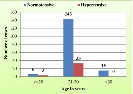
Maximum number of cases i.e.87.4% among
[image:2.595.312.546.512.678.2]normotensive and 91.7% among hypertensive group belong to 21-30 years age group. This difference was not statistically significant (Figure 1).
Figure 1: Distribution of cases according to age.
Maximum no. of cases i.e. among hypertensive group were nulliparous This difference was statistically significant (Figure 2).
6
143
15 3
33
0 0
20 40 60 80 100 120 140 160
<=20 21-30 >30
N
u
m
b
er
o
f
ca
ses
Age in years
Figure 2: Distribution of cases according to parity.
[image:3.595.51.286.71.222.2]Though higher no. of hypertensive cases belongs to urban residence, the difference between the two groups is not statistically significant (Figure 3).
Figure 3: Distribution of cases according to residence.
In present study, it was observed that among 45 patients who had beta-HCG levels >or=40,000mIU/ml, 30 developed hypertension. Whereas among 155 patients who had beta-HCG levels<40,000 mIU/ml, only 6 developed hypertension. This difference was statistically highly significant (Figure 4).
[image:3.595.51.284.305.458.2]Figure 4: Distribution of cases according to serum β HCG and hypertension.
Figure 5: Distribution of cases according to mean serum lipids and hypertension.
Maximum number of cases among the hypertensive group (83.33%) had serum TG levels >200 mg/dl whereas among the normotensive group, only25.60 % had serum TG levels >200 mg/dl. Among the normotensive group, only 5.5% of cases had serum TC levels >250 mg/dl, whereas, among the hypertensive group, 83.4% cases had levels >250 mg/dl. 25.6% of the normotensive cases had serum VLDL >40 mg/dl, whereas, a significantly higher number of hypertensive cases i.e. 75% had serum VLDL >40 mg/dl. 83.4% of hypertensive patients had serum LDL >140 mg/dl, whereas, among the normotensive group only 18.3% had serum LDL >140 mg/dl. In our study, 5.5% of the cases among normotensive group had serum HDL <40 mg/dl whereas, 16.6% of cases among the hypertensive group had serum HDL <40 mg/dl. The difference between the two groups regarding serum levels of TG, TC, LDL, VLDL and HDL was statistically significant (Figure 5).
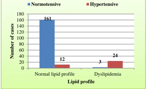
Figure 6: Distribution of cases according to serum lipid profile and hypertension.
Cases with dyslipidemia included all those patients who had abnormal values of serum triglycerides, total cholesterol and very low density lipoproteinsi.e.TG>200, TC>250 &VLDL>40 mg/dl and it was found that among the hypertensive group, 66.7% had abnormal lipid profile,
82 73 0 10 20 30 40 50 60 70 80 90
p0 p1 p2
N u m b er o f ca ses Parity Normotensive Hypertensive 77 87 15 21 0 10 20 30 40 50 60 70 80 90 100 Rural Urban N u m b er o f ca ses Residence Normotensive Hypertensive 149 15 6 30 0 20 40 60 80 100 120 140 160 <40,000 >=40,000 N u m b er o f ca ses
Serum Beta HCG Normotensive Hypertensive 161.43 204.74 120.54 48.71 32.25 238.75 250.75 157.63 47 46.01 0 50 100 150 200 250 300 M ea n ser u m l ev el s Serum lipids Normotensive Hypertensive 161 3 12 24 0 20 40 60 80 100 120 140 160 180
Normal lipid profile Dyslipidemia
[image:3.595.310.545.520.663.2] [image:3.595.51.284.576.725.2]whereas only 1.8% of cases among the normotensive group had abnormal lipid profile (p<0.00001) (Figure 6).
DISCUSSION
Hypertension in pregnancy is one of the commonest and dangerous complications causing increased fetal and maternal morbidity and mortality. Many workers have studied the association between high levels of beta HCG and dyslipidemia and hypertensive disorders of pregnancy. A total of 200 obstetric cases were studied and selected randomly from the antenatal outpatient clinics at SMGS Hospital, Jammu, Jammu and Kasmir, India. Their serum beta HCG and lipids were estimated between 14-20 weeks of pregnancy. Out of these 200 women,164 patients remained normotesive which served as controls and 36 patients turned out to be hypertensive which were termed as cases giving the prevalence rate of 18%.
Maximum no. of patients in our studty were in the age group of 21-30 years which is the peak reproductive age group in our country. 143 patients (87.4%) among the normotensive group and 33 patients (91.7%) among the hypertensive group were in this age group. However, this difference was not statistically significant.
Gokdeniz et al and Yousufnejad et al conducted a similar study in which there was no significant difference between the study group and the control group with respect to age.9,10 Maximum no. of cases in present study
i.e. 82/164 (50%) among the normotensive group and 27/36 (75%) among the hypertensive group were primigravidas indicating that primigravidas have a higher risk of developing pregnancy induced hypertension compared to multipara. The results were consistent with the finding of Yadav et al and Gohil et al where it was found that preeclampsia was significantly more common in the primigravidas.11,12
Out of 200 patients,92 were from rural area and 108 were from urban area. Among the rural women,15 developed hypertension and 77 remained normotensive whereas 21 among the urban women developed hypertension i.e.58.33% of the hypertensive group belonged to urban area and no correlation was found between the residence and the occurrence of hypertension. These findings are in contrast to the studies conducted by Gadalla et al and El-Moselhy where they found that rural residence was
significantly associated with increased risk of
preeclampsia.13,14
Among the 200 patients analysed, values of serum beta HCG ranged from 12,540 mIU/mL to 96,000 mIU/mL and it was observed that among 45 patients with beta HCG ≥40,000mIU/mL, 30 developed hypertension, whereas, among 155 patients who had levels <40,000 mIU/mL, only 6 developed hypertension. This difference was statistically highly significant (p<0.0001). Our results are consistent with that of Vidyabati et al and
Gonen et al who conducted a prospective cohort study on pregnant patients and found that women with elevated levels of beta HCG had a significantly higher risk of hypertension.4,15 Observations made in present study
were also consistent with those of Sorensen et al, Wenstrom et al, Benn et al, Yang et al and Onderoglu et al.5,6,16-18
The above association could be explained by the early placental vascular damage in preeclamptic women leading to decreased oxygen supply, which might result in increased HCG production by the hyperplastic syncytotrophoblastic cells.
Inpresent study, 128 out of total 200 women had serum triglyceride levels <200 mg/dl. Of these 128 patients, only 6 cases developed hypertension, whereas among 72 patients who had serum TG levels ≥200 mg/dl,30 cases developed hypertension. This difference was statistically highly significant (p <0.00001).
Among the normotensive group, only 9 out of 164 (5.5%) of cases had serum TC levels ≥250 mg/dl, whereas, among the hypertensive group,30 out of 36 (83.4%) cases had levels ≥250 mg/dl. This difference between the two groups regarding total cholesterol levels was highly statistically significant (p<0.0001).
25.6% of the normotensive cases had serum VLDL ≥40 mg/dl, whereas, a significantly higher number of hypertensive cases i.e. 75% had serum VLDL ≥40 mg/dl. This difference was again statistically highly significant (p <0.0001). 83.4% of hypertensive patients had serum LDL ≥140 mg/dl, whereas, among the normotensive group only 18.3% had serum LDL ≥140 mg/dl. This difference was statistically significant (p <0.0001).
In present study, 5.5% of the cases among normotensive group had serum HDL <40 mg/dl whereas, 16.6% of cases among the hypertensive group had serum HDL <40 mg/dl (p <0.03).
When we consider the lipid profile in totality taking atleast three positive parameters i.e.TG ≥200, TC ≥250 and VLDL ≥40 mg/dl, it was found that 27 out of 200 patients had abnormal lipid profile. Among the hypertensive group, 66.7% had abnormal lipid profile, whereas only 1.8% of cases among the normotensive group had abnormal lipid profile (p<0.00001).
The relationship between abnormal lipid profile and hypertension has been studied by a number of investigators with some studies showing association of some of the parameters of lipid profile and others showing association of all the parameters with hypertension.
significantly associated with the increased risk of preeclampsia.19
In the study of Jayanta De et al, the preeclampsia group was associated with a significant rise in triglycerides and VLDL cholesterol and fall in HDL concentration (p <0.05).8
In the studies of Mukherjee et al and Gohil et al, the serum TG, TC, LDL and VLDL levels were significantly higher and HDL significantly lower in preeclamptic women compared to normotensive pregnant women.20,12
CONCLUSION
It is concluded from the study that abnormally elevated maternal serum beta HCG and maternal dyslipidemia in the early second trimester i.e.14-20 weeks of pregnancy are very good non-invasive predictors of hypertensive disorders of pregnancy and the combined use of both serum beta HCG and lipid profile has even a better predictive value compared to single parameter alone.
Funding: No funding sources Conflict of interest: None declared
Ethical approval: The study was approved by the Institutional Ethics Committee
REFERENCES
1. Cunningham, Leveno, Bloom, Hauth, Gilstrap,
Wenstrom. Williams obstetrics, 23rd ed.
2008;34:706-56.
2. Lim K, Steinberg G. Preeclampsia. 2010 Available at http://emedicine.medscape.com/article/1476919-overview.
3. Dekker GA, Sibai BM. Etiology and pathogenesis of
pre-eclampsia current concepts. Am J Obstet Gynecol. 1998;179(5):1359-75.
4. Vidyabati RK, Davina H, Singh NK, Singh W
Gyaneshwar. Gyaneshwar Serum β HCG and lipid profile in early second trimester as predictors of pregnancy induced hypertension. J Obstet Gynecol India. 2010;60(1):44-50.
5. Sorensen TK, Williams MA, Zingheim RW, Hichok
D. Elevated second trimester HCG and subsequent pregnancy induced hypertension. Am J obstet Gynecol. 1993;169:834-8.
6. Wenstrom KD, Owen J, Boots LR, Dubards MB.
Elevated second trimester HCG levels in association with poor pregnancy outcome. Am J Obstet Gynecol. 1994;171:1038-41.
7. Clausen T, Djurovic S, Henriksen T. Dyslipidemia in early 2nd trimester is mainly a feature of women with early onset pre-eclampsia. Br J Obstet Gynecol. 2001;108:1081-7.
8. De J, Mukhopadhyay A, Saha PK. Study of serum lipid profile in pregnancy induced hypertension. Indian J Clin Biochem. 2006;21(2):165-8.
9. Gokdeniz R, Ariguloglu E, Bazaoglu N, Balat O. Elevated Serum HCG levels in severe pre-eclampsia. Turk J Med Sci. 2000;30:43-5.
10. Yousefnejad K, Moslemizadeh N. Serum beta HCG levels in diagnosis and management of preeclampsia. J Med Sci. 2008;8:722-7.
11. Yadav S, Gupta S. Correlation of elevated levels of maternal serum beta-hCG in pregnancy induced hypertension and pregnancy outcomes in these
patients. Indian J Pathol Microbiol. 1997
Jul;40(3):345-9.
12. Gohil TJ, Patel PK, Gupta P. Estimation of lipid profile in subjects of preeclampsia. J Obstet Gynecol India. 2011Jul;61(4):399-403.
13. Gadalla F, Abd El-Salam AF, Wassif SM.
Differential magnitude of high risk pregnancy in rural and urban communities in Sharkia governorate. Egypt J Comm Med. 1986;2(2):157-165.
14. El-Moselhy EA, Khalifa HO, Amer SM, Mohammad
KI, Abd El-Aal HM. Risk factors and impacts of
pre-Eclampsia: an epidemiological study among
pregnant mothers in Cairo, Egypt. J Am Sci. 2011;7(5):311-23.
15. Gonen R, Perez R, David M. The association between unexplained second trimester maternal serum HCG elevation and pregnancy complication. Obstet Gynecol. 1992;80:83-6.
16. Benn PA, Donna Horne BSW, Susan BBS. Elevated
second trimester maternal serum HCG alone or in combination with elevated AFP. Obstet Gynecol. 1996;87(2):217-22.
17. Feng Q, Cui S, Yang W. Clinical significance of beta HCG and human placental lactogen in serum of normal pregnancies and patients with pregnancy induced hypertension. Zhonghua Fu Chan Ke Za Zhi. 2000;35(11):648-50.
18. Onderoglu LS, Kabukcu A. Elevated second
trimester HCG level associated with adverse
pregnancy outcome. Int J Obstet Gynecol.
1997;56(3):245-9.
19. Vanden Elzen HJ, Wladimiroff JW, Cohen-overbeek
TE, de Brujin AJ, Grobbee DE. Serum lipids in early pregnancy and risk of pre-eclampsia. Br J obstet Gynecol. 1996;103:117-22.
20. Mukherjee R, Ray CD, Chakraborty C, Dasgupta S, Chaudhury K. Clinical biomarker for predicting preeclampsia in women with abnormal lipid profile: Statistical pattern approach. Systems in medicine and
biology (ICSMB), International Conference.
2010:397-401.