DESIGN OF HYDROXY XANTHONES DERIVATIVES AS ANTICANCER USING QUANTITATIVE
STRUCTURE-ACTIVITY RELATIONSHIP
EMMY YUANITA*, HARNO DWI PRANOWO, JUMINA JUMINA, MUSTOFA MUSTOFA
Department of Chemistry, Laboratory of Organic Chemistry, Universitas Gadjah Mada, Yogyakarta, Indonesia.Email: [email protected]
Received: 15 December 2015, Revised and Accepted: 06 January 2016 ABSTRACT
Objective: The objective of the research is to design a new hydroxy xanthone derivative has anticancer activity using quantitative structure-activity relationship (QSAR).
Methods: The QSAR designed new compounds were calculated by parameterized model 3 methods and analysis of multi-linear regression (MLR).
Result: The result showed that the best model as follows:
LogIC50 = –9.132 qC1 + 28.853 qC5 + 2.456 qC6 – 7.375 qC10 – 5.112 qC11 + 3.900
This result has appropriate some statistical parameters (n=24; PRESS=0.999; r2=0.782; SEE=0. 235; R=0. 885; F
cal/Ftab=4.68).
Conclusion: This Model could be used to design of halogen-substituted hydroxy xanthone scaffold and predict their inhibitory concentration (IC50) as
anticancer in the range of 0.001 - 0.484 μM.
Keywords: Anticancer, Quantitative structure-activity relationship, Xanthone.
INTRODUCTION
Cancer is the main cause of death worldwide. In 2012, there were 8.2 million to 14.1 million cases of death all over the world. The highest rate of the death is caused by an inappropriate cancer treatment. So far, the treatment requires one or more interventions such as surgery, radiotherapy, and chemotherapy [1,2]. Chemotherapy is commonly used as the treatment options. However, the low selectivity and the high toxicity of anticancer drugs compromise the beneficial treatment effects of these agents [3-5]. Therefore, development of a new chemotherapeutic agent with high selectivity and low toxicity is important to be conducted.
Xanthones, were used in this study, comprise a large number of oxygenated heterocycles planar compounds which play an important role in medicinal chemistry. Their derivatives are widely distributed in various plants, and they have a variety of biological properties such as analgesic [6], anti-oxidant [7], anti-inflammatory [8], anti-allergic [9], anti-bacterial [10], anti-tuberculosis [11], anti-fungal [12], anti-viral [13],
and also as anticancer such α-mangosteen and γ-mangosteen [14,15].
The simple structural scaffold and diverse pharmacological properties of xanthone derivatives have prompted many scientists to isolate these compounds from natural resources or synthesize them as novel drug candidates. Isolation method is not recommended because it gives a low yield of the isolated compound. Synthesis is one of the recommended ways to develop a new compound that has the best activity based on the understanding of structure modification by changing the substituents on the xanthone ring. However, this method has disadvantages over the time-consuming for “trial and error” and also cost during the synthesis.
One of the promising ways to develop a new therapeutic agent is to utilize the QSAR analysis into guiding the modification structure of the effective compounds. In the last decades, QSAR has been applied in many areas, particularly in the prediction of biological activities to save time and cost during the analysis. A QSAR equation is a mathematical equation related to the biological activity and wide variety of physical or chemical parameters. There were a lot of QSAR models mentioned in the literatures which have been successfully used for the screening
of compounds and their biological activity [16-19]. The pre-requisite for developing QSAR equations is the availability of a wide range of molecular structures and their complementary activities. Nowadays, there are some researchers conducting the computational studies on xanthones, and they believe that it is critically important.
A QSAR study conducted by Alam and Khan [20]showed the prediction
for specific targets such as HeLa cell using multi-linear regression (MLR) and optimizing their structures by MM + molecular mechanics. Their study showed that five molecular descriptors - dielectric energy, group count (hydroxyl), LogP (the logarithm of the partition coefficient between n-octanol and water), shape index basic (order 3), and the solvent-accessible surface area - were significantly correlated with anticancer activity. The other QSAR model was parameterized model 3 (PM3) methods which being used to predict a series of xanthone derivatives as antimalarial compounds and showed that hydroxyl groups could influence their activity [21]. The QSAR model of a new series of xanthone derivatives against the oral human epidermoid carcinoma (KB) cancer cell was conducted by Suphavanich et al. [22]. They reported the combination of steric, electrostatic, hydrophobic and hydrogen-bond, which was calculated using 3D QSAR models, has satisfactory correlated to the test set activities. Practically, all of the QSAR models involved molecular descriptors. In this study, we focused on to find out the electronic descriptor which could affect the anticancer activity of xanthone derivatives.
In this study, a series of xanthone derivative compound using QSAR was carried out based on the data of the inhibitory concentration (IC50, μM)
of xanthone derivatives from literatures [23-25]. The aim of this work focuses on finding establish new model which has chosen from the first step of study by selecting method of calculation of nuclear magnetic resonance (NMR) properties each model austin model 1 (AM1), PM3, Hartree-Fock (HF) level of theory, and density functional theory (DFT) methods. Based on these the best QSAR models, new compounds with highly predicted anticancer activity were theoretically designed, and they are waiting for experimental verification.
METHODS
Hardware and software
This study used a PC with Intel® Core™ i3 CPU M 350 4.54 GHz; RAM
5.00 GB. The used programs were Gaussian® 09W [26], statistical SPSS®
Release 17.0.0 [27].
Data set
The structures of xanthone derivative compounds and its anticancer activity were divided into two sets as listed in Table 1. Namely a training set (19 compounds) for generating QSAR models and a test set (5 compounds) for validating the quality of the models. The selected compound of the training set and test set is an important feature and key of any QSAR models. Hence, the treatment was taken in such a way by biological activities of all tests set compounds with the maximum and minimum range of biological activities of the training set and test set compounds. The maximum and minimum data of training and test set were compared with: (i) The maximum data of the log IC50 of
test set which should be less than or equal to the maximum value of log IC50 of training set, (ii) the minimum data of the log IC50 of test set
that should be higher or equal to the minimum value of log ic50 of the
training set [28].
Computational validation and descriptor calculation
Xanthone compounds were the first modeled computationally using AM1, PM3, HF, and DFT to obtain the most suitable calculation method and using the Gaussian package to calculate their chemical shift (NMR properties). The model with the smallest difference (PRESS value) between calculated and experimental data for each method (AM1, PM3, HF, or DFT) was chosen as the statistics of the training and test sets for QSAR calculation.
The development of QSAR model
The QSAR model was generated by the MLR Backward method using the SPSS package. This method refers to the dependent variable
ŷ (biological activity) with a number of independent variables
xi (electronic descriptors) by using linear equations. Moreover, this regression method estimates the values of the regression coefficients by applying least square curve fitting method. The model was chosen for QSAR calculation based on some statistical parameters such as r2,
standard estimation of error (SEE), F-ratio between the variance of prediction and observation activity, and PRESS (predictive residual sum
of square), where: PRESS = Σ (predicted value-observed value)2 [29] in
criteria r2 > 0.6 [30]; SEE < 0.3 [31]; F
cal/Ftab≥ 1 [32]. The validation of QSAR model
The best-selected model obtained from the previous step was used to predict the Log IC50 of the test set. The model was validated by criteria
r2
prediction > 0.5 [33].
Design and activities prediction of the new compounds
The new designed and predicted xanthone derivative compounds based on the validated data with the best inhibitory activity and has lower IC50 value was chosen as a new candidate of the anticancer compound.
RESULT AND DISCUSSION Validated method
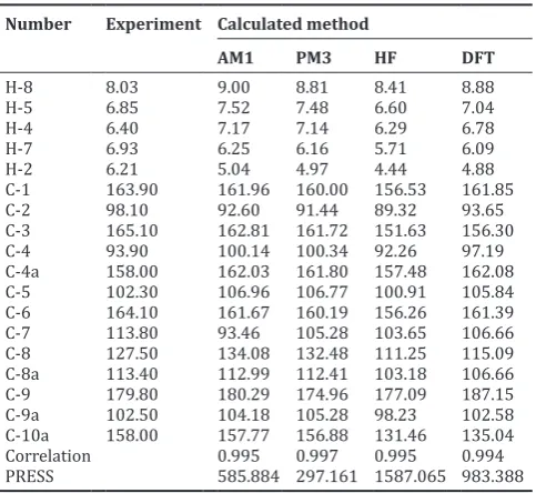
The calculation of the chemical shift from each method (AM1, PM3, HF, and DFT) was compared with the experimental measurements to get the most suitable calculation method for modeling the series of xanthones derivatives with anticancer activity. The experimental measurement showed that the PM3 method gave the smallest difference (PRESS value) between calculated and experimental result. The correlation coefficient from each model was more than 0.99 (near 1) and listed in Table 2. Therefore, PM3 method has been selected as a calculation method for further modeling of anticancer activity of xanthones derivatives.
The selection method is important to improve the accuracy because the method of calculation giving smallest differences between calculated and experimental data validated method for the selection of methods of QSAR is also done in the design of new insecticide compounds have potent of organophosphate [34] with the results of AM1 method used for the analysis of advanced QSAR.
Generation and selection of QSAR model
Multiple linear regression analysis using SPSS version 17 for Windows has been performed to obtain the best model which correlates independent variables (descriptors) to a dependent variable (biological activity). QSAR analysis of this research was done to see the relationship between electronic variable against anticancer activity. Analysis of different from that done by previous researchers [10,11] that combines electronic and molecular variables. The use of electronic parameter because the electronic charge on ring xanthone can
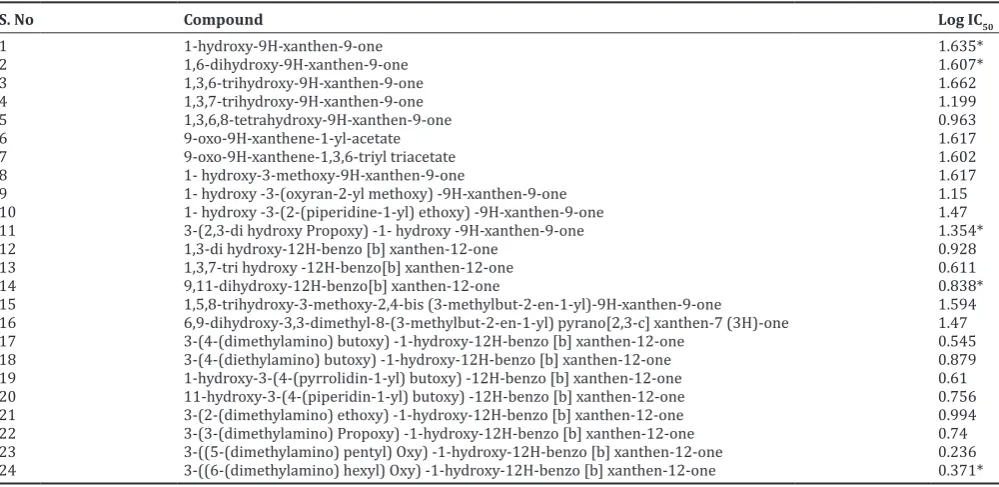
Table 1: A series of xanthone derivatives
S. No Compound Log IC50
1 1-hydroxy-9H-xanthen-9-one 1.635*
9 1- hydroxy -3-(oxyran-2-yl methoxy) -9H-xanthen-9-one 1.15
10 1- hydroxy -3-(2-(piperidine-1-yl) ethoxy) -9H-xanthen-9-one 1.47
11 3-(2,3-di hydroxy Propoxy) -1- hydroxy -9H-xanthen-9-one 1.354*
12 1,3-di hydroxy-12H-benzo [b] xanthen-12-one 0.928
13 1,3,7-tri hydroxy -12H-benzo[b] xanthen-12-one 0.611
14 9,11-dihydroxy-12H-benzo[b] xanthen-12-one 0.838*
15 1,5,8-trihydroxy-3-methoxy-2,4-bis (3-methylbut-2-en-1-yl)-9H-xanthen-9-one 1.594
16 6,9-dihydroxy-3,3-dimethyl-8-(3-methylbut-2-en-1-yl) pyrano[2,3-c] xanthen-7 (3H)-one 1.47
17 3-(4-(dimethylamino) butoxy) -1-hydroxy-12H-benzo [b] xanthen-12-one 0.545
18 3-(4-(diethylamino) butoxy) -1-hydroxy-12H-benzo [b] xanthen-12-one 0.879
19 1-hydroxy-3-(4-(pyrrolidin-1-yl) butoxy) -12H-benzo [b] xanthen-12-one 0.61
20 11-hydroxy-3-(4-(piperidin-1-yl) butoxy) -12H-benzo [b] xanthen-12-one 0.756
21 3-(2-(dimethylamino) ethoxy) -1-hydroxy-12H-benzo [b] xanthen-12-one 0.994
22 3-(3-(dimethylamino) Propoxy) -1-hydroxy-12H-benzo [b] xanthen-12-one 0.74
23 3-((5-(dimethylamino) pentyl) Oxy) -1-hydroxy-12H-benzo [b] xanthen-12-one 0.236
24 3-((6-(dimethylamino) hexyl) Oxy) -1-hydroxy-12H-benzo [b] xanthen-12-one 0.371*
describe the role or position of atoms which are the most influential on the anticancer activity. The 24 active compounds with their in vitro
inhibition concentration were randomly divided into the training set of 19 compounds and a test set of 5 compounds. 14 independent variables for electronic descriptor was described to consist of 14 atomic nets-charges (q) of C1, C2, C3, C4, C5, C6, C7, C8, C9, C11, C12, O14, and O15. Of the 15 charges is difficult to determine the most dominant nets-atom effect, so the backward method was performed to gradually eliminate the less relevant variables from the models. This procedure has finally given 8 QSAR models as listed in Table 3. From the Table 3, it was emerged that all selected models show a good correlation (r≈ 0.9)
between biological activity and the selected descriptors.
This result indicated the determination of the best model among 8 QSAR models listed in Table 3 was not adequate only by comparing the rsize, while they have similar value. Therefore, other statistical parameters such as r2, standard estimation of error (SEE<0.3), PRESS (predictive
residual sum of square) and also Fcal/Ftab (≥1) could be taken into account.
Comparison of the mentioned parameters (r2, SEE, PRESS, and F cal/Ftab)
toward the 8 models, presented in Table 3, pointed that it was also not easy to choose the best model because their value were not significantly different. However, Fcal/Ftab considered as the most noticeable parameter
which give a variety of value from 1.26 to 3.99. According to this value, model 7 and 8 were decided to be the best QSAR model because their Fcal/Ftab value was the highest among the others (>3). Furthermore, model
7 and 8 were used to search and design of new xanthones derivatives with better anticancer activity and their complete QSAR equations was, respectively, presented in Eq. 1 and Eq. 2 as below as:
Log IC50 = –9.378 qC1 + 31.283 qC5 + 3.962 qC6 -7.278 qC10 – 4.898
qC11 + 4.135 (1)
Log IC50 = 19.633 qC5 + 6.458 qC6 – 5.731 qC11 + 3.986 (2)
The two equations above can be sure yet where that will be used to design new anticancer compound xanthone, if the terms of the simplicity of electronic parameter number of influential model equation 8 or 2 the most good, but needs to be continued validation against both equations is obtained to ensure that the equation with parameters most appropriate statistics.
Validation of QSAR models
QSAR equation (1) and (2) was applied to calculate the activity of 5 test set compounds, and we could see how good these models predict the anticancer activity of the xanthone derivatives. A validation for searching the best model was performed using calculation for each equation toward the test set. The PRESS value is the amount of quadrate difference between the predicted and observed LogIC50 value, where the equation with the
smallest difference was chosen as the best equation. PRESS value from the predicted and observed LogIC50 of model 7 and 8 were listed in Table 4.
Based on the PRESS parameter, the smallest PRESS value was shown by model 7, with 0.51 in difference, compared with model 8 with 0.69 in value. This result indicated that it has a small difference of anticancer activity as IC50 values within the predicted and experimental. It could be
decided from PRESS value, that the model 7 was better than 8 as QSAR model to predict the anticancer activity and also it could give good structure of the predicted xanthone derivative compound.
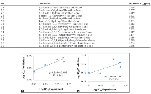
Other parameters to convince the best model was with compared their slope and correlation coefficient (r2). As seen in Fig. 1, it was determined
that r2 of model 7 and 8 were 0.861 and 0.556, respectively.
According to PRESS and r2 parameters of statistical analysis, model 7 with
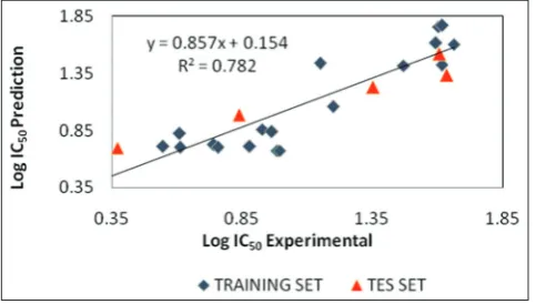
equation 1 would be the best QSAR model to generating the validation model toward the test set. Further validation for determining the best QSAR model was evaluated using enter statistical calculation. In the end, based on the calculation toward some statistical parameters, we found out the result as follows: PRESS=0.999, r2=0.7829, SEE=0. 235, R=0. 885,
FCal/FTab=4.68. Plots of the predicted versus experimental of anticancer
activity as Log IC50 values was shown in Fig. 2.
The correlation coefficient (r2) was 0.7829, it means there is 78.29%
similarity value between predicted and experimental anticancer activity. Based on statistics obtained renewal of the parameter values of the equation 2, it could be generated the best equation model of QSAR regression as follows:
Log IC = –9.132 qC1 + 28.853 qC5 + 2.456 qC6 – 7.375 qC10 – 5.112
qC11 + 3.900 (3)
Based on the Equation 3, electronic descriptors for electron charge of carbon atom in number 1, 5, 6, 10 and 11 (Fig. 3) Showed as the position with the most affect to the anticancer activity. The better anticancer activity as IC50 could be given by the more negative of the
Table 3: Statistical parameters of 8 selected QSAR models of xanthones derivatives
Model Descriptors r R2 Adjusted R SEE PRESS F
Calc/Ftab
1 qO14, qC8, qC6, qC7, qC10, qC12, qC1, qC11, qC4, qC13, qC5 0.93 0.86 0.642 0.26 0.47 1.26
2 qO14, qC8, qC6, qC10, qC12, qC1, qC11, qC4, qC13, qC5 0.93 0.86 0.686 0.24 0.47 1.58
3 qO14, qC6, qC10, qC12, qC1, qC11, qC4, qC13, qC5 0.93 0.86 0.719 0.23 0.47 1.97
4 qO14, qC6, qC10, qC12, qC1, qC11, qC4, qC5 0.92 0.85 0.735 0.22 0.50 2.32
5 qO14, qC6, qC10, qC1, qC11, qC4, qC5 0.92 0.84 0.747 0.21 0.52 2.76
6 qO14, qC6, qC10, qC1, qC11, qC5 0.91 0.82 0.734 0.22 0.60 2.98
7 qC6, qC10, qC1, qC11, qC5 0.90 0.80 0.726 0.22 0.473 3.38
8 qC6, qC10, qC11, qC5 0.88 0.78 0.717 0.23 0.74 3.99
SEE: Standard estimation of error, QSAR: Quantitative structure-activity relationship Table 2: Comparison of calculated and experimental NMR
chemical shift data (δ, ppm)
C-1 163.90 161.96 160.00 156.53 161.85
C-2 98.10 92.60 91.44 89.32 93.65
C-3 165.10 162.81 161.72 151.63 156.30
C-4 93.90 100.14 100.34 92.26 97.19
C-4a 158.00 162.03 161.80 157.48 162.08
C-5 102.30 106.96 106.77 100.91 105.84
C-6 164.10 161.67 160.19 156.26 161.39
C-7 113.80 93.46 105.28 103.65 106.66
C-8 127.50 134.08 132.48 111.25 115.09
C-8a 113.40 112.99 112.41 103.18 106.66
C-9 179.80 180.29 174.96 177.09 187.15
C-9a 102.50 104.18 105.28 98.23 102.58
C-10a 158.00 157.77 156.88 131.46 135.04
Correlation 0.995 0.997 0.995 0.994
PRESS 585.884 297.161 1587.065 983.388
log IC50. To get the most negative value of logIC50, it was ordered by
the negative net atomic charge of qC5 and qC6 and the positive net atomic charge of qC1, qC10, and qC11. From the Equation 3, qC5 and qC6 have positive value 28.853 and 2.456, respectively, so to make an electron net charge becoming negative, the C5 and C6 or neighbor’s position should be occupied with electron donating groups (EDG)
such as hydroxy and methoxy group to make the π system more
nucleophile. Meanwhile, the other positions such as C1, C10, and C11 were supposed to fill up with electron withdrawing groups (EWG) such as nitro (–NO2) and also halogen (–X) to remove the electron
density from the π system and make it less nucleophile or to make
the net charge become positive. In other word, to make the net charge become negative, it is important to consider for adding the EDG and EWG groups on the best position to predict a series of xanthones derivatives with the best activity. Based on previous reports [35], it showed that the halohydrin (bromo and chloro) xanthones where the most effective inhibitors for Topo II, similar with doxorubicin as commercial cancer therapy, which has same inhibition activity. So in this study, it has been conducted to design a new hydroxy xanthone
by modification of hydroxy xanthone scaffold with halogen groups, such as bromo, chloro and iodo, to look out their predicted anticancer activity, as listed as Table 5.
This research showed that there was a significant difference in the value of predicted Log IC50 compared with doxorubicin as commercial
cancer therapy. This result indicates that the halogen-substituted hydroxy xanthone has potential to be developed as anticancer drugs, especially for HepG2 (human liver cancer) with predicted IC50 in the
range of 0.001 - 0.484 μM, wherein it was lower than IC50 of doxorubicin
at 3.83 μM [24]. Compounds predicted to also differ from other types
of cancer as a result of the prediction by Alam and Khan [10] for HeLa cell cancer design derivatives of xanthone compounds hydroxy, methoxy, epoxy, and prenyl. While the special halogen compound bromo, and chloro is not done. Hence, the design of compound analysis results QSAR is a new xanthone derivative hydroxy compound with activity better value when compared to the values of IC50 cancer drug doxorubicin.
CONCLUSION
Study of the correlation between electronic parameters, atomic net charge, and the anticancer activity of xanthones has been performed using semi-empirical molecular orbital calculation PM-3. This study found out that the descriptors of atomic net charges: qC1, qC5, qC6, qC10, and qC11, were hypothetically active at the molecular site of xanthone derivatives and seem to be the most responsible for their pharmacological activity. The best QSAR regression equation from this study was revealed to be: Log IC50 = –9.132 qC1 + 28.853 qC5
+ 2.456 qC6 - 7.375 qC10 - 5.112 qC11 + 3. 900. n=24, PRESS=0.999,
r2=0.7829, SEE=0.235, R=0.885 and F
cal/Ftable=4.68. This QSAR model
could predict a good inhibition activity (IC50) value of
halogen-substituted hydroxy xanthone derivatives within the range
0.001-0.484 μM.
Table 5: New designed xanthones derivatives as anticancer and their predicted IC50 calculated using the best QSAR model
No. Compound Predicted IC50 (μM)
25 2,4-dibromo-3-hydroxy-9H-xanthen-9-one 0.068
26 2,4-dichloro-3-hydroxy-9H-xanthen-9-one 0.207
27 2,4-diiodo-3-hydroxy-9H-xanthen-9-one 0.019
28 4-bromo-1,3-dihydroxy-9H-xanthen-9-one 0.003
29 4-chloro-1,3-dihydroxy-9H-xanthen-9-one 0.005
30 4-iodo-1,3-dihydroxy-9H-xanthen-9-one 0.001
31 4,7-dibromo-1,3,6-trihydroxy-9H-xanthen-9-one 0.013
32 4,7-dichloro-1,3,6-trihydroxy-9H-xanthen-9-one 0.035
33 4,7-diiodo-1,3,6-trihydroxy-9H-xanthen-9-one 0.003
34 2,4-dibromo-3,5,6,7-tetrahydroxy-9H-xanthen-9-one 0.147
35 2,4-dichloro-3,5,6,7-tetrahydroxy-9H-xanthen-9-one 0.484
36 2,4-diiodo-3,5,6,7-tetrahydroxy-9H-xanthen-9-one 0.038
37 1,5-dibromo-2,3,4,6,8-pentahydroxy-9H-xanthen-9-one 0.029
38 1,5-dichloro-2,3,4,6,8-pentahydroxy-9H-xanthen-9-one 0.264
39 1,5-diiodo-2,3,4,6,8-pentahydroxy-9H-xanthen-9-one 0.036
Table 4: The comparison between predicted and experimental anticancer activity (Log IC50) of 5 test set selecting calculated by
selected model 7 and 8
Compounds
of test sets Experimental Log IC50
Predicted Log IC50
Model 7
(Eq. 1) Model 8 (Eq. 2)
1 0.37 0.70 0.75
2 0.84 0.99 0.72
11 1.35 1.21 1.37
14 1.61 1.29 1.22
24 1.64 1.12 1.02
PRESS 0.51 0.69
Fig. 1: Plot of prediction versus experiment anticancer (Log IC50) of model 7 (a) and 8 (b)
Fig. 2: Plot of predicted versus experimental anticancer activity values of model 7
Fig. 3: Structure of xanthone
ACKNOWLEDGMENTS
This work was supported by the Ministry of Research, Technology and Higher Education through Beasiswa Pendidikan Pascasarjana Dalam Negeri scholarship for the PhD program. Moreover, we would like to say thanks to the Computer Laboratory, were provided by the Austrian - Indonesian Centre (AIC), for Computational program Gaussian 09 licenses.
REFFERENCES
1. Heffeter P, Jakupec MA, Körner W, Chiba P, Pirker C, Dornetshuber R,
et al. Multidrug-resistant cancer cells are preferential targets of the new antineoplastic lanthanum compound KP772 (FFC24). Biochem Pharmacol 2007;73(12):1873-86.
2. Khonkarn R, Mankhetkorn S, Talelli M, Hennink WE, Okonogi S. Cytostatic effect of xanthone-loaded mPEG-b-p(HPMAm-Lac2) micelles towards doxorubicin sensitive and resistant cancer cells. Colloids Surf B Biointerfaces 2012;94:266-73.
3. Iranshahi M, Sahebkar A, Hosseini ST, Takasaki M, Konoshima T, Tokuda H. Cancer chemopreventive activity of diversin from Ferula diversivittata in vitro and in vivo. Phytomedicine 2010;17(3-4):269-73. 4. Tsuruo T. Molecular cancer therapeutics: Recent progress and targets in
drug resistance. Intern Med 2003;42(3):237-43.
5. Li R, Hehlman R, Sachs R, Duesberg P. Chromosomal alterations cause the high rates and wide ranges of drug resistance in cancer cells. Cancer Genet Cytogenet 2005;163(1):44-56.
6. Shi LM, Fan Y, Myers TG, O’Connor PM, Paull KD, Friend SH, et al.
Mining the NCI anticancer drug discovery databases: Genetic function approximation for the QSAR study of anticancer ellipticine analogues. J Chem Inf Comput Sci 1998;38(2):189-99.
7. Oloff S, Mailman RB, Tropsha A. Application of validated QSAR models of D1 dopaminergic antagonists for database mining. J Med Chem 2005;48(23):7322-32.
8. Meneses-Marcel A, Marrero-Ponce Y, Machado-Tugores Y, Montero-Torres A, Pereira DM, Escario JA, et al. Alinear discrimination analysis based virtual screening of trichomonacidal lead-like compounds:
Outcomes of in silico studies supported by experimental results. Bioorg Med Chem Lett 2005;15(17):3838-43.
9. Santana L, Uriarte E, González-Díaz H, Zagotto G, Soto-Otero R, Méndez-Alvarez E. A QSAR model for in silico screening of MAO-A inhibitors. Prediction, synthesis, and biological assay of novel coumarins. J Med Chem 2006;49(3):1149-56.
10. Alam S, Khan F. QSAR and docking studies on xanthone derivatives for anticancer activity targeting DNA topoisomerase IIa. Drug Des Devel Ther 2014;8:183-95.
11. Amanatie A, Jumina J, Mustofa M, Hanafi M, Armunanto R. QSAR study of xanthone derivatives as antiplasmodial agent. Indones J Chem 2010;10:357-62.
12. Suphavanich K, Maitarad P, Hannongbua S, Sutda P, Suksamrarmn S, Tantirungrotechai Y, et al. CoMFA an CoMSIA study on a new series of xanthone derivatives against the oral human epidomoid carcinoma (KB) cancer cell line. Monatash Chem 2009;140:273-80.
13. Cui J, Hu W, Cai Z, Liu Y, Li S, Tao W, et al. New medicinal properties of mangostins: Analgesic activity and pharmacological characterization of active ingredients from the fruit hull of Garcinia mangostana L. Pharmacol Biochem Behav 2010;95(2):166-72.
14. Jung HA, Su BN, Keller WJ, Mehta RG, Kinghorn AD. Antioxidant
xanthones from the pericarp of Garcinia mangostana (Mangosteen).
J Agric Food Chem 2006;54(6):2077-82.
15. Cheng JH, Huang AM, Hour TC, Yang SC, Pu YS, Lin CN. Antioxidant xanthone derivatives induce cell cycle arrest and apoptosis and enhance cell death induced by cisplatin in NTUB1 cells associated with ROS. Eur J Med Chem 2011;46(2):1222-31.
16. Nakatani K, Atsumi M, Arakawa T, Oosawa K, Shimura S, Nakahata N, et al. Inhibitions of histamine release and prostaglandin E2 synthesis by mangosteen, a Thai medicinal plant. Biol Pharm Bull 2002;25(9):1137-41.
17. Sakagami Y, Iinuma M, Piyasena KG, Dharmaratne HR. Antibacterial activity of alpha-mangostin against vancomycin resistant Enterococci (VRE) and synergism with antibiotics. Phytomedicine 2005;12(3):203-8.
18. Suksamrarn S, Suwannapoch N, Phakhodee W, Thanuhiranlert J,
Ratananukul P, Chimnoi N, et al. Antimycobacterial activity of
prenylated xanthones from the fruits of Garcinia mangostana. Chem Pharm Bull (Tokyo) 2003;51(7):857-9.
19. Kaomongkolgit R, Jamdee K, Chaisomboon N. Antifungal activity of alpha-mangostin against Candida albicans. J Oral Sci 2009;51(3):401-6. 20. Chen LG, Yang LL, Wang CC. Anti-inflammatory activity of mangostins
from Garcinia mangostana. Food Chem Toxicol 2008;46(2):688-93.
21. Matsumoto K, Akao Y, Kobayashi E, Ohguchi K, Ito T, Tanaka T,
et al. Induction of apoptosis by xanthones from mangosteen in human leukemia cell lines. J Nat Prod 2003;66(8):1124-7.
22. Aisha AF, Abu-Salah KM, Ismail Z, Majid AM. In vitro and in vivo
anti-colon cancer effects of Garcinia mangostana xanthones extract. BMC Complement Altern Med 2012;12:104.
23. Su QG, Liu Y, Cai YC, Sun YL, Wang B, Xian LJ. Anti-tumour effects of xanthone derivatives and the possible mechanisms of action. Invest New Drugs 2011;29(6):1230-40.
24. Kuete V, Sandjo LP, Ouete JL, Fouotsa H, Wiench B, Efferth T. Cytotoxicity and modes of action of three naturally occurring xanthones (8-hydroxycudraxanthone G, morusignin I and cudraxanthone I) against sensitive and multidrug-resistant cancer cell lines. Phytomedicine 2014;21(3):315-22.
25. Luo L, Qin K, Dai ZK, Gou SH. Synthesis and biological evaluation of novel benzo [b] xanthone derivatives as potential antitumor agents. J Serbian Chem Soc 2013;78:1301-8.
26. Frisch MJ, Trucks GW, Schlegel HB, Scuseria GE, Robb MA, Cheeseman JR, et al. Gaussian 09, Revision A.02. Wallingford, CT: Gaussian, Inc.; 2009.
27. Yee BJ. Chemical synthesis of 1,6 dioxygenated xanthones and their cytotoxic activities. B. Sc Degree, Faculty of Science, Universiti Tungku Abdul Rahman; 2011.
28. Jain SV, Ghate M, Bhadoriya KS, Bari SB, Chaudhari A, Borse JS. 2D, 3D-QSAR and docking studies of 1,2,3-thiadiazole thioacetanilides analogues as potent HIV-1 non-nucleoside reverse transcriptase inhibitors. Org Med Chem Lett 2012;2(1):22.
29. Podunavac-Kuzmanovic SO, Cvetkovic DD, Barna DJ. QSAR analysis of 2-amino or 2-methyl-1-substituted benzimidazoles against
Pseudomonas aeruginosa. Int J Mol Sci 2009;10(4):1670-82. 30. Golbraikh A, Shen M, Xiao Z, Xiao YD, Lee KH, Tropsha A. Rational
evidence for surface freezing in supercooled n-alkane nanodroplets. Phys Chem Chem Phys 2013;15(18):6783-95.
32. Motta LF, Almeida WP. Quantitative structure-activity relationships (QSAR) of a series of ketone derivatives as anti-Candida albicans. Int J Drug Disc 2011;3:100-17.
33. Frimayanti N, Yam ML, Lee HB, Othman R, Zain SM, Rahman NA. Validation of quantitative structure-activity relationship (QSAR) model for photosensitizer activity prediction. Int J Mol Sci
2011;12(12):8626-44.
34. Mudasir M, Wibowo YM, Pranowo HD. Design new potent insecticides of organophosphate derivatives based on QSAR Analysis. Indones J Chem 2013;13(1):86-93.