Developmental expression of chicken FOXN1 and putative
target genes during feather development
DIANA K. DARNELL*
,1, LI S. ZHANG
1, SRIDHAR HANNENHALLI
2and SERGEY Y. YAKLICHKIN*
,3 1Department of Cellular and Molecular Medicine, University of Arizona, Tucson, AZ,2Center for Bioinformatics and Computational Biology, University of Maryland, College Park, MD and 3Department of Molecular and Cellular Biology, Baylor College of Medicine, Houston TX, USA
ABSTRACT FOXN1 is a member of the forkhead box family of transcription factors. FOXN1 is crucial for hair outgrowth and thymus differentiation in mammals. Unlike the thymus, which is found in all amniotes, hair is an epidermal appendage that arose after the last shared common ancestor between mammals and birds, and hair and feathers differ markedly in their differentiation and gene expression. Here, we show that FOXN1 is expressed in embryonic chicken feathers, nails and thymus, demonstrating an evolutionary conservation that goes beyond obvious homology. At embryonic day (ED) 12, FOXN1 is expressed in some feather buds and at ED13 expression extends along the length of the feather filament. At ED14 FOXN1 mRNA is restricted to the proximal feather filament and is not detectable in distal feather shafts. At the base of the feather, FOXN1 is expressed in the epithelium of the feather sheath and distal barb and marginal plate, whereas in the midsection FOXN1 transcripts are mainly detected in the barb plates of the feather filament. FOXN1 is also expressed in claws; however, no expression was detected in skin or scales. Despite expression of FOXN1 in developing feathers, examination of chick homologs of five putative mammalian FOXN1 target genes shows that, while these genes are expressed in feathers, there is little similarity to the FOXN1 expression pattern, suggesting that some gene regulatory networks may have diverged during evolution of epidermal appendages.
KEY WORDS:
chick, forkhead, WHN, HFH11, thymus, nudee
The forkhead family transcription factor FOXN1 (formerly called Winged helix transcription factor nude, Whn (Meier et al., 1999) and HNF-3/forkhead homolog 11, Hfh11 (Segre et al., 1995) is essential for hair and thymus development in mammals. Hair production is complex and cyclic, involving many transcription factors and their targets (Lee and Tumbar, 2009; Lin et al., 2009). In the murine hair follicle FOXN1 exhibits a dynamic expression pattern that is dependent on the stage of the hair cycle. FOXN1 appears to be strongly expressed in the anagen or growth phase and its expression decreases by the telogen or quiescent phase. In the mature hair follicle FOXN1 is expressed in the hair shaft and the inner root sheath (Lee at al., 1999). In the hair shaft, FOXN1 is expressed in the cortex, which is consistent with absence of the cortex in Nude (FOXN1nu/nu) mice. Nude mice develop normal hair follicles and produce hair shafts, but the cortex fails to form most keratins so the hair does not extend. The absence of visible
*Address correspondence to: Diana K. Darnell. Department of Cellular and Molecular Medicine, 1656 E. Mabel Street - MRB324, Tucson, AZ 85724, USA.
Tel: +1-520-626-9925. Fax: +1-520-626-7600. E-mail: [email protected] or Sergey Yu. Yaklichkin. Department of Molecular and Cellular Biology, Baylor College of Medicine, One Baylor Plaza N620, Houston, Texas 77030, USA. E-mail: [email protected]
Supplementary Material (table) for this paper is available at: http://dx.doi.org/10.1387/ijdb.130023sy
Accepted: 9 January 2014. Final, author-corrected PDF published online: 17 February 2014.
ISSN: Online 1696-3547, Print 0214-6282
© 2014 UBC Press Printed in Spain
Abbreviations used in this paper: ED, embryonic day; FOX, forkhead box transcription factor.
hair is due to a coiling of hair shafts in the hair canal resulting in a failure to penetrate the epidermal layer of the skin or breaking off at the surface (Mecklenburg et al., 2001, 2004, 2005). Similarly, humans with FOXN1 mutation have congenital alopecia (hair loss), nail dystrophy and immunodeficiency (Frank et al., 1999, Pignata et al., 2009).
arose after the last shared common ancestor between mammals and birds in the late Carboniferous period, it is not surprising that proteins in these two lineages have functionally diverged (Furlong, 2005). For example, hair is characterized by a preponderance of soft alpha keratin (Eckhart et al., 2008), whereas feathers are characterized predominantly by the harder beta keratins (Glenn et al., 2008). Alpha keratins are found in the feather sheath and barb ridges but are rapidly overwhelmed by deposition of beta keratins (Alibardi, 2013). Since one function of transcription factor FOXN1 in mice is the activation of alpha keratins 2-6 (Schlake et al., 2000; Mecklenburg et al., 2004) and birds appear primarily to use beta keratin proteins in feathers, it has not been tested whether feathers express FOXN1. We previously showed that another Forkhead transcription factor, FOXE1, is transcribed in the feather filament (Yaklichkin et al., 2011), and its mammalian ortholog is implicated in hair morphogenesis (Brancaccio et al., 2004). This suggests the hypothesis that FOXN1 and homologs of its mammalian regulatory targets might also be expressed in developing feathers.
In this report we show that, whereas FOXN1 and its putative
some of the gene regulatory pathways downstream of FOXN1 in epidermal appendage growth appear to have diverged since the division between mammal and bird ancestors about 310 million years ago (Kumar and Hedges, 1998).
Results
Phylogenetic and sequence analysis of avian FOXN1 sequences
The chick FOXN1 gene encodes a 653 amino acid protein with a molecular weight of 71,957 Da. The chick FOXN1 gene shares synteny with mammalian FOXN1, thus providing unambiguous evidence for orthology. Of non-avian orthologs, chick FOXN1 pro-tein sequence overall shares the highest sequence identity, 75%, with turtle and lizard FOXN1, although the lizard protein sequence is incomplete (Fig. 1A). For comparison, Chick FOXN1 protein shares 59% and 53-55% sequence identity with opossum and placental FOXN1 (Fig. 1A). To determine the evolutionary related-ness of avian FOXN1 gene to orthologs, a phylogenetic analysis was performed using the Neighbor-joining method (Kumar et al., 2004). The phylogenetic tree shows that avian FOXN1 genes are clustered within the FOXN1 group and the closest phylogenetic relatedness is observed to the reptilian and amphibian FOXN1 genes, which is supported by the high bootstrap values (Fig. 1B).
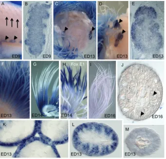
Expression of chick FOXN1 gene in developing thymus We performed whole mount in situ hybridization analysis to ex-amine FOXN1 expression during chick thymus development from stages ED1-16. No expression was detected from ED1-4.5 (not shown). In chicken, thymus is derived from the third and/or fourth pharyngeal pouch endoderm at stage ED5. At about ED9 thymus primordia detach from the pharynx and migrate to their mature locations in the neck, where they form bilateral multi-lobed glands near the jugular vein. We found that FOXN1 mRNA was strongly expressed bilaterally in ED9 thymus (Fig. 2A). A section through the ED9 thymus primordia shows that FOXN1 labeling is stronger in the prospective cortex with uniform lower intensity label in the medulla (Fig. 2B). This is consistent with the proposed function of mouse FOXN1 in regulation of keratin gene expression, which is present in medullary and cortical thymus epithelial cells during early thymus development (Rodewald, 2008; Nowell et al., 2011). As chick embryonic development progresses (stage ED13) the thymus lobes migrate laterally and expand; expression of FOXN1 persisted (Fig. 2C-D). A section through the thymus at this stage shows increased intensity of FOXN1 mRNA labeling in the cortex (Fig. 2E). This is consistent with the observation in mouse that a FOXN1 mutant phenotype is more severe in the cortical compart-ment, although both cortex and medulla are affected (Nowell et al., 2011); and expands on the published FOXN1 expression at older stages in chick: (ED18; Neves et al., 2012).
Expression of chick FOXN1 gene in developing feathers Feathers are unquestionably the most complex vertebrate epidermal appendages and the specifics of their embryology are well described (e.g., Yu et al., 2002, 2004; Alibardi, 2009, 2013). In short, a feather placode in the epidermis with an underlying dermal condensation sprouts into a feather filament with a dermal pulp separated from the epidermal sheath by the pulp epithelium. Barb Fig. 1 (left and above). Comparison of amino acid sequences of avian
FOXN1 with orthologous proteins and phylogenetic tree of FOXN1/ N4. (A) Predicted amino acid sequences of avian FOXN1 proteins aligned with orthologous protein sequences using T-coffee algorithm. The conserved forkhead DNA-binding (Kaestner et al., 2000) and transcriptional activa-tion domains (Schüddekopf et al., 1996) are underlined in blue and red, respectively. Similar amino acid residues are shaded, threshold 70%. (B)
A phylogenetic tree of the FOXN1 proteins, with FoxN4 protein sequences used as an outgroup. A neighbor-joining method was used to construct the tree topology based on the forkhead DNA-binding domain.
ridges begin to form in one side of the epidermis and are displaced laterally to create two vertical half-spirals or arcs between the base and their distal merger into the feather stem or rachis on the opposite side (downy feathers have no rachis). Each barb ridge appears as an arch in cross section with paired stacks of barb plates at its base and a growth zone at its apex, all surrounded by marginal cells. At maturity, a medullary core forms in each barb, the feather sheath is lost and the barbs separate.
Because FOXN1 is important for normal hair development in mammals, we used in situ hybridization to examine expression of FOXN1 in developing chicken feathers. FOXN1 expression was at, or barely above, background levels in initial feather buds at ED9 (Fig. 2A), but strong transient expression was observed in feather filaments at stages ED13-16 (Fig. 2 F-G, I-M). Strong expression of FOXN1 mRNA in the feather filament was detected nearly from base to apex of the feather at stage ED13 (Fig. 2F). Weaker FOXN1 label was observed in the most proximal part of the feather filament and the most distal tip. This may indicate the beginning of the maturation of the distal region of the filament. By stage ED14 FOXN1 transcripts become restricted to the proximal feather filament (Fig. 2G), which is a less differentiated and mor-phologically more active portion of the feather filament (Haake et al., 1984). To verify that the failure to hybridize in the distal filament was not due to exclusion of the color reaction or another artifact, in situ hybridizations were performed on comparable feather filaments using a probe for another transcription factor, FOXE1 (Fig. 2H; Yaklichkin et al., 2011). FOXE1 showed a complementary expres-sion pattern to that of FOXN1 in proximal/distal distribution at this stage, indicating that both proximal and distal filaments were able
to label. By stage ED16, FOXN1 mRNA was no longer detected in the feather filament (Fig. 2I, J), which can be explained by either down-regulation of FOXN1 expression in a differentiated feather filament or increased keratinization in the feather sheath, which may block FOXN1 probe penetration at this stage. Small flecks of trapped color precipitate (artifact, arrowheads Fig. 2J) indicate that probe and labeling solution were able to penetrate.
To determine specific localization of FOXN1 mRNA within the labeled feather filament, feathers were paraffin sectioned trans-versely. FOXN1 expression was evident in the proximal section of the feather filament at ED12-13 in a tight peripheral band with alternating intensity (Fig.2K) the ‘ring sheath cells’ (Haake et al., 1984). At the midpoint of the developing filament, FOXN1 mRNA was expressed within the barb plates (Fig. 2L). At the feather tips, expression was much diminished (Fig. 2M). Thus, our data sug-gest that FOXN1 may play a role in regulation of differentiation programs or morphogenesis of the feather filament.
Expression of FOXN1 and its putative target genes in cutaneous appendages
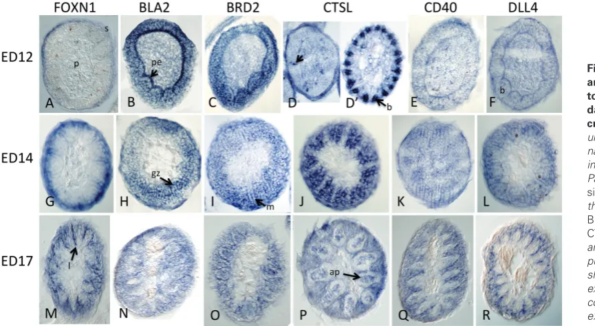
Putative downstream target genes of FOXN1 have been identified in mouse thymocytes (Nowell et al., 2011) and human embryonic stem cells (hESNet). In addition to regulating keratins (e.g., Meck-lenburg et al., 2004), the regulation of MHC Class II genes, BRD2 (Bromodomain-containing gene 2), CTSL (Cathepsin L, a lysosomal protease), CD40 (TNF-related receptor), and DLL4 (delta-like gene 4) was positively correlated with FOXN1 dosage (Nowell et al., 2011; hESnet). To determine if these components represent a gene regulatory network that is conserved in other FOXN1-expressing Fig. 2. Expression of chick FOXN1 in developing thymus and feathers.(A)FOXN1expression (arrow-heads) in ED9 chick neck showing one side of bilateral, lobular expression in the thymus glands. Early feather buds (arrows) and skin show no expression. (B) A section through the thymus lobe in panel A showing expression throughout the thymus with increased intensity in the cortex. (C) Bilateral FOXN1 expres-sion (arrowheads) in ED13 chick neck looking down from above after decapitation. Expression in feathers filaments can be seen peripherally. (D) Another ED13 embryonic neck from the side after removal of the skin showing two lobes of the thymus (arrowheads).
of chick homologs of these genes in feathers and other epidermal appendages. Chick BLA2 represented the MHC Class II genes, while other chick gene symbols are similar to mouse. Comparisons of expression in whole mount and feather section are shown in Figures 3 and 4. Our section photos were taken near the base of the feather filament where barb ridges are first forming on one side (Fig. 4. ED12), or at the proximodistal level corresponding to the
of the potential targets exhibited expression in feather filaments and overlapped in expressing regions with FOXN1, none of the patterns were highly similar to FOXN1 in either whole mounts (Fig. 3) or cross sections of feathers (Fig. 4). FOXN1 expression was not detected as extensively in wing feathers or skin at ED12 (Fig. 3A), whereas the other five genes were strongly expressed (Fig. 3 B-F). At ED14, four of the putative target genes were expressed in
skin whereas FOXN1 and BLA2 were not. Feather expression was similar but not identical between FOXN1 and the putative target genes for body feathers at ED12-17 (compare Fig. 3 rows ED12 skin, ED14 skin and ED17 skin). In ED14 feet (scales and nails) the difference in expression of these genes was more pronounced. FOXN1 mRNA was expressed in nails but not in scales (Fig. 3E’, E”). BLA2 and BRD2 were expressed in scales not nails (Fig. 3F’, F”, G’, G”). CTSL was expressed in scales and nails (Fig. 3H’, H”), whereas CD40 (Fig. 3 I’, I”) and DLL4 (Fig. 3 J’, J”) were ex-pressed in neither scales nor nails. FOXN1 and CTSL expression were similar in nails. We conclude that all of the tested candidate target genes for FOXN1 are expressed in some avian epidermal appendages; however, direct FOXN1 regulation is not sufficient to explain all of their expression patterns.
To investigate specific localization of FOXN1 mRNA and these five candidate genes within feather filaments, hybridized feathers were paraffin sectioned transversely. At ED12 at the base of the feather where barb plates were just beginning to form, FOXN1 transcripts were predominantly localized in the feather sheath (Fig.4A), and more distally in the ends of the marginal plates (not shown) whereas transcripts of the five FOXN1 candidate targets did not share this pattern (Fig.4 B-F). For example, at ED12 BLA2 (and somewhat less so BRD2) mRNA was expressed in the pulp epithelium and within proximal barb plates. CTSL mRNA was ubiquitously expressed with stronger intensity in the sheath and distributed cells (Fig. 4D, arrow), whereas slightly more distally strong expression was uniquely seen in the barbule plates outside the growth zone (Fig. 4D’), a pattern not shared with any of the other probes. DLL4 and CD40 mRNA were weakly expressed in the outside layer of the feather sheath and in the pulp epithelium (Fig. 4 E,F). At ED13-14, in a more distal part of the filament, the overlap in expression patterns was more similar but with identifi-able differences (Fig. 2L, 4G-L). FOXN1 was still expressed in the sheath, and also in the lateral barb plates and marginal cells.
Labeling of BLA2 appeared stronger in the barb ridge (Fig. 4H), BRD2 labeled lateral barb plates, medial barb ridges and marginal cells equally (Fig. 4I) whereas CTSL was strongly expressed in the barb plates (Fig. 4J). Expression of CD40 and DLL4 persisted fairly ubiquitously at this level (Fig. 4 K,L) with the exception of the dermal pulp for DLL4. By ED17 the strongest FOXN1 label was in a thin line of unknown fate between the barbs, with barb and marginal cells labeling but no label in sheath, medullary cells of the axial plate or pulp (Fig. 4M). A similar labeling pattern was ob-served for the other genes with the exception of the inter-marginal line, and that BRD2 and CTSL also labeled in the axial plate of the barbs (Fig. 4 N,R). Based on these data, the putative FOXN1 target genes show dynamic and interesting expression patterns in feather compartments. However, the expression of each of the genes is distinctly different from that of FOXN1 at similar stages and proximodistal levels.
Discussion
In the present study, we confirm that chick FOXN1 is expressed in the developing thymus and demonstrate its expression in some epidermal appendages, suggesting a homologous, evolutionarily conserved role for FOXN1 across amniotes. We have identified a novel expression pattern of FOXN1 in embryonic feathers. Chick FOXN1 initially is expressed along the length of the feather filament, with the exception of the base and tip. As the filament elongates between ED 12 and 17, FOXN1 expression becomes restricted to the proximal end of the feather. Although others have published differences in proximal-distal gene expression (e.g., Yu et al., 2002, Chodankar et al., 2003), to our knowledge FOXN1 and FOXE1 (Yaklichkin et al., 2011), the two FOX genes we have characterized are the only known differentially and dynamically expressed genes along the proximal-distal axis of the elongating feather filament.
The murine ortholog of FOXN1 regulates expression of specific Fig. 4. Expression of FOXN1 and putative FOXN1 regula-tory targets in embryonic day 12, 14 and 17 feather cross sections. Photo col-umns are labeled with the gene name for the in situ probe, rows indicate the days of incubation. Panels show the results of in situ hybridization of probes for the genes FOXN1 (A,G,M), BLA2(B,H,N),BRD2(C,I,O),
CTSL(D,D’,J,N),CD40(E,KQ)
soft alpha-keratins and feathers form predominantly from hard beta-keratins, it is possible FOXN1 could be modulating expression of feather-specific keratins. Feather-specific beta-keratin expres-sion coincides with differentiation of the barb ridges (Haake et al., 1984, König and Sawyer 1985). Images of feather filaments in the Haake paper indicate the proximal to distal pattern of beta-keratin expression correlates well with the expression pattern of FOXN1 shown here (Fig. 2). Specifically, beta-keratin expression is initiated at stage 38 (ED12) and expressed in the ring sheath cells similar to our image in Fig. 2K, whereas more distal expression is similar to Fig. 2L. Patterns of alpha-keratins also correlate with FOXN1 expression (Alibardi, 2013). Based on these observations and the expression of both mouse and chick FOXN1 in the nails and thymus, other keratinized epidermal structures, chick FOXN1 may regulate expression of both alpha and beta-keratins.
Finally, expression of FOXN1 and the other genes investi-gated in the feather raises more questions than it answers about the evolution of FOXN1 function, considering that the feather is thought to have evolved from scutate scales of reptiles. Loss and gain of function studies will be required to establish a precise role for FOXN1 transcriptional regulation in epidermal evolution and development.
Materials and Methods
Sequence, data and phylogenetic analysis
The protein sequences used in the analysis were obtained from the NCBI database (ncbi.nlm.nih.gov) and Ensemble database 69 (ensemble. org). The accession numbers of FOXN1/N4 protein sequences analyzed are provided in Supplementary Table S1. Multiple sequence alignments were constructed using T-COFFEE, version 7.7.1. (tcoffee.vital-it.ch/cgi-bin/Tcoffee/tcoffee_cgi/index.cgi). Aligned FOXN1/N4 sequences were edited using the alignment editor BioEdit 7.0.4.1. (mbio.ncsu.edu/bioedit/ page2.html). A phylogenic tree for FOXN1/N4 proteins was generated based on the forkhead DNA-binding domain sequences (~100 residues). A neighbor-joining method was used to construct the tree topology. The phylogenetic tree was converted into a cladogram using MEGA 4 (Kumar
et al., 2004) (metameme.sdsc.edu).
Incubation and isolation of chicken tissues
Fertilized white leghorn eggs (Gallus gallus) were incubated in a forced-draft incubator at approximately 38ºC with high humidity. Slight variations in temperature between incubators or over the course of the experiment make staging approximate due to long incubation times, therefore days of incubation are given. For older embryos, neck/thorax and skin/feathers were isolated prior to fixation with paraformaldehyde.
Molecular cloning and in situ hybridization with gene probes
To clone chicken FOXN1 cDNA sequences for the in situ probe template, mRNA was isolated from day 5 chicken embryos using the Qiagen RNeasy Mini Kit (cat. number: 74104) and cDNA synthesized using standard pro-cedures. Template for probe generation was generated by PCR with the GC-rich PCR System Kit (cat. number: 12 140 306 001), Roche Applied Science; 34 cycles of annealing at 50ºC and the following primers: forward primer,
5’-TTATAAAAGCTTGCGGCCGCAGAATATCACTACCCCTAC-CAAAGGATTGC-3’; reverse primer: 5’-GCTCTAGAAATTAACCCTCAC-TAAAGGGCCTCCCGCCTCCCCAGAAGGAAACATTG-3’. Chick FOXN1
sequences are in bold, the T3 RNA polymerase binding site is italicized. The PCR generated template was sequenced to confirm identity (NCBI Accession number: XM_415816.3, 904-1622 bp). A 719 nt RNA probe of
FOXN1 was produced using T3 polymerase and in situ hybridizations were
performed using the GEISHA mRNA Detection Protocol (geisha.arizona.
as previously described (Yaklichkin et al., 2011). Probe for MHC Class II gene, BLA2 (NCBI Acc. #: NM_001245061) was generated from GEISHA plasmid W34 with Not I using T7 polymerase. Probe for BRD2 was gen-erated from plasmid ChEST826a15 (NM_001030674.1) with NotI and T3 polymerase. Probe for Cathepsin L, CTSL2 (NM_001168009) was gener-ated from GEISHA plasmid S12 with NotI and T7 polymerase. Probe for
CD40 (TNFRSF5, NM_204665) was generated by PCR: Forward primer:
5’- CTGACAAGCAGTATGAGCACAAGG -3’, Reverse primer:
5’-ATTA-ACCCTCACTAAAGGGGAAGTCCACCTCCTCGGG-3’, where the bold
sequence allows for directional amplification of the 666 nt probe using the T3 polymerase. Probe DLL4 (XM_421132) was generated from plasmid pgf2n.pk001.e7 (www.chickest.udel.edu) using NotI and T3 polymerase. Probe sequences and additional images are available at http://geisha. arizona.edu/geisha.
Aknowledgments
We are thankful to Tatiana Yatskievych and Terry Sesepasara for tech-nical assistance. This work was supported by NIH grant GM085226 and NIH NICHD grant P41HD064559.
References
ALIBARDI L (2009). Cornification of the pulp epithelium and formation of pulp cups in downfeathers and regenerating feathers. Anat Sci Int. 84: 269-279. ALIBARDI L (2013) Immunolocalization of alpha-keratins and feather beta-proteins
in feather cells and comparison with the general process of cornification of the skin of mammals. Ann Anat 195: 189-198.
BRANCACCIO A, MINICHIELLO A, GRACHTCHOUK M, ANTONINI D, SHENG H, PARLATO R, DATHAN N, DLUGOSZ AA, MISSERO C (2004). Requirement of the forkhead gene Foxe1, a target of sonic hedgehog signaling, in hair follicle morphogenesis. Hum Mol Genet 13: 2595-2606.
CHODANKAR R, CHANG CH, YUE Z, JIANG TX, SUKSAWEANG S, BURRUS LW, CHUONG CM AND WIDELITZ RB (2003). Shift of Localized Growth Zones Con-tributes to Skin Appendage Morphogenesis: Role of the Wnt/b-catenin Pathway.
J. Invest Dermatol 120: 20-26.
ECKHART L, VALLE LD, JAEGER K, BALLAUN C, SZABO S, NARDI A, BUCH-BERGER M, HERMANN M, ALIBARDI L, TSCHACHLER E (2008). Identification of reptilian genes encoding hair keratin-like proteins suggests a new scenario for the evolutionary origin of hair. Proc Natl Acad Sci USA 105: 18419-18423. FRANK J, PIGNATA C, PANTELEYEV AA, PROWSE DM, BADEN H, WEINER L,
GAETANIELLO L, AHMAD W, POZZI N, CSERHALMI-FRIEDMAN PB, AITA VM, UYTTENDAELE H, GORDON D, OTT J, BRISSETTE JL, CHRISTIANO AM (1999). Exposing the human nude phenotype. Nature 398: 473-474.
FURLONG RF (2005). Insights into vertebrate evolution from the chicken genome sequence. Gen Biol 6: 207.
GLENN TC, FRENCH JO, HEINCELMAN TJ, JONES KL, SAWYER RH (2008). Evolutionary relationships among copies of feather beta ({beta}) keratin genes from several avian orders. Integr Comp Biol 48: 463-475.
HAAKE AR, KÖNIG G, SAWYER RH (1984). Avian feather development: relationships between morphogenesis and keratinization. Dev Biol 106: 406-413.
hESNet: Human Embryonic Stem Cell Transcription Network www.wanglab.ucsd. edu/star/hESnet/gene.jsp?gene=FOXN1; accessed Dec. 4, 2013.
KAESTNER KH, KNOCHEL W, MARTINEZ DE (2000). Unified nomenclature for the winged helix/forkhead transcription factors. Genes Dev 14: 142-146.
KÖNIG G, SAWYER RH (1985). Analysis of morphogenesis and keratinization in transfilter recombinants of feather-forming skin. Dev Biol 109: 381-392. KUMAR S, HEDGES SB (1998). A molecular timescale for vertebrate evolution.
Nature 392: 917-920.
KUMAR S, TAMURA K, NEI M (2004). MEGA3: Integrated software for Molecular Evolutionary Genetics Analysis and sequence alignment. Brief Bioinform 5: 150-163. LEE D, PROWSE DM, BRISSETTE JL. (1999) Association between mouse nude gene expression and the initiation of epithelial terminal differentiation. Dev Biol. 208: 362-374.
cycling. Semin Cell Dev Biol 23: 906-916.
LEE YH, WILLIAMS A, HONG CS, YOU Y, SENOO M, SAINT-JEANNET JP (2013). Early development of the thymus in Xenopus laevis. Dev Dyn 242: 164-178. LIN KK, KUMAR V, GEYFMAN M, CHUDOVA D, IHLER AT, SMYTH P, PAUS R,
TAKAHASHI JS, ANDERSEN B (2009). Circadian Clock Genes Contribute to the Regulation of Hair Follicle Cycling. PLoS Genet 5: e1000573.
MA D, WANG L, WANG S, GAO Y, WEI Y, LIU F (2012). FOXN1 maintains thymic epithelial cells to support T-cell development via mcm2 in zebrafish. Proc Natl
Acad Sci USA 109: 21040-21045.
MECKLENBURG L, NAKAMURA M, SUNDBERG JP, PAUS R (2001). The nude mouse skin phenotype: the role of FOXN1 in hair follicle development and cycling.
Exp Mol Pathol 71: 171-178.
MECKLENBURG L, PAUS R, HALATA Z, BECHTOLD LS, FLECKMAN P, SUNDBERG JP (2004). FOXN1 is critical for onycholemmal terminal differentiation in nude (FOXN1) mice. J Invest Dermatol 123: 1001-1011.
MECKLENBURG L, TYCHSEN B, PAUS R (2005). Learning from nudity: lessons from the nude phenotype. Exp Dermatol 11: 797-810.
MEIER N, DEAR TN, BOEHM T (1999). Whn and mHa3 are components of the genetic hierarchy controlling hair follicle differentiation. Mech Dev 89: 215-221. NEVES H, DUPIN E, PARREIRA L, LE DOUARIN NM (2012). Modulation of Bmp4
signalling in the epithelial-mesenchymal interactions that take place in early thymus and parathyroid development in avian embryos. Dev Biol 361: 208-219.
NOWELL CS, BREDENKAMP N, TETÉLIN S, JIN X, TISCHNER C, VAIDYA H, SHERIDAN JM, STENHOUSE FH, HEUSSEN R, SMITH AJ, BLACKBURN CC (2011). FOXN1 regulates lineage progression in cortical and medullary thymic epithelial cells but is dispensable for medullary sublineage divergence. PLoS
Genet 7:e1002348.
PIGNATA C, FUSCO A, AMOROSI S (2009). Human clinical phenotype associated with FOXN1 mutations. Adv Exp Med Biol 665: 195-206.
RODEWALD HR (2008) Thymus Organogenesis. Annu Rev Immunol 26: 355-388. SCHLAKE T, SCHORPP M, MAUL-PAVICIC A, MALASHENKO AM, BOEHM T (2000). Forkhead/winged-helix transcription factor Whn regulates hair keratin gene ex-pression: molecular analysis of the nude skin phenotype. Dev Dyn 217: 368-376. SCHÜDDEKOPF K, SCHORPP M, BOEHM T (1996). The whn transcription factor encoded by the nude locus contains an evolutionarily conserved and function-ally indispensable activation domain. Proc Natl Acad Sci USA. 93: 9661-9664. SEGRE JA, NEMHAUSER JL, TAYLOR BA, NADEAU JH, LANDER ES (1995).
Positional cloning of the nude locus: genetic, physical, and transcription maps of the region and mutations in the mouse and rat. Genomics 28: 549-559. YAKLICHKIN SY, DARNELL DK, PIER MV, ANTIN PB, HANNENHALLI S (2011).
Accelerated evolution of 3’avian FOXE1 genes, and thyroid and feather specific expression of chicken FoxE1. BMC Evol Biol 11: 302.
YU M, WU P, WIDELITZ RB, AND CHUONG C-M (2002) The morphogenesis of feathers. Nature 420: 308-312.
Int. J. Dev. Biol. (2005) 49: 137-142 http://dx.doi.org/10.1387/ijdb.041959js
How and when the regional competence of chick epidermis is established: feathers vs. scutate and reticulate scales, a problem en route to a solution
Fabrice Prin and Danielle Dhouailly Int. J. Dev. Biol. (2004) 48: 137-148
5 yr ISI Impact Factor (2011) = 2.959
http://dx.doi.org/10.1387/ijdb.15272378
Integument pattern formation involves genetic and epigenetic controls: feather arrays simulated by digital hormone models
Ting-Xin Jiang, Randall B Widelitz, Wei-Min Shen, Peter Will, Da-Yu Wu, Chih-Min Lin, Han-Sung Jung and Cheng-Ming Chuong
Int. J. Dev. Biol. (2004) 48: 117-135 http://dx.doi.org/10.1387/ijdb.15272377
Chick-embryo culture techniques employed at Karnatak University in Dharwad, India, for studying cellular and molecular aspects of morphogenesis
Sohan P Modak
Int. J. Dev. Biol. (2003) 47: 165-170 http://dx.doi.org/10.1387/ijdb.12705665
Early chick embryos in vitro