Two different vestigial like 4 genes are differentially expressed
during Xenopus laevis development
MARÍA-GUADALUPE BARRIONUEVO
1, MANUEL J. AYBAR*
,1, 2and CELESTE TRÍBULO*
,1,21Instituto Superior de Investigaciones Biológicas (INSIBIO, CONICET-UNT) and 2 Instituto de Biología “Dr. Francisco D. Barbieri”, Facultad de Bioquímica, Química y Farmacia, Universidad Nacional de Tucumán, San Miguel de Tucumán, Tucumán, Argentina
ABSTRACT The vestigial gene (vg) was first characterized in Drosophila and several homologues were identified in vertebrates and called vestigial like 1-4 (vgll1-4). Vgll proteins interact with the transcription factors TEF-1 and MEF-2 through a conserved region called TONDU (TDU). Vgll4s are characterized by two tandem TDU domains which differentiate them from other members of the vestigial family. In Xenopus two genes were identified as vgll4. Our bioinformatic analysis demon-strated that these two genes are paralogues and must be named differently. We designated them as vgll4 and vgll4l. In situ hybridization analysis revealed that the expression of these two genes is rather different. At gastrula stage, both were expressed in the animal pole. However, at neurula stage, vgll4 was mainly expressed in the neural plate and neural folds, while vgll4l prevailed in the neural folds and epidermis. From the advanced neurula stage onward, expression of both genes was strongly enhanced in neural tissues, anterior neural plate, migrating neural crest, optic and otic vesicles. Nevertheless, there were some differences: vgll4 presented somite expression and vgll4l was localized at the skin and notochord. Our results demonstrate that Xenopus has two or-thologues of the vgll4 gene, vgll4 and vgll4l with differential expression in Xenopus embryos and they may well have different roles during development.
KEY WORDS:
ectoderm, Vgll4, Vgll4l, TONDU
Vestigial (vg) genes are expressed in invertebrates and
verte-brates, and have been shown to be involved in a variety of devel-opmental processes. In Drosophila, Vestigial (Vg) interacts with the transcription factor Scalloped (Sd) to regulate myogenesis and wing development (Kim et al., 1996, Simmonds et al., 1998). This binding specifically activates numerous target genes in the cell fate determination process (Halder et al., 1998, Simmonds
et al., 1998). In vertebrates, several vg homologues have been
identified. They are called vestigial like 1-4 (vgll1-4) (Chen et al., 2004, Faucheux et al., 2010, Maeda et al., 2002). In zebrafish, two orthologues of mammalian vgll2 named as vgll2a and vgll2b (Johnson et al., 2011, Mann et al., 2007) were described. Recently, were also identified two vgll4 orthologues designated as vgll4 and
vgll4l (Melvin et al., 2013).
The vgll genes encode proteins that have a conserved region called TONDU (TDU) motif (Maeda et al., 2002, Vaudin et al., 1999). Vgll proteins were shown to physically interact with Scal-loped homologues such as Transcriptional Enhancer Factor-1 (TEF-1 also known as TEAD-1) (Gunther et al., 2004) and Myocyte
www.intjdevbiol.com
*Address correspondence to: Celeste Tríbulo or Manuel J. Aybar. INSIBIO (CONICET-Universidad Nacional de Tucumán), Chacabuco 461, T4000ILI – San Miguel de
Tucumán, Tucumán, Argentina. Tel: +54-381-4107214. E-mail: [email protected] ; [email protected]
Accepted: 13 May 2014. Final, author-corrected PDF published online: 30 September 2014.
ISSN: Online 1696-3547, Print 0214-6282
© 2014 UBC Press Printed in Spain
Abbreviations used in this paper: vg, vestigial gene; vgll, vestigial-like; vgll4l: vestigial
like 4-like; TDU, tondu region; TEF, transcriptional enhancer factor.
Enhancer Factor-2 (MEF-2) (della Gaspera et al., 2009, Maeda
et al., 2002). Unlike other members of the Vgll family that have a
single interaction domain TDU, Vgll4 has two tandem TDU motifs in its carboxyl-terminal domain suggesting that Vgll4 might form a bridge between TEF-1 and MEF-2 transcription factors (Chen
et al., 2004, Faucheux et al., 2010).
In Xenopus a vgll4 gene was identified and its expression pat-tern was described only for late midneurula stage embryos. The expression was localized in the epidermis, olfactory placodes and neural crest cells (Faucheux et al., 2010). However, a detailed and comparative analysis of its early expression pattern during
Xenopus embryogenesis is still lacking. We identified a new vgll4 paralogue and found it to be different from the previously
described (Faucheux et al., 2010). We called this paralogue as
vgll4l based on it similarity with zebrafish vgll4l. The bioinformatic
presence of the two TDU motifs (Chen et al., 2004, Faucheux et
al., 2010), and new phylogenetic features of the Vgll family. In
addition, the comparative analysis of the expression pattern of both genes in different developmental stages of Xenopus laevis embryos revealed undescribed and interesting distinct domains of expression for each gene.
Results and Discussion
Identification and sequence analysis of vgll4 and vgll4l in Xenopus laevis
Xenopus vestigial like 4 (vgll4) gene was first cloned in 2002
by Klein et al., (Accession Number: BC123267) and it was initially
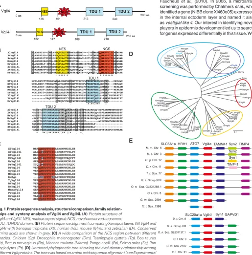
Fig. 1. Protein sequence analysis, structural comparison, family relation-ships and synteny analysis of Vgll4 and Vgll4l. (A) Protein structure of Vgll4 and Vgll4l. NES, nuclear export signal; NCS, novel conserved sequence;
TDU 2
XlVgll4 SLHASHLYG-IPPLAVDQPLALTKNSMDASRSMG---IVPPMERQQNRPSVITCAPASNR XtVgll4 SLHASHLYG-IPPLAVDQPLALTKNSMDASRSMG---IVPTMERQQNRPSVITCAPASNR HsVgll4 SLHGSHLYTSLPSLGLEQPLALTKNSLDASRPAGLSPTLTPGERQQNRPSVITCASAGAR MmVgll4 SLHGGHLYASLPSL-MEQPLALTKNSSDTGR---SAVERQQNRPSVITCASAGAR DrVgll4 SLVGGHHLY-MPSLALDQPLALTKN-MDSSRSMGISPTASPVERQQNRPSVITCAPANNR XlVgll4l PAKSHLERPEPASSPEEQPLALVKRVVKPSEQAQ----LSPALLQQMRPSVITCISRQKP XtVgll4l PTKAAMERPVPAASPDEQPLALVKRAQKPSEQAQ----PSPALLQQMRPSVITCVSRQKP DrVgll4l SPTHLIPSPVFSSPVMDEPLALIKKPRPEPEKTE--SQNKATTQIQMRPSVITCVSSASR . .. ::**** *. . . * ******* . XlVgll4 NCSLSHCPTVTHSGCASAAPGNFRRPSNTTTACDPVVEEHFRRSLGKNYKE---PEPVAN XtVgll4 NCSLSHCP-VTHSGGVSAVPGSFRRPSNTTTACDPVVEEHFRRSLGKNYKE---PEPVAN HsVgll4 NCNLSHCP-IAHSGCAAPGPASYRRPPSAATTCDPVVEEHFRRSLGKNYKE---PEPAPN MmVgll4 NCNLSHCP-IAHSGCSAPGSASYRRPPSATATCDPVVEEHFRRSLGKNYKE---PEPAPN DrVgll4 NCNLSHCT-GSHNGCSPGLNASYRRASNSNTACDPVIEEHFRRSLGKNYKE---PEPVTN XlVgll4l ---VVSLPTEAANPSRACVTKGLSKTPLSDVEEHFQRSL--AHKCPVVPRPTPT XtVgll4l ---VAQPPTEAANPSRTCVTKGLSRTPSPDVEDHFQRSLALAHKRPIAPRPIPT DrVgll4l ---STKQDCCNHSTAVSKHSYDHVEEHFQRSLGINYHR--- . . :*:**:*** :: XlVgll4 -SVSITGSVDDHFAKALGDTWLQIKAAKDGGPSSPESASRRGQ-SPPSAHMMNHNHSPSV XtVgll4 -SVSITGSVDDHFAKALGETWLQIKAAKDGGPSSPESASRRGQ-SSPSAHMMNHNHSPSV HsVgll4 -SVSITGSVDDHFAKALGDTWLQIKAAKDGASSSPESASRRGQPASPSAHMVSHSHSPSV MmVgll4 -SVSITGSVDDHFAKALGDTWLQIKAAKDSASSSPESASRRGQPASPTAHMVSHSHSPSV DrVgll4 -SVSITGSVDDHFAKALGETWLQIKAK--GGSSSPD---ASPNTHMVNHNHSPSL XlVgll4l SQSSSNISVEDHFSKALGSKWLLIRAAADSPSSPDGRSKSLF--- XtVgll4l PQSSS-ISVEEHFSKALGSKWLLIRAAADSPSSPEGRGRRLV--- DrVgll4l -ATSISVSVDDHFAKALGDKWLQLKASSSSCHSSSSSSSSSPPSSPTFIHSPGYSPKRAR * **::**:****..** ::* . *.
TDU, TONDU domain. (B) Protein sequence alignment comparing Xenopus laevis (Xl) Vgll4 and Vgll4l with Xenopustropicalis (Xt), human (Hs), mouse (Mm), and zebrafish (Dr). Conserved Amino acids are shown in grey. (C) A wide comparison of the NCS region between different species. Chicken (Gg), Drosophila melanogaster (Dm), Taeniopygiaguttata (Tg), Bos taurus
(Bt), Rattus norvegicus (Rn), Macaca mulatta (Mama), Pongo abelii (Pa), Salmo salar (Ss), Pan troglodytes (Pt). (D) Unrooted phylogenetic tree showing the evolutionary relationship among different Vgll proteins. The tree was based on amino acid sequence alignment (see Experimental Procedures for details). (E) Analysis of conserved syntenic regions containing Vgll4 and Vgll4l loci in Xenopus tropicalis (X.t.), zebrafish (Danio rerio, D.r.), puffer fish (Takifugu rubripes, T.r.),
stickleback(Gasterosteus aculeatus, G.a.), medaka(Oryzias latipes, O.l.), tilapia(Oreochromis niloticus, O.n.), cod (Gadus morhua, G. m.), human (H.s.), mouse (M.m.), and chicken (G.g.) genomes. Genes are represented as boxes and arrows indicate the orientation of the transcription unit. Boxes with the same color indicate orthologue genes. The genomic regions representations are not to scale to avoid complexity.
were interested in the vestigial like 4 gene described by Chalmers et al., 2006; NIBB clone Xl460o05). We first sequenced and analyzed this clone in silico (Accession number: KF963131) and found it to be different from the sequence named as vestigial like 4 at the NCBI Nucleotide database (Klein et al., 2002) (Accession number: BC123267). The Xl460o05 clone showed high identity with a sequence called rexp52 (90% identity) (Acc. Number: DQ096895.1), an uncharacterized gene with differential expression found in a large-scale, semi-automated whole mount in situ hybridization screening performed in Xenopus laevis (Pollet et al., 2005). Thereby, currently there are two sequences named as vestigial like
4. Notwithstanding, our bioinformatics analysis of these
sequences demonstrated that they are two different genes so they should have different names. Thus, we propose in Xenopus laevis the name vestigial like 4 (vgll4) for the first cloned gene (Accession number: BC123267) and
vestigial like 4-like (vgll4l) for the second (Accession
number: KF963131).
In Xenopus tropicalis only a sequence corresponding to vgll4 was found into the Ensembl genomic information. We extended our in silico analysis to multiple sequences databases and this led us to find a sequence that corre-sponds to Xenopus tropicalis vgll4l (Accession number: KJ690263, Fig. 1B). This gene was found in the Gurdon Institute Xenopus tropicalis EST Database (Transcript name 1012072090).
In zebrafish two sequences of 282 (Accession number: NP998440) and 266 amino acids (Accession number: NP001073467) were identified and named as vgll4 and
vgll4l, respectively (Melvin et al., 2013). This finding
could be extended to other bony fishes such as puffer fish, stickleback, tilapia and cod. According to their phy-logenetic relationship (Fig. 1D) and sequence similarity (Fig. 1B) Xenopus vgll4 is the orthologue of fish vgll4 and
Xenopus vgll4l is the orthologue of fish vgll4l.
The protein sequence analysis showed that Xenopus
laevis Vgll4 has the same sequence that the previously
described Vgll4 (Klein et al., 2002, Faucheux et al., 2010). The bioinformatic analysis of Xenopus laevis and Xenopus
tropicalis Vgll4l protein sequence allowed us to identify
the two TDU motifs that characterize Vgll4 co-factors (Fig. 1A,B), and the putative NES (nuclear export signal) motif. When we compared Vgll4 and Vgll4l with Vgll4s from other species, we found an undescribed conserved region that we identified as NCS (novel conserved sequence) (Fig. 1 A,C). This region is unique in vgll4 genes and probably can act as a feature that differentiates Vgll4s from other members of the Vgll family and could interact with other protein components. A phylogenetic tree based in amino acid sequences indicate that Vgll genes of different spe-cies can be organized into four different groups, Vgll1, 2, 3 and 4 (Fig. 1D). This phylogenetic analysis is in agreement with Faucheux et al., (2010) and Koontz et al., (2013), who demonstrated that Vgll1-3 are related to Drosophila Vg, while all Vgll4s are related to the recently identified
Drosophila orthologue, Tgi. A divergence between Vgll4
and Vgll4l is reflected in the extent of conservation of the
Fig. 2. RT-PCR analysis of vgll4 gene expression in embryos and adult tissues. RT-PCR was performed on total RNA extracted from embryos at different embryonic stages, adult tissues and internal and external layers of animal caps. ef1alfa was used as loading control. (A) Temporal expression of vgll4 and vgll4l throughout develop-ment. (B) Analysis of vgll4 and vgll4l expression in adult tissues. (A,B) Quantifications of gel are shown; the results are expressed as Relative Intensity (sample/ef1alfa X 10). (C)vgll4 and vgll4l expression in isolated layers of the animal cap ectoderm.
0
heart liver gut muscle eye skin lung vgll4 vgll4l
St. 17 Controlcaps Innerlayer Outerlayer
other hand, strong Vgll4 homology was observed in TDU domains, which are completely conserved between human, mouse, zebraf-ish, Xenopus tropicalis and Xenopus laevis (Fig. 1B), as well as for NES and NCS regions (Fig. 1C). This high conservation in specific regions allows us to designate Vgll4l as a paralogue of Vgll4.
We analyzed the evolution of the vestigial-like genes in verte-brate genomes by synteny analysis. The genomic databases (i.e. Ensembl) provided us with sufficient information on the chromo-protein sequence: Vgll4 is more similar than Vgll4l to mammalian
Vgll4s (Fig. 1D) and it also shows a high similarity with Xenopus
tropicalis Vgll4 (92%). Xenopus laevis Vgll4 has 73% identity to
human and 70% to mouse Vgll4 proteins. By contrast, Xenopus
laevis Vgll4l is only 31% identical to mouse and 33% to human
Vgll4 and Xenopus tropicalis Vgll4l is 42% identical to human Vgll4. Furthermore, Xenopus laevis Vgll4 and Vgll4l are 40% identical while Xenopus tropicalis Vgll4 and Vgll4l are 30% identical. On the
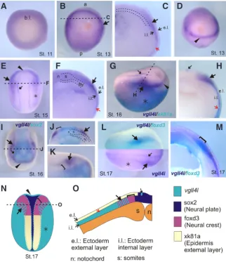
Fig. 3. Spatio-temporal expression pattern of vgll4l during early development by in situ hybridization.(A) Vegetative view. (B, E) Dorsal view, anterior to the top. (D, I) Anterior view, dorsal to the top, anterior to the front. (G) Lateral view anterior to the right. (L) Dorsal view, anterior to the right. (C, F, H, J, M) Transversal sections. (K) Higher magnification view of the anterior region of the embryo shown in I. Black dashed lines in B, E, G, I indicate the positions of the cuts made to give transversal-sections in C, F, H, J. (G, H) Double in situ hybridization for vgll4l (purple) and xk81a (turquoise). (I-K) Double in situ hybridization forvgll4l (purple) and
sox2 (turquoise). (L) (upper half) Double in situ hybridization for vgll4l (purple) and foxd3 (tur-quoise), (M) cross- sectioned embryo labeled by double in situ hybridization for vgll4l (purple) and foxd3 (turquoise). (E, G, L) Asterisk, vgll4l expression in the prospective epidermis. (B, C, E, F, H-L) Large black arrow, vgll4l neural folds expression. (C, F, H) Red arrow, vgll4l expression in the internal layer of the ectoderm. (E, G, H, I, L) Small black arrow, vgll4l row expression surrounding neural plate. (J, K, M, N) Brackets, vgll4l neural folds gap of expression. (N, O) Schematic diagrams summarizing the expression of vgll4l and neural plate (sox2), neural crest (foxd3) and epidermal (xk81a) markers. (N) Dorsal view. (O) Transversal section. References: a, anterior. b.l., blastopore lip. e.l., external layer. en, endoderm. i.l., internal layer. n, notochord. p, posterior. s, somites.
G
B
C D
E
F
H
I
J
K
L
A
M
N
O
some regions containing the orthologues of X.
laevis vgll4 to perform such analysis. As shown in
Fig. 1E, vgll4 and vgll4l are remarkably conserved between X. tropicalis, zebrafish (D. rerio), puffer fish (T. rubripes), medaka (O. latipes), stickleback (G. aculeatus), tilapia (O. niloticus) and cod (G.
morhua). In the case of X. tropicalis vgll4l the
genomic information available is limited but shows that this gene is syntenic with other species, since its neighbor synapsin-1 (syn-1) could be identified (Fig. 1E). Curiously, in chicken, mouse, and hu-man genomes only the vgll4 occurred, no vgll4l paralogue was identified. These finding suggest that the chromosomal regions containing vgll4 and vgll4l genes have been conserved for at least 340-390 million years, the expected divergence time for amphibian and amniota lineages (Blair and Hedges, 2005). Our results from phylogenetic and syntenic analyses support the hypothesis that in fishes and amphibians two different vgll4 genes are present.
Analysis of vgll4s expression in Xenopus embryos
First, we assessed the temporal expression pro-file of vgll4s by reverse transcriptase-polymerase reaction (RT-PCR) (Fig. 2A). Our findings agree with what has been reported for vgll4 (Faucheux
et al., 2010). This gene was maternally expressed
and its expression remains constant throughout development (Fig. 2A). vgll4l was also maternally expressed and the transcripts were continuously detectable until after the hatching stage (Fig. 2A). In addition, we analyzed vgll4s expression in adult tissues. vgll4 was observed in all the tissues ana-lyzed with exception of the skin (Fig. 2B). Previous reports showed that vgll4 was expressed at a similar level in all the tissues analyzed (Faucheux
et al., 2010). The expression we found for vgll4
is different from vgll4l, which was expressed at a roughly similar level in all tissues analyzed but showed a higher expression in skin (Fig. 2B). This not tissue-restricted expression of vgll4s is coincident with vgll4 expression in human that was detected in the heart, kidney and brain as well as in other tissues at lower levels (Chen et al., 2004).
the epidermis (Chalmers et al., 2006). We performed a detailed analysis of vgll4l expression at different stages comparing it with different neural and epidermal marker genes. Our results showed that in early gastrula vgll4l was widely expressed in the animal hemisphere (Fig. 3A). vgll4l expression was decreased in the dorsal and posterior ectoderm during gastrulation. In neurula, stage 13 onwards, it was expressed surrounding the neural plate anteriorly and laterally (Fig. 3B, black arrow; Fig. 3D, black arrowhead). Trans-versal sections revealed that vgll4l expression is restricted to the internal layer of the ectoderm (Fig. 3C, red arrow). At neurula stage the expression is restricted to the anterior and lateral neural folds (Fig. 3E, arrowhead and black arrow) and displays an expression pattern that in the ectodermal domain overlaps with the epidermal marker xk81a (Fig. 3G, asterisk). Transversal sections confirmed that vgll4l expression is located in the inner layer of the ectoderm compared with xk81a, which is expressed in the external layer (Fig. 3 H,O). Besides, we performed a RT-PCR determination of vgll4l in explants samples dissected from the internal and external layers of the epidermis. This analysis showed a clear vgll4l expression in the internal but not in the external layer (Fig. 2C) that correlates with the internal layer marker deltaNp63 expression (Tribulo et
al., 2012). Double in situ hybridization showed that vgll4l anterior
expression corresponds to the anterior neural fold because it is complementary and does not overlap with the neural plate territory expressing sox2 marker (Fig. 3 I,N, arrowhead). The expression of vgll4l in the lateral neural folds shows a gap between its ex-pression and sox2 (Fig. 3 J,K,N, brackets) and partially overlaps with the neural crest marker foxd3 (Fig. 3 L,M,N,O, black arrow and brackets), demonstrating that vgll4l expression is located in the most ventral region of the prospective neural crest. Moreover,
vgll4l expression extends laterally beyond foxd3 expression into the
contiguous territory that corresponds to the pre-placodal ectoderm (Fig 3 E,G,I, black arrowhead and black arrow). Two rows of vgll4l expression that extend surrounding the limit between neural plate and neural crest can also be seen (Fig. 3 E,G,H,L,N,O, small black arrow). Transversal sectioning demonstrated that this expression is the only one located in the external layer of the ectoderm (Fig. 3 H,O, small black arrow).
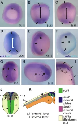
Our comparison between vgll4l and vgll4 revealed significant differences between their expressions. At gastrula stage both genes are expressed in the animal pole but vgll4 is more strongly expressed in the dorsal region of the embryo (Fig. 4A). At early neurula vgll4 is expressed in the anterior and posterior region of the neural plate but not in the middle region (Fig. 4 B,C, black ar-rowhead). The comparison between vgll4 and sox2 demonstrate that at stage 13 there is not vgll4 in the middle region of the neural plate (Fig. 4D). Since stage 16 vgll4 expression is located in the neural plate (small black arrowhead) and in the neural folds (arrow) (Fig. 4 E,G,J). It also presents a weak expression in the epidermis (Fig. 4 F,H,J, asterisk). Transversal sections confirmed that vgll4 is expressed in the neural plate, neural folds and epidermis of midneurula embryos (stage 17, Fig. 4 H,I,K) and also showed a faint expression in the mesoderm (Fig. 4I). Similarly to vgll4l, this gene presents expression in the internal layer of the ectoderm (Fig. 2C, 4I). The onset of vgll4 expression was detected earlier than in Faucheux et al., (2010) that have shown its expression since stage 16 onward. Our results extend the initial findings that reported expression of vgll4 in epidermis and neural crest (Facheux et al., 2010) but not in the neural plate.
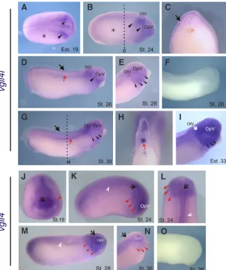
At advanced neurula (Stage 19), vgll4l continued to be expressed at the neural crest and epidermis (Fig. 5A). At tailbud stage, vgll4l expression was located at the migrating cephalic neural crest (Fig. 5B), principally in the mandibular stream (Fig. 5 B,D; arrowhead) and continued to be located in the internal layer of the epidermis (Fig. 5 A-C; asterisk and black arrow). When the development progresses, the main expression of vgll4l is located in tailbud skin
Fig. 4. Spatio-temporal expression pattern of vgll4 during early develop-ment by in situ hybridization. (A) Vegetative view. (B-D, G) Dorsal view, anterior to the top. (E, F) Lateral view, dorsal to the left, anterior to the top. (H) Lateral view, dorsal to the top, anterior to the right. (I) Transversal section. Black dashed line in H indicates the position of the cut made to give cross-section in I. (D) Double in situ hybridization for vgll4 (purple) and sox2 (turquoise). (F) Double in situ hybridization for vgll4 (purple) and xk81a
(turquoise). (B-D) Arrowheads, anterior and posterior vgll4 expression in the neural plate. Brackets, lack of expression in the middle neural plate. (F, H) Asterisk, weak vgll4 expression in the prospective epidermis. Arrow, neural folds vgll4 expression. Arrowhead, neural plate expression. (J, K) Schematic diagrams summarizing the expression of vgll4l and neural plate (sox2), neural crest (foxd3) and epidermal (xk81a) markers. (J) Dorsal view. (K) Transversal section. References: b.l., blastopore lip. e.l., external layer. en, endoderm. i. l.: internal layer. n, notochord. s, somites.
(Fig. 5 D,G; black arrow) and branchial arches (Fig. 5 E,G,I; black arrowheads). It is also expressed in the notochord (red arrow) and otic vesicle (Fig. 5 E,G,H). The expression of vgll4 in these stages was consistent with previous reports (Faucheux et al., 2010). At stage 19 it can be observed at the neural plate, preferentially in the anterior region and in the neural crest that is starting to migrate (Fig. 5J). At stage 24 vgll4 is observed at migrating neural crest (red arrowheads), prospective brain (black small arrow) and somites (white arrowhead) (Fig. 5 K,L). At later stages, the expression is located in the eye primordium and branchial arches and persists at
somites and brain (Fig. 5 M,N). Although vgll4 and vgll4l expression at these stages have some coincidences they are not expressed in exactly the same territories. vgll4l remains expressed in the epidermis and later in the derived skin while vgll4 do not present expression in this tissues. Nevertheless, both are expressed in the branchial arches and central nervous system. To the best of our knowledge, these are the first results that differentiate vgll4 from
vgll4l gene expression in Xenopus.
In zebrafish vgll4l is expressed in the epidermis since early development. During somitogenesis the expression is located in
Fig. 5. Expression of vgll4l and vgll4 at advanced neurula and tailbud stages. (A-I) vgll4l
expression. (J-O) vgll4 expression. (A) Dorsal view, anterior to the right. (B, D, F, G, K, M, O) lateral view, anterior to the right. (C, H) Transversal section. Black dashed lines in B and G indicate the positions of the cuts made to give cross-section in C and H. (E, I, N) Higher-magnification views of head region. (J) Anterior view, dorsal to the top. (L) Dorsal view, anterior to the top. (F) Negative control, vgll4l sense probe. (A-B) Asterisk, vgll4l epidermal expression. (A) Ar-rowhead: vgll4l neural folds expression. (B,D) Arrowhead, vgll4l expression in the migrating neural crest. E, G, I: Arrowhead, vgll4l branchial arches expression. (C) Black arrow, vgll4l
expression in the internal layer of the ectoderm. (D, G) Black arrow, vgll4l skin expression. (D, G, H) Red arrow, vgll4l notochord expression. (O) Negative control, vgll4 sense probe. (J-N) Arrow, vgll4 expression in the prospective brain. (J-L) Red arrowhead, vgll4 expression in the migrating neural crest. (M-N) Red arrowheads, vgll4 branchial arches expression. (K, L, M) White arrowhead, vgll4 somites expression. n, notochord. OpV, optic vesicle. OtV, otic vesicle.
G
O
B
C
D
E
F
H
I
J
K
L
A
M
N
the epidermis and in the neural plate border. At later stages (24-48 hpf) vgll4l remains located at the epidermis and also is expressed in the nose, otic vesicle, pharyngeal pouches and lateral line (Thisse et al., 2001). vgll4 in chicken is expressed in the caudal notochord and in the migratory neural crest cells (Rabadan et al., 2013). In mammals
vgll4 expression was assessed only in adult
tis-sues and its expression was high in the heart, kidney and brain (Chen et al., 2004). However, there are not reports about vgll4 expression in early mammalian development.
Regulation of vgll4s expression
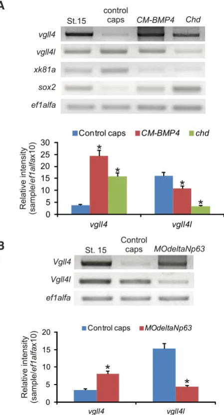
It was determined in Xenopus that early ecto-dermal cell pattern is regulated by a BMP4 sig-naling pathway. BMP4 is essential for epidermal specification while low or absent BMP4 activity results in neural specification (Wilson and Hem-mati-Brivanlou, 1995). Due to the complex vgll4 and vgll4l expression pattern in the ectoderm we analyzed whether BMP4 was necessary for vgll4s expression. To assess that, we decreased the levels of BMP4 expressing a dominant-negative truncated BMP4 (CM-BMP4) or chordin (chd) (Montero-Balaguer et al.), both antagonist of BMP4 signaling. RT-PCR analysis of explanted animal caps injected with CM-BMP4 or chd mRNA revealed an up-regulation of sox2 consistent with the neuralization effect and a decrease in the epi-dermal marker xk81a. In this context we observed an increase in vgll4 expression and a decrease in vgll4l (Fig. 6A). Previous results demonstrated that vgll4 expression was stimulated in a dose dependent manner by activin, but not by FGF or BMP4 signals (Faucheux et al., 2010).
We also analyzed the effect of deltaNp63, a gene that is regulated in a BMP4-dependent man-ner and that is required during the development of early epidermis (Tribulo et al., 2012). We carried out a loss of function in vitro experiment by microinjec-tion of an antisense morpholino oligonucleotide
MOdeltaNp63. RT-PCR performed on animal caps
showed that the inhibition of deltaNp63 increased the level of vgll4 and down regulated significantly the expression of vgll4l (Fig. 6B). These results together with the expression analysis of vgll4 and
vgll4l suggest that these genes participate in the
vgll4 could be participating during neural development and vgll4l
during epidermis and neural crest development.
Recent studies demonstrated that vgll4l is involved in the cra-niofacial development of zebrafish embryos. Morpholino knock-down of vgll4l produced a loss of neural crest derived cartilages suggesting an important role of this gene during zebrafish neural crest specification and survival (Melvin et al., 2013). According to vgll4l expression pattern and preliminary functional analysis in
Xenopus there is a strong possibility that this gene also participates
in neural crest development. However, further studies are needed to fully understand the role of this gene.
It was demonstrated in mammals that vgll4 participates in cardiac myocytes differentiation (Chen et al., 2004). Besides, biochemi-cal studies demonstrated that vgll4 is able to bind to inhibitor of apoptosis proteins (IAPs) playing a role in the apoptotic pathway as an apoptotic promoter (Jin et al., 2011). Nevertheless, in human embryonic stem cells (hESCs) vgll4 was identified as a positive regulator of survival. It was shown that the overexpression of
vgll4 in hESCs decreases death and enhances colony formation
(Tajonar et al., 2013).
Recently, an orthologue of the mammalian Vgll4 that was called Tgi/SdBP and has the two TDU domains was identified in Drosophila. In this model Tgi/SdBP suppresses tissue growth participating in the Hippo pathway by interfering with targets transcription (Guo
et al., 2013, Koontz et al., 2013).
Considering the complex functions observed for vgll4s genes in other model organisms, it could be relevant to explore the dif-ferential functions that Xenopus vgll4 and vgll4l could have during development and organogenesis.
Materials and Methods
Phylogenetic analysisSequences were extracted from NCBI and aligned with the Clustal W program. Phylogenetic analyses were performed with the follow-ing sequences: Xenopus laevis: XlVgll1 NP_001182314, XlVgll2 NP_001080827, XlVgll3 BP689609, XlVgll4 AAI23268 (BC123267), XlVgll4l (KF963131); Xenopus tropicalis: XtVgll1 XP_002932640, XtVgll2 NP_989178, XtVgll3 NP_001072251, XtVgll4 NP_001072615, XtVgll4l KJ690263; Homo sapiens: HsVgll1 AAH03362; HsVgll2 NP_872586, HsVgll3 EAW68870, HsVgll4 NP_001121691; Mus musculus: MmVgll1 EDL42164.1, MmVgll2 EDL05072.1, MmVgll3 NP_082848.1, MmVgll4 EDK99520; Danio rerio: DrVgll1 XM_681743, DrVgll2a NM_001025486, DrVgll2b NP_001028267, DrVgll3 XP_002663398, DrVgll4 NP_998440, DrVgll4l NP_001073467; Gallus gallus: GgVgll1 XP_001234166.1, GgVgll2 ACN54257, GgVgll3 XP_416671, GgVgll4 NP_001025764; Oryzias latipes: OlVgll2 ENSORLP00000015665, OlVgll3 ENSORLG00000015881, OlVgll4 ENSORLT00000005638, OlVgll4l ENSORLT00000016519;
Oreochronis niloticus: OnVgll2a ENSONIG00000015743, OnVgll2b
EN-SONIP00000004388, OnVgll3 ENSONIG00000004268, OnVgll4 ENSO-NIP00000018031, OnVgll4l ENSONIP00000013686; Takifugu rubripes: TrVgll2a ENSTRUG00000008567, TrVgll2b ENSTRUP00000030470, Trvgll3 ENSTRUG00000004178, TrVgll4 ENSTRUP00000006707, TrVgll4l ENSTRUP00000019716; Gasterosteus aculeatus: GaVgll2a ENSGACP00000007733, GaVgll2b ENSGACG00000009615, GaVgll3 ENSGACG00000006678, GaVgll4 ENSGACT00000009758, GaVgll4l ENS-GACT00000015849; Gadus morhua: GmVgll2a ENSGMOP00000012209, GmVgll2b ENSGMOP00000020185, GmVgll3 ENSGMOP00000000452, GmVgll4 ENSGMOT00000011483, GmVgll4l ENSGMOT00000006944;
Drosophila melanogaster: DmVg AAF58444, DmTgi CG10741 (SdPB
NP_648658); Taeniopygia guttata: TgVgll4 XP_002187401; Bos taurus: BtVgll4 DAA16844; Rattus norvegicus: RnVgll4 EDM02166; Macaca
mu-latta: MamaVgll4 NP_001181777; Pongo abelii: PaVgll4 NP_001125047, Salmo salar: SsVgll4 NP_001134829; Pan troglodytes: PtVgll4 JAA19895.
The phylogenetic tree was drawn using the Phylodendron application (http://iubio.bio.indiana.edu/treeapp/treeprint-form.html). Syntenic analysis was performed using Blast tools in Ensembl genome databases. X. laevis
vgll4 and vgll4l were used as queries.
Embryo collection
Xenopus laevis embryos were obtained by stimulating adult male and
female specimens with 400 IU and 800 IU of chorionic gonadotropin (HCG, Elea Lab., Argentina), respectively. Fertilized eggs were obtained after natural single-pair mating and were staged according to the Nieuwkoop
Fig. 6. Regulation of vgll4 and vgll4l expression in the ectoderm. One-cell stage embryos were injected with CM-BMP4 or chd mRNA (A) or MOdeltaNp63(B). Stage 15- control embryos were not injected. At stage 9, animal caps were dissected. Total RNA was isolated from stage 15 embryos and treated and control caps and the expression of vgll4
and vgll4l was analyzed by RT-PCR. ef1alfa was used as loading control. Quantification of gels is shown; the results are expressed as Relative Intensity (sample/ef1alfaX10). Differences were considered statistically significant at P < 0.001(*).
0
St.15 controlcaps CM-BMP4 Chd
and Faber developmental table (1967).
RNA isolation and RT-PCR expression analysis
Total RNA was isolated from whole embryos, adult tissues and animal caps using Trizol reagent (Invitrogen) according to manufacturer’s instruc-tions. cDNAs were synthesized by M-MLV reverse transcriptase (Promega, USA) with oligo dT15 priming from 3 mg total RNA extracted from embryos
at different stages. PCRs were performed with Taq Pegasus (PB-L, Argen-tina) and ef1alfa was used as a loading control. The primers used were:
vgll4 5´-CAACAACATCGCTATTCTCTGC-3´ and
PCR amplification, DNA contamination controls, and quantification of gels were performed as previously described (Tribulo et al., 2012). RT-PCR was performed twice with a pool of 20 embryos each one. Quantita-tion of PCR bands was performed using ImageJ software (NIH, USA) on 8-bit greyscale JPG files. Measures were made six different times and an average was taken to performer the graphics. Values were normalized to the ef1alfa levels from the same sample and expressed for comparison as relative intensities (sample/ef1alfaX10).
In situ hybridization
vgll4 cDNA was obtained from Open Biosistems, clone
MXL1736-99822090, BC123267=Vestigial like 4 and vgll4l was obtained from NIBB, clone Xl460o05. Antisense probes containing digoxigenin-11-UTP or fluorescein-12-UTP were prepared, hybridized and stained for vgll4 (Fau-cheaux et al., 2010), vgll4l (Chalmers et al., 2006), sox2, xk81a, foxd3 by
in vitro transcription. Specimens were prepared, hybridized and stained
as described (Tribulo et al., 2012). For transversal sections embryos were cut using eyebrow knives or a scalpel.
Microinjection and animal cap assay
Xenopus laevis deltaNp63 morpholino antisense oligonucleotide (delt-aNp63MO) was synthesized as described (Tribulo et al., 2012). CM-BMP4
and Chordin (chd) were donated by Dr. K. W. Cho (Hawley et al., 1995) and cDNAs were linearized and transcribed as indicated in Tribulo et al., 2004. Embryos were microinjected with deltaNp63MO, CM-BMP4 or chd mRNA and animal caps were dissected out from them using eyebrow knives as described (Aguero et al., 2012, Tribulo et al., 2004).
Acknowledgements
We wish to thank Dr. Naoto Ueno for the donation of vgll4l clone. We specially thank to Ms. Virginia Mendez for proofreading. This investiga-tion was supported by grants from ANPCyT-Foncyt to M.J.A. (PICT2012-1224, PICT2013-1686) and to C.T. (PICT2013-0219), and by grants from CIUNT to M.J.A. (PIUNT 26/D506).
References
AGUERO, T.H., FERNANDEZ, J.P., LOPEZ, G.A., TRIBULO, C. and AYBAR, M.J. (2012). Indian hedgehog signaling is required for proper formation, maintenance and migration of Xenopus neural crest. Dev. Biol. 364: 99-113.
BLAIR, J.E., HEDGES, S.B. (2005). Molecular phylogeny and divergence times of Deuterostome animals. Molec. Biol. Evol. 22: 2275-2282.
CHALMERS, A.D., LACHANI, K., SHIN, Y., SHERWOOD, V., CHO, K.W. and PAPALOPULU, N. (2006). Grainyhead-like 3, a transcription factor identified in
a microarray screen, promotes the specification of the superficial layer of the embryonic epidermis. Mech. Dev. 123: 702-718.
CHEN, H.H., MULLETT, S.J. and STEWART, A.F. (2004). Vgl-4, a novel member of the vestigial-like family of transcription cofactors, regulates alpha1-adrenergic activa-tion of gene expression in cardiac myocytes. J. Biol. Chem. 279: 30800-308006. DELLA GASPERA, B., ARMAND, A.S., SEQUEIRA, I., LECOLLE, S., GALLIEN,
C.L., CHARBONNIER, F. and CHANOINE, C. (2009). The Xenopus MEF2 gene family: evidence of a role for XMEF2C in larval tendon development. Dev. Biol. 328: 392-402.
FAUCHEUX, C., NAYE, F., TREGUER, K., FEDOU, S., THIEBAUD, P. and THEZE, N. (2010). Vestigial like gene family expression in Xenopus: common and divergent features with other vertebrates. Int. J. Dev. Biol. 54: 1375-1382.
GUNTHER, S., MIELCAREK, M., KRUGER, M. and BRAUN, T. (2004). VITO-1 is an essential cofactor of TEF1-dependent muscle-specific gene regulation. Nucleic
Acid. Res. 32: 791-802.
GUO, T., LU, Y., LI, P., YIN, M.X., LV, D., ZHANG, W., WANG, H., ZHOU, Z., JI, H., ZHAO, Y. et al., (2013). A novel partner of Scalloped regulates Hippo signaling via antagonizing Scalloped-Yorkie activity. Cell Res. 23: 1201-1214.
HALDER, G., POLACZYK, P., KRAUS, M.E., HUDSON, A., KIM, J., LAUGHON, A. and CARROLL, S. (1998). The Vestigial and Scalloped proteins act together to directly regulate wing-specific gene expression in Drosophila. Genes Dev 12: 3900-9. HAWLEY, S.H., WUNNENBERG-STAPLETON, K., HASHIMOTO, C., LAURENT,
M.N., WATABE, T., BLUMBERG, B.W. and CHO, K.W. (1995). Disruption of BMP signals in embryonic Xenopus ectoderm leads to direct neural induction.
Genes Dev 9: 2923-2935.
JIN, H.S., PARK, H.S., SHIN, J.H., KIM, D.H., JUN, S.H., LEE, C.J. and LEE, T.H. (2011). A novel inhibitor of apoptosis protein (IAP)-interacting protein, Vestigial-like (Vgl)-4, counteracts apoptosis-inhibitory function of IAPs by nuclear sequestration.
Biochem. Biophys. Res. Commun. 412: 454-459.
JOHNSON, C.W., HERNANDEZ-LAGUNAS, L., FENG, W., MELVIN, V.S., WILLIAMS, T. and ARTINGER, K.B. (2011). Vgll2a is required for neural crest cell survival during zebrafish craniofacial development. Dev. Biol. 357: 269-281.
KIM, J., SEBRING, A., ESCH, J.J., KRAUS, M.E., VORWERK, K., MAGEE, J. and CARROLL, S.B. (1996). Integration of positional signals and regulation of wing formation and identity by Drosophila vestigial gene. Nature 382: 133-138. KLEIN, S.L., STRAUSBERG, R.L., WAGNER, L., PONTIUS, J., CLIFTON, S.W. and
RICHARDSON, P. (2002). Genetic and genomic tools for Xenopus research: The NIH Xenopus initiative. Dev. Dynamics 225: 384-391.
KOONTZ, L.M., LIU-CHITTENDEN, Y., YIN, F., ZHENG, Y., YU, J., HUANG, B., CHEN, Q., WU, S. and PAN, D. (2013). The Hippo effector Yorkie controls normal tissue growth by antagonizing scalloped-mediated default repression. Dev. Cell 25: 388-401.
MAEDA, T., CHAPMAN, D.L. and STEWART, A.F. (2002). Mammalian vestigial-like 2, a cofactor of TEF-1 and MEF2 transcription factors that promotes skeletal muscle differentiation. J. Biol. Chem. 277: 48889-48898.
MANN, C.J., OSBORN, D.P. and HUGHES, S.M. (2007). Vestigial-like-2b (VITO-1b) and Tead-3a (Tef-5a) expression in zebrafish skeletal muscle, brain and notochord.
Gene Exp. Patt. 7: 827-836.
MELVIN, V.S., FENG, W., HERNANDEZ-LAGUNAS, L., ARTINGER, K.B. and WIL-LIAMS, T. (2013). A morpholino-based screen to identify novel genes involved in craniofacial morphogenesis. Dev. Dynamics 242: 817-831.
MONTERO-BALAGUER, M., LANG, M.R., SACHDEV, S.W., KNAPPMEYER, C., STEWART, R.A., DE LA GUARDIA, A., HATZOPOULOS, A.K. and KNAPIK, E.W. (2006). The mother superior mutation ablates foxd3 activity in neural crest progenitor cells and depletes neural crest derivatives in zebrafish. Dev.
Dynam-ics 235: 3199-212.
POLLET, N., MUNCKE, N., VERBEEK, B., LI, Y., FENGER, U., DELIUS, H. and NIEHRS, C. (2005). An atlas of differential gene expression during early Xenopus embryogenesis. Mech. Dev. 122: 365-439.
RABADAN, M.A., USIETO, S., LAVARINO, C. and MARTI, E. (2013). Identification of a putative transcriptome signature common to neuroblastoma and neural crest cells. Dev. Neurobiol. 73: 815-827.
SIMMONDS, A.J., LIU, X., SOANES, K.H., KRAUSE, H.M., IRVINE, K.D. and BELL, J.B. (1998). Molecular interactions between Vestigial and Scalloped promote wing formation in Drosophila. Genes Dev. 12: 3815-3820.
D.E., ELLEDGE, S.J. and MELTON, D.A. (2013). VGLL4 is a Novel Regulator of Survival in Human Embryonic Stem Cells. Stem Cells.
TAKAHASHI, N., TOCHIMOTO, N., OHMORI, S.Y., MAMADA, H., ITOH, M., INAMORI, M., SHINGA, J., OSADA, S. and TAIRA, M. (2005). Systematic screening for genes specifically expressed in the anterior neuroectoderm during early Xenopus development. Int. J. Dev. Biol. 49: 939-951.
THISSE, B., PFLUMIO, S., FÜRTHAUER, M., LOPPIN, B., HEYER, V., DEGRAVE, A., WOEHL, R., LUX, A., STEFFAN, T., CHARBONNIER, X.Q. THISSE, C. (2001) Expression of the zebrafish genome during embryogenesis (NIH R01 RR15402).
ZFIN Direct Data Submission (http://zfin.org).
TRIBULO, C., AYBAR, M.J., SANCHEZ, S.S. and MAYOR, R. (2004). A balance between the anti-apoptotic activity of Slug and the apoptotic activity of msx1 is required for the proper development of the neural crest. Dev. Biol. 275: 325-42.
TRIBULO, C., GUADALUPE BARRIONUEVO, M., AGUERO, T.H., SANCHEZ, S.S., CALCATERRA, N.B. and AYBAR, M.J. (2012). DeltaNp63 is regulated by BMP4 signaling and is required for early epidermal development in Xenopus. Dev.
Dynam. 241: 257-269.
VAUDIN, P., DELANOUE, R., DAVIDSON, I., SILBER, J. and ZIDER, A. (1999). TONDU (TDU), a novel human protein related to the product of vestigial (vg) gene of Drosophila melanogaster interacts with vertebrate TEF factors and substitutes for Vg function in wing formation. Development 126: 4807-4816.
Vestigial like gene family expression in Xenopus: common and divergent features with other vertebrates
Corinne Faucheux, François Naye, Karine Tréguer, Sandrine Fédou, Pierre Thiébaud and Nadine Thézé Int. J. Dev. Biol. (2010) 54: 1375-1382
http://dx.doi.org/10.1387/ijdb.103080cf
Differential expression of two TEF-1 (TEAD) genes during Xenopus laevis development and in response to inducing factors
5 yr ISI Impact Factor (2011) = 2.959
François Naye, Karine Tréguer, Fabienne Soulet, Corinne Faucheux, Sandrine Fédou, Nadine Thézé and Pierre Thiéboud
Int. J. Dev. Biol. (2007) 51: 745-752 http://www.ijdb.ehu.es/web/paper/072375fn
Msx1 and Msx2 have shared essential functions in neural crest but may be dispensable in epidermis and axis formation in Xenopus
Deepak Khadka, Ting Luo and Thomas D. Sargent Int. J. Dev. Biol. (2006) 50: 499-502
http://dx.doi.org/10.1387/ijdb.052115dk
BMP signalling in craniofacial development
Xuguang Nie, keijo Luukko and Paivi Kettunen Int. J. Dev. Biol. (2006) 50: 511-521
http://dx.doi.org/10.1387/ijdb.052101xn
Differential regulation of Dlx gene expression bya a BMP morphogenetic gradient
Ting Luo, Mami Matsuo-Takasaki, Jae H Lim and Thomas D Sargent Int. J. Dev. Biol. (2001) 45: 681-684