MP-03.01
A single dose of intraoperative antibiotics is sufficient to prevent urinary tract infection during ureteroscopy
Chew, Ben H.1; Flannigan, Ryan1; Kurtz, Michael P.2; Arsovska, Olga1;
Paterson, Ryan F.1; Eisner, Brian2; Lange, Dirk1
1Urologic Sciences, University of British Columbia, Vancouver, BC, Canada; 2Urology, Harvard Medical School, Boston, MA, United States
Introduction and Objectives: AUA Best Practice Guidelines for uretero-scopic stone treatment recommend antibiotics coverage for less than 24 hours after the procedure. The purpose of this study was to evaluate if the rate of post- operative urinary tract infection (UTI) differed in patients receiving a single dose of antibiotics pre-operatively compared to those patients who also received post-operative antibiotics.
Methods: A retrospective review was performed of consecutive patients at two institutions, University of British Columbia and Massachusetts General Hospital, Harvard. All patients were given a single dose of antibiotics prior to ureteroscopic stone treatment. A subset of patients were also given post-operative antibiotics ranging in time and selection of antibiotic. Patients who displayed symptoms of infection had a urine culture performed for speciation and antibiotic sensitivity.
Results: Eighty one patients underwent ureteroscopy for renal calculi. Patients with pre and post operative antibiotics were compared to those receiving only pre-operative antibiotics. Eight (9.9%) patients in total (2 from pre-operative antibiotic and 6 from the pre and post-operative antibiotic group, P=0.219) developed UTI’s in the post-operative period. Surgical factors such as ureteral access sheath, bilateral procedures, use of basket or laser was not associated with rates of infection or whether the surgeon prescribed post-operative antibiotics. Risk factors such as pre-operative stenting, nephrostomy tubes, and foley catheters did not differ between groups or predispose patients to post operative infections.
Conclusions: Our data suggests that post-operative antibiotics do not decrease infection rates following ureteroscopic stone treatment, even among patients with risk factors for infection. A single pre operative dose is sufficient.
MP-03.02
Predictors of fluoroscopy time during percutaneous nephroli-thotomy: Impact of postgraduate urology trainees and S.T.O.N.E. nephrolithometry score
Noureldin, Yasser1,2; Elkoushy, Mohamed1; Andonian, Sero1
1Urology, McGill University Health Centre, Montreal, QC, Canada; 2Urology, Benha University Hospital, Benha University, Benha, Egypt
Objectives: To determine predictors of Fluoroscopy Time (FT) during Percutaneous Nephrolithotomy (PCNL) and assess the impact of urology Post-Graduate Trainees (PGTs) and S.T.O.N.E. Nephrolithometry Score. Methods: A prospective review of patients undergoing PCNL between 2010 and 2013 at a tertiary health care centre was performed. Patients’ demographics, stone characteristics, including S.T.O.N.E. Nephrolithometry Score, and operative data were compared among PGTs. Predictors of FT were determined using univariate and multivariate models.
Results: A total of 103 PCNLs were assisted by 10 PGTs from Post-Graduate Years (PGY) 4 and 5 [37 (35.9%) and 66 (64.1%) cases, respectively)]. Sixty percent of patients were males with a mean age of 55.2±1.5 years and a mean BMI of 26.4±0.5 kg/m2. The mean S.T.O.N.E score was 7.7±0.1, with tubeless PCNL in 53 (51.5%) cases. The mean FT was 120±5 seconds, mean operative time was 102±3.5 minutes and mean length of hospital stay was
4.2±0.34 days. The overall stone-free rate was 72.8%. PGY-5 trainees used significantly less FT than PGY-4 trainees (115±6 vs. 130±7 sec; p=0.04). FT significantly correlated with the number of involved calyces (r= 0.24, p= 0.02), number of punctures (r=0.6, p=0.01), number of tracts (r=0.4; p=0.01), and operative time (r=0.4, p=0.01). In addition, cases with esti-mated blood loss (EBL) <250 mL were associated with significantly less FT than those with blood loss >250 mL (109±5.1 vs. 148.2±10.9 sec; p=0.001). On multivariate analysis, the number of punctures, EBL and operative time were found to be independent predictors for FT. However, there was no correlation of FT with S.T.O.N.E. Nephrolithometry Score (r=0.16; p=0.1). Conclusions: Fluoroscopy Time significantly increased with lower PGY level, higher number of punctures and tracts, more EBL, and longer opera-tive time. However, the number of punctures, EBL and operaopera-tive time were the only independent predictors of prolonged FT during PCNL.
MP-03.03
Which is better? Guy’s vs. S.T.O.N.E. Nephrolithometry Scoring Systems in predicting stone-free status post-Percutaneous Nephrolithotomy
Noureldin, Yasser1,2; Elkoushy, Mohamed1; Andonian, Sero1
1Urology, McGill University Health Centre, Montreal, QC, Canada; 2Urology, Benha University Hospital, Benha University, Benha, Egypt
Introduction and Objectives: Both Guy’s (GSS) and S.T.O.N.E. Nephrolithometry Scoring Systems have been recently validated as pre-dictors of stone-free status post-Percutaneous Nephrolithotomy (PCNL). Therefore, the aim of the present study was to compare the accuracy of the GSS and S.T.O.N.E. scoring systems in predicting PCNL outcomes. Methods: After obtaining ethics approval, medical records of patients under-going PCNL between 2009 and 2013 at a tertiary stone referral centre were retrospectively reviewed. GSS and S.T.O.N.E. Nephrolithometry scores were calculated. Logestic regression analysis and ROC curves were generated to compare accuracy of each scoring system.
Results: A total of 185 PCNLs were reviewed. Mean patient age was 55.2±14 years with mean body mass index of 27.8±6.2 kg/m2 and mean operative time of 98.7±34.2 minutes. The overall stone-free rate was 71.9% with complication rate of 16.2%. When compared with patients with residual fragments, stone-free patients had significantly lower Guy’s Score (2.7 vs. 2; p p<0.001) and S.T.O.N.E. Nephrolithometry score (8.3 vs. 7.4; p=0.004). Logestic regression analysis showed that both GSS and S.T.O.N.E. Nephrolithometry Scoring Systems were significantly associated with stone-free status, (OR=0.4, [95%CI 0.2-0.5]; p<0.001) and (OR=0.7, [95%CI 0.5-0.8]; p=0.001), respectively. Furthermore, both scoring systems were significantly associated estimated blood loss (p=0.01 and p=0.005). There was good correlation between both scoring systems and operative time (r=0.3, p<0.001 and r=0.4, p<0.001) and length of hospital stay (r=0.2, p=0.001 and r=0.3, p=<0.001). However, there were no significant asso-ciations between both scoring systems and post-operative complications (p=0.7 and p=0.6). The ROC curve revealed that the GSS is more accu-rate than the S.T.O.N.E. Nephrolithometry score as a predictor of post-PCNL stone-free status (AUC=0.75 [95% CI 0.7-0.8 vs. AUC=0.63 [95% CI 54-72]), respectively.
Conclusions: Although both scoring systems are suboptimal in prediction of stone-free rates post-PCNL, the Guy’s Scoring System seems to be more accurate than the S.T.O.N.E Nephrolithometry Scoring System. Other factors not included in the current scoring systems may be involved in predicting stone-free rates post-PCNL.
MP-03.04
Leisure time physical activity, smoking and risk of recent symp-tomatic urolithiasis
Soueidan, Michael1; J. Bartlett, Susan1; Noureldin, Yasser1; E. Andersen,
Ross1; Andonian, Sero1
1Urology, McGill University Health Centre, Royal Victoria Hospital,
Montreal, QC, Canada
Introduction and Objectives: Urolithiasis is a recurrent condition. Although prolonged bed rest can contribute to stone formation, it remains unclear how physical activity affects urolithiasis. The aim of this study was to explore relationships between Leisure Time Physical Activity (LTPA) and other lifestyle factors associated with recent symptomatic urolithiasis (≤ 6 months).
Methods: Surveys were administered to a convenience sample of patients attending urology clinics and queried stone history, smoking, diet and supplements. The International Physical Activity Questionnaire assessed LTPA for walking (WALK), moderate (MOD) and vigorous (VIG) activity. Regression analysis was used to identify associations between risk factors and recent symptomatic urolithiasis.
Results: Out of 167 patients with a mean (SD) age of 56.7 (14.3) years and BMI of 27.3 (5.3) kg/m2, 62% were male, 78% white and 34% reported recent symptomatic stones. There were no significant differences between patients with and without symptomatic urolithiasis in terms of age, sex, race, BMI, or dietary risk factors. Only 33% of patients met physical activity guidelines with 24% and 18% reporting adequate MOD and VIG, respectively. LTPA did not differ significantly between patients with and without stones. In symptomatic stone formers, MOD (rho=0.28), VIG (rho=0.37) and total LTPA (rho=0.33) were positively associated with water intake (p<0.05), though no relationships were evident in non-stone formers. Those with recent symptomatic urolithiasis were more likely to smoke (21% vs. 7%, p=0.021). After controlling for sex, diet and LTPA, recent symptomatic stone formers had 5.6 times the odds (95% CI: 1.6-19.4) of smoking.
Conclusions: Most urology patients do not appear to meet physical activity guidelines. Current smoking was a potent predictor of recent symptomatic stones. Urolithiasis patients need to be routinely screened for smoking status and referred for cessation assistance.
MP-03.05
Distal ureteroscopy with conscious sedation for ureteric calculi: 10 year experience
Kroczak, Tadeusz J.1; Kaler, Kamaljot1; Patel, Premal1; Al Essawi, Turki2 1Department of Surgery, Section of Urology, University of Manitoba,
Winnipeg, MB, Canada; 2Urology, Saad Specialist Hospital, Al-Khobar,
Saudi Arabia
Introduction and Objectives: Distal ureteroscopy for urolithiasis extrac-tion is a common procedure that is generally preformed with spinal or general anesthesia. We retrospectively reviewed all distal uretroscopy preformed for ureteric stone extraction with conscious sedation at our institution over a ten year period to determine its efficacy and safety. Methods: A retrospective chart review was preformed of all distal ure-teroscopy preformed for calculus removal from April 2004 to April 2014. Patient characteristics, analgesic requirement, tolerability, procedure time, stone size and composition, method of stone extraction, success rate and complications were collected. Fishers exact test and unpaired t-test were used for data analysis.
Results: Three hundred and thirty one procedures were preformed between April 2004 and April 2014. Mean age was 53.84 years old (median, 54; range, 17-88) with 164 males (50.5%) and 157 females (49.5%). We had a success rate of 97% with 10 complications reported. Two patients had a Clavien classification of Grade3a with the remain-der Grade 1. Mean procedure time was 25 minutes (median, 24; range, 3-65). Mean analgesics requirement was 189μg of fentanyl (median, 200; range, 50-400) and 4mg of midazolam (median, 3; range, 0-8). 268 patients (80.9%) tolerated the procedure well with only 7 having poor tolerability (2%). The most common stone composition was calcium oxalate monohydrate. When comparing females to males, females were found to require less fentanyl (p = 0.0001), midazolam (p = 0.0001) and
found the procedure more tolerable (p = 0.0231). When calculi greater than 5mm were compared those less than 5mm, there was no statistically significant difference in success rate, procedure time, analgesic require-ment, tolerability, or complications.
Conclusion: Various different analgesic options exist with distal ureteros-copy for urolithiasis extraction. Using conscious sedation is safe and efficacious. To our knowledge this is the first report to show that stones larger than 5mm can be safely and effectively treated with the use of conscious sedation during this procedure.
MP-03.06
‘Electronic Stent Register’: Future solution for forgotten stent
Brodie, Andrew1; Moghul, Masood1; Akhter, Waseem1; Kalsi, Jas1 1Urology, Frimley Health NHS Hospital, Slough, United Kingdom
Introduction: JJ stent insertion is a common urological procedure world-wide. The majority of patients have their stents removed or replaced within the requisite timeframe; however, a minority are lost to follow up. Forgotten stents are posing a significant pecuniary burden on the NHS. We carried out an audit to assess efficacy and efficiency of an electronic stent register in minimizing forgotten stents.
Methodology: Retrospective data collection from January 1st 2014 to March 31st 2014. We compared the patient entries in the theatre log books of three separate theatres with the electronic stent register. We re-audited it from 1st August to 31st October to close the loop and to assess the change in practice.
Results: 67 stents were inserted, 11 of which were performed as an emer-gency procedure. Majority of these cases; 50 (75%); were recorded on stent register while 17 cases were missed. Main reasons of missed stents were untrained surgeons in using electronic stent register and antegrade stent insertion by radiologist who do not have access to electronic stent register. All of the missed stents cases were either removed or replaced urgently uneventfully. None of the missed stents cases suffered any major morbidity. During the re-audit time period 105 stents inserted, there was an improvement noted on electronic stent register entry (89, 85%). 16 patients were not entered because they had stent insertions in radiology department but they were booked for TCI of removal/ /further uretero-scopic procedure.
Conclusion: We concluded that an electronic stent register is helpful for follow up which will prevent the possible complications and cost. We also introduced robust teaching sessions for new trainees in updating stent register. We are also in touch with IT department to update our stent register as per BAUS stent register. Where regular reminders are generated and posted to the responsible person, which have reduced complications regarding forgotten stent.
MP-03.07
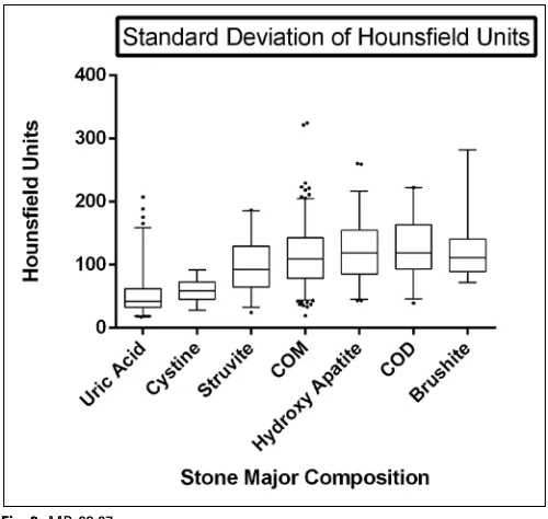
Mean and standard deviation of Hounsfield units on pre-opera-tive computed tomography allow confident prediction of urinary stone composition
Tailly, Thomas1; Larish, Yaniv3; Nadeau, Brandon R.2; Olvera-Posada,
Daniel1; Alenezi, Husain1; Violette, Philippe1; Glickman, Leonard3;
Amann, Justin2; Denstedt, John1; Razvi, Hassan1
1Urology, Schulich School of Medicine and Dentistry, London, ON,
Canada; 2Radiology, Schulich School of Medicine and Dentistry, London,
ON, Canada; 3Urology, Lenox Hill Hospital, New York, NY, United States
Introduction and Objectives: The chemical composition of a urinary stone may influence its management. Previous attempts at identifying stone composition based on mean Hounsfield Units (HUm) on computed tomography (CT) have had varied success. We aimed to evaluate the additional use of the standard deviation of HU (HUsd) to more accurately predict stone composition in a large multi-center population.
in 90, Apatite (A) in 58, Struvite (S) in 35, Brushite (B) in 27, Cystine (C) in 25 and CO dihydrate (COD) in 22 of the patients. UA stones had significantly lower and B stones significantly higher HUm than any other stone composition. COM, COD and A could not be differentiated by HUm or HUsd. Although C and S stones have a similar HUm, HUsd was significantly different. UA and C stones both have similarly low HUsd, but are significantly different from all other stone types (Graph 1&2). HUm has relatively good predictive accuracy for UA, C, COM, and B stones (AUC= 0.975, 0.724, 0.743, and 0.882 respectively) with cutoff HUm values <588, <738, >763, and >1235 respectively. HUsd has the most predictive accuracy for UA (AUC = 0.866) and C stones (AUC = 0.763) with cutoff HUsd values of <68 and <88 respectively (Fig. 1, Fig. 2). Combining HUm and HUsd cutoffs increases the positive predictive value for most stone types.
Conclusions: Both mean and standard deviation of HU may help pre-dict stone composition. This is the first report of CT data aiding in the prediction of Brushite stones. The HUsd allows differentiation of Cystine and Struvite stones. COM cannot be differentiated from COD or Apatite.
MP-03.08
The effect of stone prevention counseling at the initial consulta-tion on 24-hour urine collecconsulta-tion results (“clinic effect”)
Alzahrani, Tarek M.1; Ghiculete, Daniela1; Pace, Kenneth T.1; Lee, Jason
Y.1; Honey, R. John D.1
1Urology, St. Michael’s Hospital, University of Toronto, Toronto, ON,
Canada
Introduction and Objectives: 24-hour urine collections are integral to long-term management of patients with recurrent urolithiasis and iden-tifying treatable metabolic risks for urolithiasis (stone prevention). At our institution, patients bring a 24-hour urine collection to the initial assessment for extracorporeal shockwave lithotripsy (SWL); they are then given general dietary advice and brochures about stone prevention before knowing the initial 24-hour urine results. Our objective was to determine if there are differences in 24-hour urine parameters between the first 2 consecutive samples collected if stone prevention counseling did or did not occur at the time of the initial 24-hour collection, reflecting a pos-sible “clinic effect”.
Methods: We reviewed data for 24-hour urine collections for two groups of new patients: ones who had stone prevention counseling at the time of the initial 24-hour collection and ones who did not. Patients were
included if they had at least two complete collections, not more than 6 months apart. The rates of abnormal 24-hour urine collection param-eters (e.g. low urine volume, hypercalciuria, hypocitraturia, hyperoxaluria) were determined by comparing the percentage of normal and abnormal results between the two samples.
Results: 79 patients had counseling at initial presentation and 146 did not. Baseline urinary lithogenic risk factors were similar in both groups. There was a significant improvement in the urine volume in the repeated samples in both groups (P < 0.01). The rate of abnormal oxalate and citrate levels in patients who had counseling decreased in the repeated collections but did not reach statistical significance (P = 0.064 and 0.052 respectively); this change was not seen in patients who did not get stone prevention counseling.
Conclusions: First 24- hour urine collections prior to counseling are important to pick up some abnormalities such as low fluid intake or hyperoxaluria that could be missed in the second sample due to the “clinic effect”. Improvements in the 24-hour urine parameters can be noticed even after simple general dietary advice at the initial consultation prior to the results of the 24-hour urine parameters are known.
MP-03.09
Perinephric fat distribution and anatomical considerations when performing percutaneous nephrolithotomy in obese patients
Alzahrani, Tarek M.1; Ghiculete, Daniela1; Garbens, Alaina1; Pace,
Kenneth T.1; Honey, R. John D.1
1Urology, St. Michael’s Hospital, University of Toronto, Toronto, ON,
Canada
Introduction and Objectives: Knowledge of renal and retroperitoneal anatomy is essential to performing percutaneous nephrolithotomy (PCNL) with minimal patient morbidity. The kidneys are oblique in reference to the sagittal, coronal, and axial planes of the body with the lower pole more anterior and freely mobile, accounting for the difficulty in puncture as the lower pole may move away from the needle and dilators dur-ing PCNL. Our objective is to study the difference in the distribution of perinephric fat and its impact on the skin – calyx length and renal angle during PCNL in obese patients.
Methods: We reviewed 38 consecutive abdominal CT scans done at our institution with 76 kidneys included. We performed several measurements in relation to the anatomy of the kidneys including: Renal Angle, upper pole to skin (UPS), lower pole to skin (LPS), upper pole to muscle (UPM)
- representing the upper pole perinephric fat - and lower pole to muscle (LPM). We excluded patients with missing demographic data such as body mass index (BMI) or any abnormality on CT that would change the position of the kidney in the retroperitoneum such as large renal cysts. Results: The mean BMI was 27.5 ± 7.5. The UPS distance was signifi-cantly higher in the obese patients (BMI>30) compared to the non-obese (82.5 ± 21.9, 56.8 ± 11; P value <0.01), the same was noticed in the LPS distance (134.4 ± 37.6, 93.3 ± 18.2; P value <0.01). The perinephric fat distribution measured by the UPM and LPM were both significantly higher in the obese population (P value <0.01 for both) but the obese patient had significantly more fat surrounding the lower pole (P value = 0.011), which caused the lower pole to shift more anterior in the obese population changing the renal angle from 22.2 ± 8 to 30.6 ± 9.7 on the sagittal access (P value <0.01).
Conclusion: There is a significant change in the perinephric fat distribu-tion in the obese patients which is more pronounced in the lower pole making lower pole access longer and at a more difficult angle in these patients. This supports our recommendation for an upper pole approach in the obese patient.
MP-03.10
Changing patient position can eliminate arrhythmia developing during shock wave lithotripsy (SWL)
Alzahrani, Tarek M.1; Ghiculete, Daniela1; Pace, Kenneth T.1; Honey, R.
John D.1
1Urology, St. Michael’s Hospital, University of Toronto, Toronto, ON,
Canada
Introduction and Objectives: SWL has a low complication rate. While serious side effects are rare arrhythmias such as ventricular tachycar-dia may occur and require cessation of treatment. The etiology of these arrhythmias is poorly understood, but is most likely related to the relative position of the stone and the heart. This study examines the effect of rotat-ing the patient 15 to 20 degrees when an arrhythmia occurs.
Methods: 1369 patients were prospectively evaluated for arrhythmias dur-ing SWL. The initial patient position was dependent on the location of the stone and the size of the patient. Stones on either side may be treated with the patient flat supine. More frequently, the patients are rotated about 15 degrees with the opposite side to the stone elevated. With stones on the right the patient would be right posterior oblique (RPO) and for left sided stones left posterior oblique (LPO). If a sustained arrhythmia developed, treatment was held for 2 minutes and then recommenced. If the arrhyth-mia returned, as occurred in all 20 patients, the patient was rotated to the flat, RPO or LPO position, angling the shock wave away from the heart. Results: 20 patients developed significant arrhythmias during SWL. Arrhythmias occurred more frequently in patients with a lower BMI (P<0.01) and in those with right-sided stones (P=0.035). After rotation no further arrhythmias were seen in 13 patients. Gated ESWL was only needed in only 3 of the 20 patients after changing their position. Conclusions: Changing the position of the patient by rotating the patient by 15 to 20 degrees may eliminate arrhythmias that develop during SWL allowing the continuation of the SWL treatment without the need to perform a gated SWL.
MP-03.11
Ambulatory bilateral tubeless PCNL: Is it safe and feasible?
Kokorovic, Andrea2; Fuoco, Michael B.1; Wilson, James W.1; Beiko, Darren
T.1
1Urology, Queen’s University, Kingston, ON, Canada; 2Urology, Dalhousie
University, Halifax, NS, Canada
Introduction and Objectives: Bilateral tubeless percutaneous nephro-lithotomy (PCNL) has been reported to be safe and effective in select patients. Although outpatient PCNL has been recently been shown to be safe and effective in a series of 50 patients, it requires further study before urologists embrace same day discharge following PCNL. The objective of this study is to report our early experience in performing bilateral PCNL on a completely outpatient basis, assessing its safety.
Methods: A review of all outpatient tubeless PCNL cases between March 2007 and May 2014 at a single Canadian centre was performed, including collection of preoperative, intraoperative and postoperative data. Strict preoperative, intraoperative and postoperative criteria were used in the selection of candidates for outpatient bilateral PCNL: no intraoperative complications including significant bleeding or collecting system per-foration; postoperative hemodynamic stability; adequate pain control; reliable patient with supportive family.
Results: Forty patients underwent ambulatory PCNL during the study period, of which 4 patients underwent bilateral ambulatory tubeless PCNL. Mean maximum stone diameter was 3.5 cm and 5 of the 8 renal units contained staghorn calculi. All 4 patients were discharged home on the same day with a mean hospital stay of 186 minutes or 3hrs 6min. The mean narcotic requirement was 70.5mg of oral morphine equiva-lents. Importantly, there were no postoperative complications, emergency room visits, hospital readmissions, ancillary procedures or deaths. Conclusions: This small series represents the largest series of outpatient bilateral PCNL to date. In very carefully selected patients, bilateral PCNL on a completely ambulatory basis appears safe and may be feasible. Further research on outpatient bilateral PCNL is required prior to wide-spread adoption by urologists.
MP-03.12
Flank percussion in the semi-prone position can relocate stones prior to percutaneous nephrolithotomy or laparoscopic pyelo-plasty
Althunayan, Abdulaziz M.1; Barrett, Keith P.1; Honey, R. John D.1 1Urology, St. Michael’s Hospital, University of Toronto, Toronto, ON,
Canada
Introduction: Stones in remote calyces may be difficult to access. During percutaneous nephrolithotomy (PCNL) or robot assisted laparoscopic pyeloplasty (RLP), renal stones within the calyces require flexible neph-roscopy and the use of a nitinol stone basket for removal. During RLP this tends to be complex and time consuming on occasions. In dilated systems, stone may be mobile, as suggested by a rounded shape, making their relocation a possibility. Relocation of calyceal stones to the renal pelvis facilitates and simplifies their extraction. The objective of this study is to describe a technique for manually relocating stones to the renal pelvis prior to PCNL and RLP.
Methods: Patients undergoing either PCNL or RLP who were known to have remote calyceal calculi were identified pre-operatively. Under gen-eral anesthetic, prior to placing them in the prone position for PCNL, or lateral for RLP, we place the patients on their side with the affected kidney superiorly and rotated 45˚ anteriorly to position the renal pelvis in a dependent position. This rotation also reduces the effect of the more posterior and medial lie of the upper pole calyces. Manual percussion is then performed over the kidney to dislodge the calyceal stones into the renal pelvis. C-arm fluoroscopy and nephroscopy was then used to confirm movement of all stones out of the calyces into the renal pelvis during PCNL, and flexible nephroscopy alone during RLP.
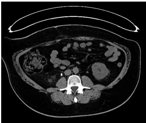
Results: We have now used this technique in 2 patients at the time of RLP and 3 patients at the time of PCNL. All patients had dilated collecting systems with smooth, round or pebble-shaped stones. All of the stones moved into the renal pelvis with this technique (images will be provided with the presentation).
UP-03.01
Ureteric stricture following ureteral access sheaths in the mod-ern era: How rare is it?
Althunayan, Abdulaziz M.1; Ghiculete, Daniela1; Lee, Jason Y.1; Ordon,
Michael H.1; Honey, R. John D.1; Pace, Kenneth T.1
1Urology, St. Michael’s Hospital, University of Toronto, Toronto, ON,
Canada
Introduction and Objectives: Ureteral access sheaths (UAS) can aid ure-teroscopy by facilitating multiple passes of the ureteroscope, maximizing irrigation drainage, and reducing intra-renal pressures. However insertion of the access sheath may induce ureteral ischemia, cause iatrogenic ure-teric injury, and could ultimately lead to ureure-teric stricture. In this study, we aim to evaluate the stricture rate following ureteroscopy both with and without the use of UAS.
Methods: We performed a retrospective chart review of consecutive ureteroscopies performed at our center (a tertiary referral center for endourology) between April 2012 and April 2014 to treat ureteric and renal calculi. The primary outcome was the development of new hydro-nephrosis three months following successful ureteroscopy, not due to an obstructing stone. Patients without follow-up renal ultrasound (US) or CT scan 3 months post-ureteroscopy were excluded. Data on age, sex, size of the stone, location of the stone, stone density, stone-free rate (SFR), time of the procedure, pre-op stenting, post-op stenting, use of the UAS, size of the UAS, length of the UAS, stone analysis, and imaging details were recorded. Baseline and outcome variables were compared with ANOVA and Chi-square analysis where appropriate using SPSS statistical software. Results: 203 patients were eligible. A UAS was used in 121 (59.6%) patients; 103 (85.1%) for renal stones and 18 for proximal ureteric stones. There was no significant difference in baseline or demographic data. None of the patients developed new hydronephrosis or developed a ureteric stricture, and none required endoureterotomy.
Conclusions: UAS use during ureteroscopy for renal and ureteric stones is both safe and effective. Even with routine use of 11.5F and 14F UAS, ureteric stricture rates are very low (zero in this series), suggesting that significant ureteric injury is rare with proper technique and case selection.
UP-03.02
Preoperative patient and stone characteristics associated with a prolonged length of hospital stay following tubeless PCNL
Barrett, Keith P.1; Ghiculete, Daniela1; Ordon, Michael H.1; Lee, Jason
Y.1; Pace, Kenneth T.1; Honey, R. John D.1
1Urology, St. Michael’s Hospital, University of Toronto, Toronto, ON,
Canada
Introduction and Objectives: The adoption of tubeless percutaneous nephrolithotomy (tPCNL) has decreased patient discomfort and length of hospital stay following PCNL. Ambulatory PCNL (hospital stay ≤ 24 hours) is now common, but identifying patients who are likely to require longer hospital stays remains a challenge. The objective of this study is to identify preoperative patient and stone characteristics associated with a required of hospital stay beyond post-operative day 1 (POD 1) following tPCNL. Methods: A database of patients undergoing PCNL at our institution between 2011 and 2014 was reviewed, and cases of tubeless PCNL were identified. Length of stay (LOS) was defined as the period from the time operating room entry until the time of discharge. Preoperative variables evaluated included: age, gender, BMI, ASA, hemoglobin, cre-atinine, bacteriuria, and stone characteristics. Patients were divided into those discharged on POD 1 (Group 1) and those discharged beyond POD 1 (Group 2).
Results: 128 cases of tPCNL were identified during the study period. Mean LOS was 37.7 hours (range = 14.0 to 316.4 hours). 100 patients were discharged POD 1 (Group 1 = 78.8%) with a mean LOS of 24.6 hours (SD ± 3.5 hours), and 28 patients were discharged later than POD 1(Group 2 = 21.8%) with a mean LOS of 84.5 hours (SD ± 58.3 hours). Pre-operative patient characteristics associated with a prolonged hospital stay included BMI (27.9 ± 4.7 vs. 30.4 ±7.7, p = 0.04), ASA 3 & 4 (47% vs. 67.9%, p = 0.05), anemia (27% vs. 53.6%, p < 0.01), creatinine (82.6 ± 30.2 vs. 106.3 ± 72.9 μmol/L, p = 0.01), and bacteriuria (10.1% vs. 37.0%, p < 0.01). Of the stone characteristics, only stone area was
significantly associated with a prolonged hospital stay 491.4 ± 459.4 vs. 1043.9 ± 983.6 mm2, p <0.01).
Conclusion: Both preoperative patient and stone characteristics can be used to identify patients likely to require a prolonged hospital stay fol-lowing tubeless PCNL. This information may be useful in counseling patients and in pre-operative planning.
UP-03.03
A new and simple intraoperative technique to radiographically determine appropriate ureteric stent length
Barrett, Keith P.1; Honey, R. John D.1
1Urology, St. Michael’s Hospital, University of Toronto, Toronto, ON,
Canada
Introduction: It is important to choose the appropriate length of ureteric stent for every patient. Stents that are too long increase stent-related bladder symptoms and patient discomfort, and stents that are too short may migrate. Patient height is often used to choose stent length, but this is not a reliable predictor of ureteric length. The objective of this study is to describe a new and simple intraoperative technique to determine appropriate stent length using fluoroscopic measurements.
Methods: The charts of 5 patients who underwent ureteric length mea-surement and subsequent stent placement were reviewed. During cys-toscopy, the tip of the cystoscope was positioned at the ureterovesical junction (UVJ). A metal BB was then affixed with adhesive tape to the skin over the UVJ. At the end of the case, an angiographic catheter with radiopaque markings at 1, 5, 10, 15, 20, and 25cm was used to perform a retrograde pyelogram and its tip positioned at the ureteropelvic junc-tion (UPJ). Ureteric length was determined by measuring the distance from the UPJ to the BB at the UVJ using the catheter markers. A length of stent closest to the measured ureteric length was then placed. Stents were deemed of appropriate length if the proximal coil was in the renal pelvis and the distal coil in the bladder with no excess stent length. Results: All 5 patients underwent successful radiographic measurement of ureteric length using the new technique. In 4 cases the stent was left to a tether. When the length of stent placed matched the radiographi-cally measured ureteric length, stent length was found to be appropriate in all cases. In 2 cases, stents longer than the radiographically measured ureteric length were placed, and in both cases stent length was not felt to be appropriate due to excess length in the bladder.
Conclusion: This study describes a simple intraoperative technique to radiographically determine ureteric length. This technique is helpful in choosing appropriate stent lengths for individual patients.
UP-03.04
Tubeless percutaneous nephrolithotomy with retrograde teth-ered stent
Barrett, Keith P.1; Ghiculete, Daniela1; Ordon, Michael H.1; Lee, Jason
Y.1; Pace, Kenneth T.1; Honey, R. John D.1
1Urology, St. Michael’s Hospital, University of Toronto, Toronto, ON,
Canada
Introduction and Objectives: The adoption of “tubeless” percutane-ous nephrolithotomy (tPCNL) has decreased the morbidity of PCNL by eliminating the need for nephrostomy and nephroureterostomy tubes, in exchange for a ureteric stent. However, the need to perform a cystoscopic procedure to remove this stent remains a source of inconvenience and discomfort for patients. The objective of this study was to present our experience with tPCNL using retrograde stenting with a tether. We com-pared rates of stent related complications to those of a similar cohort of tPCNL patients in whom an un-tethered stent was placed.
visits, adjunctive procedures for renal drainage, and urinary tract infec-tions (UTI) diagnosed in the week following discharge were compared. Results: A total of 239 patients underwent tPCNL during the study period (Group 1 = 38, Group 2 = 193). Pre-operative and intra-operative char-acteristics were similar between the groups. No premature stent removals occurred in Group 1. In Group 2 there were 13 premature stent removals (6.7%), leading to 6 ER presentations (3.1%), and 1 patient requiring stent replacement (0.5%). UTIs were identified in the week following discharge in 5.3% of Group 1 and 7.3% of Group 2 (p = 0.67).
Conclusions: tPCNL with a retrograde tethered stent appears to be a safe, with a low risk of stent related complications or need for adjunctive procedures when compared to stents without tethers.
UP-03.05
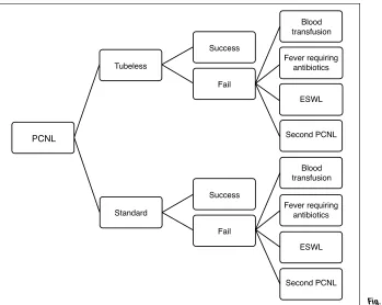
Percutaneous nephrolithotomy: A cost-effective analysis of tube-less and standard procedures
Akerman, Jason P.1; Tu, Hin Yu Vincent1; Hoogenes, Jen1; Lambe, Shahid
A.1; Matsumoto, Edward D.1
1McMaster Institute of Urology, Hamilton, ON, Canada
Introduction and Objectives: Percutaneous nephrolithotomy (PCNL) is the established gold standard for treatment of large renal stones. Traditionally, a nephrostomy tube was left post-operatively to tamponade bleeding along the tract, allow for urinary drainage, and facilitate a second pro-cedure if necessary. In appropriately selected patients, tubeless PCNL has emerged as a safe and effective alternative. The purpose of our study was to assess whether the tubeless method of PCNL is cost-effective when compared to standard PCNL.
Methods: A decision tree was constructed to calculate the costs of achiev-ing a complication free-outcome in standard and tubeless PCNL (Fig. 1). A comprehensive literature review was performed to determine the rates of common post-operative complications. Each complication was graded using the validated Revised Clavien classification. Costs of the surgical procedures were obtained from the Ontario Case Costing Initiative and the Ontario Schedule of Benefits. Drug costs were obtained through the Ontario Drug Benefit Formulary. A willingness to pay threshold of $3000 was used and adopted from drug-eluding stent literature.
Results: Tubeless PCNL was more cost-effective than standard PCNL at $10171.09 with a complication grade of 0.29 compared to $12673.62 and 0.35. Overall complication rates in the tubeless and standard PCNL groups were 12.8% and 15.6% respectively. Sensitivity analysis demon-strated that tubeless PCNL remained cost effective as long as the cost of the procedure remained below $12000 and the cost of standard PCNL remained above $9000.
Conclusions: In appropriately selected patients, tubeless PCNL is more cost effective than standard PCNL in achieving a complication free out-come at a WTP of $3000. A shorter average length of hospitalization and reduced cost of OR supplies among tubeless patients led to lower upfront costs of the primary procedure. Due to comparable complication rates in the two groups, tubeless PCNL remained the more cost-effective option when post-operative events were accounted for.
UP-03.06
Contemporary imaging practice patterns after ureteroscopy for stone disease
Chaparala, Hemant 1; Mohamed, Omar1; Monga, Manoj1; Sivalingam,
Sriharan1
1Urology, Glickman Urological and Kidney Institute, Cleveland Clinic,
Cleveland, OH, United States
Introduction and Objectives: Routine imaging following ureteroscopy for treatment of renal/ureteral calculi continues to be a topic of debate. However, with the increasing focus on healthcare costs and quality, judi-cious use of diagnostic imaging to optimize outcomes while minimizing resource utilization is a priority. We sought to identify post-ureteroscopy imaging practices amongst experienced urologists.
Materials and Methods: A REDcap questionnaire was sent to urologists in North America. The questionnaire surveyed demographic data, clinical volume, and imaging preferences post-ureteroscopy. Additionally, we sur-veyed the extent to which stone, anatomic, and procedure-related factors influenced these preferences. The likelihood of altering clinical practice and the desire for specific imaging guidelines were also assessed. The Interquartile range (IQR) was utilized as a measure of median consensus, with a lower IQR denoting increased agreement.
PCNL
Blood transfusion
Fever requiring antibiotics
ESWL
Second PCNL
Blood transfusion
Fever requiring antibiotics
ESWL
Second PCNL Fail
Success Fail Success Tubeless
Standard
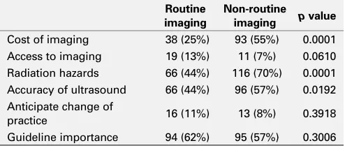
Results: Three hundred twenty-two urologists completed the question-naire. The mean number of years in practice was 18±10; 82% of respon-dents performed more than 5 ureteroscopic stone procedures monthly. Routine postoperative imaging was obtained by 48% of participants as follows: US (47%), KUB (17%), CT (4%), IVP (2%), and KUB + US (30%). Urologists who did not routinely image patients were more concerned about cost (55% vs. 25%, p= <0.0001), radiation exposure (69% vs. 44%, p= <0.0001), and diagnostic inaccuracy of US (57% vs. 44%, p= <0.02) (Table 1). These urologists were also less likely to have completed an Endourology fellowship (7% vs. 23%, p= <0.0001).The most compelling predictors of obtaining postoperative imaging were post-op pain and fever (median 5, IQR 1), residual stones (median 5, IQR 1), ureteral perforation (median 5, IQR 2), and presence of a solitary kidney (median 4.5, IQR 2) (Fig. 1). Conclusions: Currently, about 50% of urologists who regularly perform ureteroscopic stone procedures obtain post-op imaging. Imaging prefer-ences were guided by the presence of residual fragments, ureteral perfora-tion, solitary kidney, and postoperative pain or fever.
UP-03.07
Urinary uric acid does not correlate with increased cardiovas-cular risk and myocardial infarction
Kartha, Ganesh K.1; Chaparalla, Hemant1; Calle, Juan C.1; Monga, Manoj1;
Sivalingam, Sriharan1
1Urology, Cleveland Clinic, Cleveland, OH, United States
Introduction and Objective: Recent studies have shown a correlation with elevated serum uric acid and myocardial infarction (MI) risk, specifi-cally as an indicator of poor prognosis post MI. In addition, studies have demonstrated that adverse cardiovascular events are more common in individuals with nephrolithiasis. However, urine uric acid levels may be elevated even with normal serum uric acid (UA). We sought to identify
an association between urinary uric acid (UUA) levels and cardiovascular risk in our stone population.
Methods: 7410 stone patients with available 24h urine UA were strati-fied into three groups: UUA < 700, UUA 700-1000 and UUA > 1000 (mg/day). Clinical and demographic data at the time of 24h urine and the prevalence of myocardial infarction was identified and compared between the three groups.
Results: Patients with elevated UUA were more likely to have hyperten-sion, diabetes, elevated BMI, low HDL and of male gender. There was no statistical difference in the prevalence of MI between groups stratified by UUA level (4.2% vs. 3.8% vs. 5.0%, p=0.5939). When stone patients with MI were matched to stone patients without MI by age, gender, BMI, smoking history, diabetes and hypertension there was no difference in mean UUA (569.2mg vs 544.1mg, p=0.6051) or urine pH levels (5.9 vs 5.9, p=0.7376).
Conclusion: In our kidney stone population, elevated UUA is not an independent risk factor for MI, although it is positively associated with car-diovascular risk factors such as male gender, elevated BMI, hypertension, diabetes, and low HDL and may be and indicator of metabolic syndrome.
UP-03.08
Updated economic analysis on ureteric stenting after uncompli-cated ureteroscopic laser lithotripsy for urolithiasis: Is stenting necessary?
Tu, Hin Yu Vincent1; Matsumoto, Edward D.1
1Department of Urology, McMaster Institute of Urology, Hamilton, ON,
Canada
Introduction and Objectives: Routine ureteral stenting after uncompli-cated ureteroscopy and laser lithotripsy (ULL) is debatable. Purported advantages include preventing ureteral obstruction and facilitating stone passage; disadvantages include stent colic, potential stent migration and subsequent cystoscopic stent removal. The purpose of our study is to determine whether stenting after uncomplicated ULL is safe and eco-nomically sound.
Methods: A decision tree was developed to estimate the costs of ULL with and without stenting in achieving a complication free outcome (CFO) and quality-adjusted life days (QALD) over a one month period. Complications were defined as any unplanned hospital visits within the first four postop-erative weeks including emergency room visits, admissions to hospital or surgeries. They were graded according to the validated Revised Clavien classification. Complication rates and utility values were derived from recent literature. Costs of surgical procedures were obtained from the Ontario Case Costing Initiative. Univariate and probabilistic sensitivity analyses were performed to determine the robustness of our model. Results: ULL without stenting was more cost-effective than ULL with stenting in achieving a CFO. ULL with and without stenting cost $3828 and $3248 respectively to achieve an overall complication grade of 0.06 and 0.15 (p<0.0001) respectively. Overall postoperative complication rates in the stented versus non-stented groups were 2.6% and 6.5% respectively. ULL with and without stenting generated 25.2 and 26.2 QALDs over one month respectively. Probabilistic sensitivity analysis reinforced ULL without stenting as the most cost-effective strategy at a willingness-to-pay (WTP) threshold of less than $6000/QALY. ULL without
0 1 2 3 4 5
IQR Range Median Response
Recurrent Stone Ectopic Kidne
y
UPJ Obstruction Solitary Kidney Ureteral Perforatio
n
Post-Op Fever Post-Op Pain Residual Stone
Table 1. UP-03.09.
Parameter Helical Control p value
Number 15 30 — Age (y) 58.47 52.37 NS BMI (kg/m2) 28.74 27.82 NS
Unscheduled visits 20% (3/15) 20% (6/30) p=1.000
Analgesic use (morphine equivalent mg)
4.4±7.9 16.75±18.31 **p=0.0035
Visual Analog Scale
Score 13.85±13.68 14.29±11.58 p=0.791 Fig. 1. UP-03.06.
Table 1. UP-03.06. Factors discouraging routine imaging & expectations of future change
Routine imaging
Non-routine imaging p value
Cost of imaging 38 (25%) 93 (55%) 0.0001 Access to imaging 19 (13%) 11 (7%) 0.0610 Radiation hazards 66 (44%) 116 (70%) 0.0001 Accuracy of ultrasound 66 (44%) 96 (57%) 0.0192 Anticipate change of
stenting remained the most dominant strategy in our cost-utility analysis at generating QALDs across all WTP thresholds.
Conclusions: After uncomplicated ULL, not stenting was more cost-effec-tive and yielded more QALDs than stenting over a one month period at reasonable WTP thresholds. Our study demonstrated that despite the increased postoperative complication rates associated with non-stented ULL, it was still the preferred treatment strategy after uncomplicated ULL.
UP-03.09
Patients with Helical ureteral stents require significantly less analgesics than control stents
Chew, Ben H.1; Harriman, David I.1; Lange, Dirk1; Arsovska, Olga1;
McDougall, Elspeth1; Paterson, Ryan F.1
1Department of Urologic Sciences, University of British Columbia,
Vancouver, BC, Canada
Introduction and Objectives: Ureteral stent symptoms are experienced by patients with stents. Softer materials, adjuvant drugs, drug-eluting stents, and new stent designs have been attempted to reduce stent symptoms. There has not been any clear advantage in any one stent or new technol-ogy. The HelicalTM stent, a spirally cut stent made of proprietary Percuflex
material was designed to conform better to the shape of the ureter and move with the patient. We prospectively sought to compare unscheduled visits, analgesic use and pain scores in patients who received a Percuflex Helical ureteral stent following ureteroscopy compared to a historical control group.
Methods: Fifteen (n=15) patients scheduled to undergo ureteroscopy for the treatment of kidney stones were consented for the study. A Percuflex Helical ureteral stent was inserted after successfully treating the urinary stone. The control group consisted of thirty (n=30) patients, from a pre-vious (ketorolac-eluting) ureteral stent study utilizing the same protocol and clinical monitoring forms who received a regular Percuflex ureteral stent. The control patients were age and sex-matched to the study patients. The primary study outcomes were to compare unscheduled visits and the number of pain pills consumed. Secondary outcomes included compari-son of Visual Analog Scale pain scores.
Results: There were no differences in the gender, age, or stone charac-teristics (Table 1). Both groups underwent retrograde ureteroscopy using holmium: YAG laser lithotripsy and successful stone fragmentation. There was a significant reduction in the amount of analgesics required in the Helical group compared to controls (p=0.0035) to achieve similar VAS scores. There was no difference in unscheduled visits (20%).
Limitations of this study include its small size, non-randomized approach
and control group from a previous study. However, this current study fol-lowed the exact protocol of the previous study so that the control group could be adequately compared.
Conclusions: Patients who received a Percuflex Helical ureteral stent required significantly less analgesics than those who received a Percuflex stent and both had equivalent pain scores. The spirally cut Helical stent appears to improve patient comfort compared to the regular Percuflex stents.
UP-03.10
Ultra low dose CT-KUB to detect kidney stones with 50% less radiation: Is the plain radiograph obsolete?
Chew, Ben H.2; McLaughlin, Patrick D.1; Paterson, Ryan F.2; McDougall,
Elspeth2; Nugent, James1; Rowley, V. Allen1; Buckley, Jean1; Zwirewich,
Charles1
1Department of Urologic Sciences, University of British Columbia,
Vancouver, BC, Canada; 2Department of Radiology, University of British
Columbia, Vancouver, BC, Canada
Introduction and Objectives: At our institution, Kidney-Ureter-Bladder (KUB) radiographs are performed immediately prior to shockwave litho-tripsy (SWL). Conventional low dose CT-KUBs (effective dose of 2.2-3.0 mSv) are only performed if stones are not visible on KUB. Recent advances in CT detector design and image reconstruction algorithms have made sub-milliSievert ultra-low dose CT (ULDCT) acquisition feasible, but the diagnostic perfomance of these exams has not yet been reported. We sought to compare the radiation dose and diagnostic performance of ULDCT to KUB in patients prior to SWL.
Methods: Patients enrolled in this study received both a KUB radiograph and an ULDCT prior to SWL. Radiation exposure parameters were recorded and both examinations were read in random order by blinded radiologists to determine the correlation between the two modalities. Results: To date, 51 patients (M:F, 34:17) with a mean age of 56.2 ± 13.8y were enrolled. The effective radiation dose was significantly lower with ULDCT (0.28 ±0.10 mSv) compared to KUB (0.50±0.10 mSv, p=0.014). The number of stones seen on KUB was 1.59±1.27 vs. 1.92±01.51 for ULDCT (p=0.35). Measurement of stone size was equiva-lent between ULDCT (6.47±3.34mm) and KUB (6.98±3.41mm, p=0.455). In 3 cases, ULDCT localized ureteral stones that were not visible on KUB. Please see figures for ULDCT compared to standard dose CT (Fig. 1, Fig. 2).
Conclusions: Sub-milliSievert ULDCT delivers 43% less radiation than a plain KUB radiograph and was equivalent in detecting the number and
size of stones. In some cases, it helped localize stones prior to SWL better than KUB. In future, ULDCT may replace KUB as it delivers less radiation with more information. This study is ongoing.
UP-03.11
A prospective randomized study comparing Silodosin and Alfuzosin in management of ureteral fragments after renal SWL
Dawod, Tamer1; Shabana, Waleed1; Telb, Mohamed 1; Shahin, Ashrf1;
Eladl, Mahmoud1
1Urology Departement, Faculty of Medicine, Zagazig University, Zagazig,
Egypt
Objective: To compare the safety and efficacy of Silodosin and Alfuzosin in fragment clearance after successful shock wave lithotripsy (SWL) for renal stones.
Patient and Methods: Between September 2013 and June 2014, patients underwent successful SWL (residual fragments of <5 mm) for single renal stone <2 cm were enrolled in this prospective randomized study. Patients were stratified into 3 groups, 31 patients each. Group I received an anal-gesic anti-inflammatory. Group II with Silodosin 8 mg daily was added, While Alfuzosin 10 mg was prescribed instead. The treatment was contin-ued for 4 weeks. Patients’ demographics, stone free rate, need of analgesia and occurrence of complications were recorded and statistically analyzed. Results: Group II and III had significantly higher stone free rates compared to group I, 64.9%, 91.3% and 87.6% respectively (p< 0.05). There was a statistically insignificant difference among groups II and III (p> 0.05). The need for additional analgesia and incidence of steinstrasse were significantly less in group II than in the other two groups.
Conclusion: Both Silodosin and Alfuzosin enhance clearance of stone fragments after SWL for renal stone. Silodosin is better to reduce pain attacks and incidence of steinstrasse.
UP-03.12
Safety and efficacy of using the stone cone and N-TrapⓇ in
ureteroscopic lithotripsy for ureteric stones
Shabana, Waleed1; Dawod, Tamer1; Telb, Mohamed 1; Shahin, Ashrf1;
Eladl, Mahmoud1
1Urology Department, Faculty of Medicine, Zagazig University, Zagazig,
Egypt
Introduction and Objectives: To assess safety and efficacy of using stone cone and the N-Trap® to avoid stone retropulsion during ureteroscopic lithotripsy for ureteric stone.
Material and Methods: This retrospective comparative study included 439 patients subjected to ureteroscopic lithotripsy for single ureteric stone from February 2011 till January 2014. Diagnosis of unilateral calculus w as confirmed by computed tomography in all cases. The patients were divided according to the device used to avoid stone retropulsion during pneumatic lithotripsy into three groups. Group I (n=156) had no instruments used, group II (n=140) in whom the stone cone was applied and group III (n=143) with the N-Trap® was used. Patient demographics, stone criteria, operative time and complications as well as success (complete stone disintegration without upward migration) rate were reported and statistically analyzed. Results: The mean ± SD of maximal stone length was 9.8 ± 1.7, 10.3 ± 2.0 and 9.7 ± 1.5 in groups I, II and III respectively. Use of stone cone or N-Trap® did not significantly increase either operative time or complications. Retropulsion rate was 10.2% (16/156), 2.1% (3/140) and 3.4%(4/143) in groups I, II and III respectively with statistically significant difference (p >0.05).
Conclusions: The stone cone and N-Trap® have high success rate in preventing stone retropulsion during ureteric pneumatic lithotripsy. Both devices do not significantly increase operative time or complications when used cautiously.
UP-03.13
Percutaneous renal biopsies: Complications and outcomes in conjunction with R.E.N.A.L nephrotomy score
Patel, Premal1; Kaler, Kamaljot1; Pruthi, Deepak1; Kirkpatrick, Iain2;
Radulovic, Dejana2; McGregor, Thomas B.1
1Section of Urology, University of Manitoba, Winnipeg, MB, Canada; 2Division of Diagnostic Imaging, University of Manitoba, Winnipeg, MB,
Canada
Introduction and Objectives: Effectiveness and safety of percutaneous renal biopsies in the management of patients with renal masses has been widely studied. Despite this, the tool has failed to gain widespread accep-tance. Our study aims to assess predictors, complications, and outcomes of percutaneous renal biopsies. In addition, the contribution of anatomic complexity using the R.E.N.A.L nephrometry score and skin-to-tumor distance as adjunct variables.
Methods: A database was constructed of patients undergoing percutane-ous renal biopsies at our institution. Patient age, gender, biopsy results, complications, renal mass size, laterality, and if applicable, final surgi-cal pathology was extracted from patient charts. R.E.N.A.L. nephrometry score was also calculated for each tumor by a senior urology resident and a urology attending. Fishers exact test was used for statistical analysis. Results: From January 2012 to January 2015, a total of 56 patients under-went percutaneous renal biopsies. Mean patient age was 68.2 (45-89) with 40 males and 16 females. Fifty-one (91%) patients had no compli-cations. 2 patients developed a peri-renal hematoma, 1 patient with a peri-tumor hemorrhage and 1 patient developed an abdominal cellulitis. One patient required admission to hospital due to a complication. Of the 56 biopsies, 17 (30%) were insufficient, 6 (11%) demonstrated benign pathology, and 33 (59%) were malignant. Of the 33 malignant biopsies, 19 (58%) of these provided histologic sub-type and 8 (24%) provided Fuhrman grading. With regards to predictors of a diagnostic biopsy, we found no association with R.E.N.A.L. nephrometry score, tumour later-ality, skin-to-tumour distance, and tumour size. Within our follow-up period, 18 patients underwent surgery. Fifteen (83%) of these patients had adequate tissue on biopsy with all results demonstrating accurate histology as compared to final surgical pathology.
Conclusion: Percutaneous renal biopsies appear to be safe as demon-strated in our cohort with minimal complications. Our study demonstrates that 1/10 patients could potentially avoid surgery with a preoperative biopsy. Further, R.E.N.A.L nephrotomy score provided no additional infor-mation in terms of predicting adequate tissue for pathology.
UP-03.14
Safety and efficacy of percutaneous nephrolithotomy for the treatment of pediatrics renal stones
Alomar l., Mohammad A.1; Tawhary, Mousa1
1Surgery, Consultant Urologist, King Saud University, Riyadah, Saudi
Arabia
Introduction: We present our experience with pediatrics PCNL for renal calculi in children under 18 years in Saudi Arabia.
Methods: All patients undergoing PCNL at our institution between May 2005 and October 2014 were reviewed. Demographics, surgical details and post-operative follow-up information were obtained to identify stone free rates and complications.
Results: PCNL was performed in 31 renal units in 24 patients (mean age: 7.6 years). The mean stone diameter was 37 mm (range: 16–55mm). 22(71%) procedure required single puncture and 9(29%) required two tracts. We used subcostal, supracostal and both access in 51.6%, 22.6%, 19.4% respectively in our series. Overall, 20 (64.5%) staghorn stones and 11 (35.5%) single stones were treated. Our stone free rate 80.6% following initial PCNL and 90.3% at three months. ancillary procedure used were URS and SWL. There was significant bleeding encountered in two procedure and required a blood transfusion. One patient developed urosepsis and treated with course of antibiotics. Two patients complicated with hydrothorax, one of them required chest drain.
UP-03.15
Compliance of cystinuria patients with medical management
Alomar A., Mohammad A.1
1Surgery, Consultant Urologist, King Saud University, Ryadah, Saudi Arabia
Purpose: Cystinuria is an autosomal recessive disorder of dibasic amino acid transport in the kidney that leads to an abundance of cystine in the urine. This molecule is poorly soluble in urine and it is prone to crystal-lization and stone formation at concentrations above 300 mg /l. patients compliancy with Medical treatment inform of increasing urine volumes, alkalinization and thiol medications that decrease the availability of free cystine in urine are the main way to prevent stones recurrence. Materials and Methods: We retrospectively reviewed patients with cys-tinuria underwent PCNL at King Khalid university hospital in the period between 2006 to 2014. Stone free rate , Medical therapy, stone recur-rence rates, compliance with medications and the results of metabolic evaluations via 24-hour urine collections were reviewed.
Results: We identified 23 patients there age between 18 months to 67 years. Male represented 74 % of those with cystinuria. Those patients underwent PCNL for 34 renal units. Our stone free rate was 85%. Stone recurrence rate was 72%. Overall compliance with medical recommen-dations was very poor, none of those patients had recurrence maintained or achieved urine cystine less than 300 mg./l or urine volume more than 2.5 liters per day.
Conclusions: Patient’s compliance with medical management is the main target for prevention of recurrence in cysteineuric patients, patients educa-tion and special clinic for cystineuric patients may help in this. Addieduca-tional investigation is needed to determine the optimal medical therapy for preventing recurrence and regrowth of cystine stones.
UP-03.16
Stone imaging options: Survey on the current practice of endou-rology society members
Alenezi, Husain1; Nott, Linda1; Olvera-Posada, Daniel1; Tailly, Thomas1;
Pautler, Stephen E.1; Razvi, Hassan1; Denstedt, John1
1Department of Urology, University of Western Ontario, London, ON,
Canada
Objective: We describe the practice variability of Endourologists in choos-ing different stone imagchoos-ing modalities.
Methods: An online survey was sent to the Endourology society members by the society administration office. We collected the participants’ demo-graphic data on the type, level and country of practice. The survey focused on elucidating the preferred imaging modality for stone disease in different scenarios and testing the knowledge on estimated radiation doses for com-mon stone imaging modalities. Fisher’s exact test was utilized as appropriate. Results: In total, 159 urologists from 35 different countries responded. Of the respondents, 92.4% were staff, 66.5% had an academic practice and 98.7% were treating stone disease. Only 69.6% were aware of the local radiation protocol at their institution, while 43% stated that they don’t know the International Commission on Radiological protection recom-mendation for yearly occupational radiation exposure limit.
Tables 1 and 2 show the participants’ selections of preferred imaging modalities and estimated radiation doses, respectively.
North American urologists were less likely to select high radiation imaging (CT KUB and CT urography), for follow-up of recurrent stone formers, in comparison to other regions (2.9% vs. 23.6%, P<0.0001), while they were more likely to know the correct estimated dose of 2-film KUB (70.6% vs. 53.9%, P=0.025).
Academic urologists were more likely to know the correct estimated radiation dose of low dose CT KUB (35.2% vs. 20.4%, P=0.045) and had a trend to know the correct estimated dose of CT KUB (28.6% vs. 15.4%, P=0.050) in comparison to non-academic urologists.
Conclusion: The results show wide variation in the current practice of Endourologists in imaging stone disease and a significant lack of knowledge
Table 1. UP-03.16.
Initial evaluation of patients with renal colic Follow-up of patients post
shock wave lithotripsy Follow-up of patients post
ureteroscopy Follow-up of patients post PCNL 13.2%n=21 20.8%n=33 1.2%n=2 25.3%n=40 n=138.2% 27.8%n=44 3.1%n=5 158 Follow-up of recurrent stone
formers
Data show the preferred imaging modality in different stone disease scenarios by endourologists.
Table 2. UP-03.16.
mSV I don’t know Total answers
2-film x-ray KUB 61.1%n=96 5.7%n=9 0 0 0 33.1%n=52 157 Conventional CT KUB (no
contrast)
regarding the radiation doses between the participants. Our results suggest a need for developing stone imaging guidelines and integration of radia-tion protecradia-tion protocols in continuous educaradia-tion programs for urologists.
UP-03.17
In vitro assessment of antilithiatic property of Terminalia arjuna on calcium oxalate crystallization and its cytoprotective role on renal epithelial cells
Mittal, Amisha A.2; Tandon, Simran S.1; Singla, Surender S.3; Tandon,
Chanderdeep C.1
1Amity Institute of Biotechnology, Amity University, Noida, India; 2Department of Biotechnology and Bioinformatics, Jaypee University
of Information Technology, Solan, India; 3Department of Biochemistry,
Panjab University, Chandigarh, India
Introduction and Objectives: Urolithiasis is a multifactorial disease and remains a public health problem around the world. Of all types of renal stones, calcium oxalate (CaOx) is the most common composition formed in the urinary system of the patients with urolithiasis. The present study is aimed at evaluating the antilithiatic properties of the Tris-Cl extract of Terminalia arjuna (T. arjuna) bark.
Methods: The antilithiatic activity of Tris-Cl extract of T. arjuna was inves-tigated on nucleation, aggregation and growth of the CaOx crystals as
well as it’s protective potency was tested on oxalate induced cell injury of NRK-52E renal epithelial cells. It’s cytoprotective effect was determined by trypan blue exclusion assay of cell viability. Also, In vitro antioxidant activity of Tris-Cl extract of T. arjuna bark was also determined using spectrophotometric method.
Results: The Tris-Cl extract of T. arjuna exhibited inhibition of 50.5 ± 3.1 %, 43.9 ± 1.6 % and 35.51 ± 4.1 % of CaOx crystal nucleation, aggregation and growth at a concentration of 200 μg/ml in a dose-dependent manner respectively. When NRK-52E cells were injured by exposure to 1 mM oxalate for 24 hours, the cell viability was reduced from 100 % to 39.08 ± 5.33 %. The Tris-Cl extract prevented the cells from injury caused by oxalate resulting in increased cell viability from 44.03 ± 3.66 % to 63.75 ± 3.26 % in a dose-dependent manner from 5 μg/ml to 40 μg/ml respec-tively. Also, the percentage of DPPH free radicals inhibition ranged from 5.14 % at 5 μg/ml to 88.43 % at 100 μg/ml with an IC50 at 51.72 μg/ml.