Case Report
1
A Clinical Challenge & Solution: Detecting Cracks
2
in Teeth Using PTR-LUM-The Canary System; A Case
3
Report
4
Stephen Abrams 12,*
5
1 Cliffcrest Dental Office Toronto Ontario Canada E Mail: [email protected]
6
2 Quantum Dental Technologies Toronto, Ontario Canada;
7
* Correspondence: E mail; [email protected]; Tel.: +01-416-523-8453 (F.L.)
8
Received: date; Accepted: date; Published: date
9
Abstract: Detecting cracks in teeth is a clinical challenge. Patients may complain of diffuse pain on
10
chewing, pain, at times, on temperature change and pain that occurs episodically. Common
11
diagnostic tools such as radiographs and visual examination may not detect cracks. This case
12
report shows how PTR-LUM in The Canary System can detect cracks in teeth not seen with other
13
devices. In this clinical situation, the crack involved a portion of the mesial and distal surfaces of
14
a mandibular second molar.
15
Keywords: marginal ridge staining, enamel crack, detecting cracks in teeth, PTR-LUM
16
1. Introduction
17
Detecting cracks in teeth is one of the more challenging clinical situations. The “Cracked Tooth
18
Syndrome” was described over 55 years ago [1} and clinicians still struggle to detect cracks early
19
(before a part of the tooth fractures) and to provide appropriate therapy [2]. Patients usually
20
present with vague symptoms such as acute pain on mastication of grainy tough foods and sharp
21
brief pain on cold or hot. These findings relate to cusp fracture but there can also be other
22
symptoms associated with a crack or fracture such as slight to severe pain consistent with
23
irreversible pulpitis, or pulpal necrosis [3]. Periapical and bitewing radiographs usually cannot
24
image the crack or fracture. So the dilemma is how does one detect and then manage cracks and
25
fractures in teeth?
26
27
What are the predisposing factors to cracked teeth? A number of papers indicate that cracked
28
teeth were associated with intra-coronal restorations and frequently found in mandibular molars
29
[4-7]. The most commonly identified etiologic factor was the design of the cavity preparations.
30
Large restorations, inappropriate use of pins, restorations encroaching upon the marginal ridges or
31
undermining the marginal ridges are some of the other factors. Selection of restorative materials
32
may also be a factor. Bonded restorations may possibly reduce the incidence of cracks or fractures.
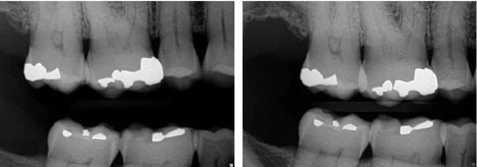
33
Bruxism and other parafunctional habits, wear, malocclusion, steep cuspal inclines or deep occlusal
34
grooves were also considered as pre-disposing factors [8-11]. Cracks can also occur in intact teeth
35
with no restorations. One study found that 28% of the longitudinal fractures occurred in teeth with
36
no restorations [12] while another study of 154 cases found that 60.4% had no restorations and a
37
further 29% had only Class I restorations. [13]
38
39
The clinical challenge is how to detect cracks in teeth? Patients may present complaining of
40
pain on chewing but not consistently. The pain may only occur when loading teeth in a certain
41
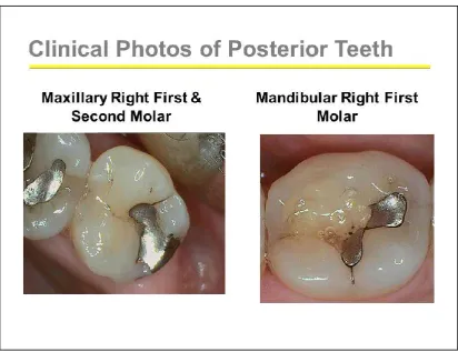
position. There may be pain on temperature change. Clinically, the teeth may look intact [2]. The
42
marginal ridges may appear stained but there are no grooves or cracks. Radiographs may not show
43
any interproximal defects or caries.
44
45
There are a number of new caries detection systems on market and they may provide some
46
additional clinical information. Since caries involves the degradation of the tooth structure in
47
response to acids released by oral bacteria one may consider using a device that can detect and
48
measure changes in the tooth structure. If the device can detect these structural changes it could be
49
used to detect and measure changes caused by parafunction or trauma which would compromise
50
the structural integrity of the tooth. The Canary System was used as one of the diagnostic tools for
51
detecting cracks in this case report. The Canary System is a laser-based caries detection system that
52
uses energy conversion technology called PTR-LUM, to image and examine teeth [14]. This case
53
study found that The Canary System could detect cracks and caries in a tooth which were not seen
54
visually or with a radiograph.
55
2. Case Report
56
This 61 year old female has been a patient in our group practice for over 20 years. During this
57
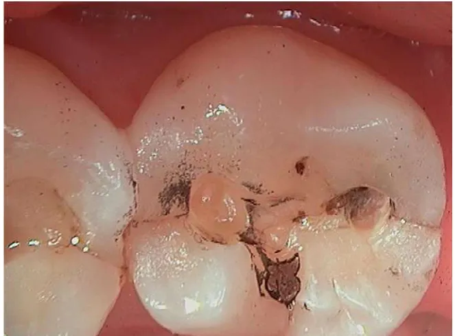
time there has been no evidence of bruxism or parafunction. Caries risk has been low and no
58
restorations have been placed or replaced on the right side of the maxilla or mandible for over ten
59
years. She had been seen on a regular basis for re-care appointments over the last twenty years.
60
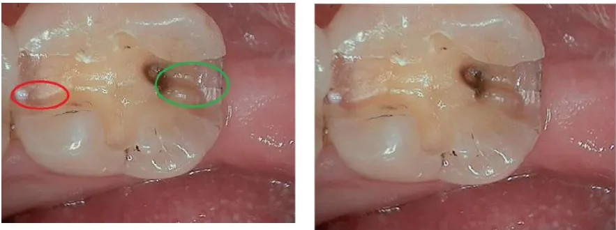
These re-care visit included visual examination, bitewing radiographs as required, scaling and
61
prophylaxis. Four months ago, the patient began to notice pain periodically on chewing on the
62
mandibular right posterior area.
63
64
Radiographs taken three months prior to pain occurring did not indicate any caries or cracks on
65
the mandibular first and second molar or on the distal surface of the mandibular right second
66
bicuspid. The maxillary first molar had a large amalgam restoration but there was no pain on hot,
67
cold, percussion or occlusal loading. The amalgam restorations on the mandibular molar teeth
68
appeared very shallow.
69
70
71
Figure 1 Bitewing Radiographs of the Right Posterior Teeth taken 3 months before pain
72
occurred
73
74
On examination, the pain on chewing or percussion, appeared to be focused on the mesial
75
portion of the mandibular second molar and distal portion of the mandibular right first molar.
76
There were no periodontal pockets greater than 3 mm. in depth and no bleeding on probing. There
77
was some mild pain on cold on the mesial contact area but no pain on cold on the distal surface of
78
the mandibular secod molar.
79
80
The image in Figure 2 shows the clinical condition of the occlusal surface of the mandibular
81
second molar. There were three shallow amalgams placed in the central area of the occlusal
82
surface. There were stained grooves on both the mesial and distal marginal ridges. There was a
83
that there was caries on the distal aspect of the tooth and visual examination did not show any open
85
lesion or staining on the distal surface.
86
87
88
Figure 2 Occlusal view of the Mandibular Right Second Molar
89
90
Visual inspection of the maxillary right first molars and mandibular right first molar did not
91
show any evidence of cracks or caries. The margins of the amalgam restorations (see Figure 3) were
92
intact. There was no pain on cold
93
94
95
Figure 3 Occlusal View of Molar Teeth on the Right Side
96
The Canary System was used to scan and examine the occlusal surfaces of the mandibular right
98
second molar. The intra-oral camera image was used to record the location of the various readings.
99
Canary Numbers above 20 indicate that the crystal structure of the tooth is not intact. The Canary
100
examination of this tooth indicated that the marginal ridges and central pit all have caries and or
101
cracks.
102
103
104
105
Figure 4: Canary Scan of the Occlusal Surface of the Mandibular Right Second Molar. The
106
Canary Scale indicates that there are defects in the crystal structure of the tooth in three locations
107
requiring treatment.
108
109
110
111
Figure 5 Initial Removal of the Amalgam shows cracks on both marginal ridges and some
112
cracks or caries in the central pit.
113
116
117
Figure 6 Image taken after removal of the amalgam and preparation of the proximal boxes.
118
There are still cracks present on the mesial and distal proximal boxes.
119
120
With the findings from The Canary System examination it was decided to remove the
121
amalgams on the mandibular second molar and examine the interior of the preparation. If the
122
cracks were minimal, we would place a bonded composite restoration and monitor the tooth. Upon
123
removal of the amalgams cracks were found on the mesial (red circle) and distal proximal boxes.
124
The crack on the distal appeared to be much more extensive (Figure 6 green circle). There was also
125
some demineralized dentin beneath the amalgam on the distal and some stain from the amalgam
126
(indicated with the green circle). On removal of the amalgam in the central pit, we noted a small
127
crack and leakage around the amalgam margin.
128
129
With the amalgam restorations removed and cracks identified, we decided to place a bonded
130
composite restoration. In our clinical experience, with the size and orientation of the cracks, this
131
should help to retain the integrity of the clinical crown and eliminate the pain on chewing and
132
temperature change..
133
134
The preparation was completed and a bonded composite restoration was placed. The
135
occlusion was adjusted on the tooth so that loading was directed over the long axis and there was
136
minimal loading on the lingual cusps. This composite restoration has been placed 9 months and the
137
patient has had no pain on temperature change or on chewing. Upon reviewing the clinical data we
138
suspected that parafunction had caused of these cracks. The patient has now agreed to wear a
139
mandibular flat plane bite splint while sleeping. We also continue to monitor the tooth as it may
140
require an onlay or full coverage restoration in the future if symptoms return or The Canary System
141
indicates a change in the integrity of the tooth or restoration margin.
142
143
4. Discussion
144
Detecting caries and cracks is a clinical challenge. Caries and cracks have similar clinical
145
outcomes: the degradation and destruction of tooth structure. At times, the symptoms may not be
146
indicative of the presence of a crack.[2] As we have seen in this case report, bitewing radiographs
147
may not be able to image small micro-fractures. Clinicians then need to find caries detection
148
devices that detect defects, cracks and caries. These devices should be able to image, record and
149
measure defects in the tooth structure.
150
151
Fluorescence is one method that is being used for detection of caries and other defects in teeth.
152
Fluorescence is simply the emission of light from an object that has absorbed light at a specific
153
wavelength. This is the core technology in SOPROLIFE (Acteon), Spectra (Air Techniques) and
154
the tooth surface. The literature indicates that the glow or fluorescence is from one or more of the
156
following clinical conditions, whether or not caries or cracks are present. [16-21]
157
bacterial porphyrins (bacterial breakdown product) [18],
158
stain,
159
tartar,
160
food debris.
161
Another concern with fluorescence is that it does not penetrate beneath the tooth surface due to
162
scattering of light from stain, plaque, organic deposits and surface features such as pits and fissures.
163
[22, 23] Studies have also demonstrated poor correlation between DIAGNOdent and other
164
fluorescence devices readings with caries lesion depth. [14, 20, 24-26] This indicates that
165
fluorescence based devices may not be able to detect or monitor cracks or defects in teeth. When a
166
crack occurs near a restoration then the fluorescence or glow from the restoration may impede the
167
ability of the device to detect any information from the enamel surface.
168
169
The Canary System uses pulses of laser light to detect caries and defects in tooth structure and
170
can measure defects and caries around and beneath restorations and through sealants. Pulses of
171
laser light are shone on the tooth and the laser light is converted to heat (Photothermal Radiometry
172
or PTR) and light (luminescence or LUM) which are emitted from the tooth surface when the laser is
173
modulated. These harmless pulses of laser light allow a clinician to examine sub-surface caries and
174
crystal structure defects, up to 5 mm. below the surface [27, 28]. Carious lesions and cracks modify
175
the thermal properties (PTR) and glow (LUM) from the healthy teeth [29]. As a lesion or crack
176
enlarges, there is a corresponding change in the signal as the heat is confined to the region with
177
crystalline disintegration (dental caries or crack); PTR increases and LUM decreases. As
178
remineralization progresses and enamel prisms begin to reform their structure, the thermal and
179
luminescence properties begin to revert back in the direction of healthy teeth [30-33]
180
181
The Canary Number (ranging from 0 – 100) is created from an algorithm combining the PTR
182
and LUM readings and is directly linked to the status of the enamel or root crystal structure [33, 34].
183
Cracks or caries will cause an increase in the Canary Number. A Canary Number of less than 20
184
indicates a healthy tooth surface. Any Canary Number above 20 indicates a defect in the tooth
185
structure [35]. In this clinical situation, the Canary Numbers were above 50, indicating large cracks
186
or extensive cracks along the marginal ridges and caries in the central pit. These cracks were found
187
upon preparation of the tooth for the restoration.
188
189
5. Conclusions
190
Detecting cracks in teeth is a clinical challenge. A good clinical history and an accurate caries
191
detection system such as The Canary System can provide the clinician with the tools needed to
192
detect cracks and caries. Once found, the cracks can be treated with the placement of direct
193
restorations or full coverage restorations depending upon the extent of the crack and other clinical
194
risk factors. One also needs to assess the cause of the cracks and how best to prevent them from
195
occurring in the future.
196
197
198
Conflicts of Interest: Dr. Stephen Abrams is the co-founder of Quantum Dental Technologies, which has
199
developed The Canary System. He has not received any compensation for this article.
200
201
References
202
203
1. Cameron C.E. Cracked-tooth syndrome. JADA, 1964;68:405-411.
204
2. Banerji, S. Mehta, S. B. Millar, B. J. Cracked tooth syndrome. Part 1: aetiology and diagnosis. Br Dent J
205
3. Hasan, S. Singh, K. Salati, N. Cracked tooth syndrome: Overview of literature. Int J Appl Basic Med Res,
207
2015, 5:164-168
208
4. Cameron C.E. The cracked tooth syndrome: additional findings. JADA, 1976;93:971–5.
209
5. Bader J.D, Martin J.A, Shugars D.A. Preliminary estimates of the incidence and consequences of tooth
210
fracture. JADA 1995;126:1650–4.
211
6. Rosen H. Cracked tooth syndrome. J Prosthet Dent 1982;47:36–43.
212
7. Bader J.D, Martin J.A, Shugars D.A. Incidence rates for complete cusp fracture. Community Dent Oral
213
Epidemiol 2001;29:346–53.
214
8. Braly B.V, Maxwell E.H. Potential for tooth fracture in restorative dentistry. J Prosthet Dent
215
1981;45:411–4.
216
9. Gher M.E Jr, Dunlap R.M, Anderson M.H, Kuhl L.V. Clinical survey of fractured teeth. JADA
217
1987;114:174–7.
218
10. Homewood CI. Cracked tooth syndrome–incidence, clinical findings and treatment. Aust Dent J
219
1998;43:217–22.
220
11. Ratcliff S, Becker IM, Quinn L. Type and incidence of cracks in posterior teeth. J Prosthet Dent
221
2001;86:168–72
222
12. Seo D-G, Young-Ah Y, et al. Analysis of Factors Associated with Cracked Teeth JOE 2012;38 (3): 288 -
223
292
224
13. Roh BD, Lee YE. Analysis of 154 cases of teeth with cracks. Dental Traumatology 2006; 22: 118–123
225
14. Abrams S.H, Sivagurunathan, K., Silvertown, J.D., Wong, B., Hellen, A., Mandelis, A., Hellen, W.M.P.,
226
Elman, G. I., Mathew, S.K., Mensinkai, P.K., Amaechi, B.T. Correlation with Caries Lesion Depth of
227
The Canary System, DIAGNOdent and ICDAS II. Open Dent J 2017;11:679-89.
228
15. Rechmann P. Featherstone JD. Caries detection using light-based diagnostic tools. Compend Contin
229
Educ Dent. 2012;33(88):582 - 93
230
16. Lussi A IS, Pitts N, Longbottom C, Reich E. Performance and reproducibility of a laser fluorescence
231
system for detection of occlusal caries in vitro. Caries Res. 1999;33(4):.261–266
232
17. Lussi A HR, Paulus R. DIAGNOdent: an optical method for caries detection. J Dent Res. 2004;83(Spec.
233
No. C):C80 - C83.
234
18. Verdonschot EH vdVM. Lasers in dentistry 2. Diagnosis of dental caries with lasers Ned Tijdschr
235
Tandheelkd. 2002;109(4):122 - 26.
236
19. König K FG, Hibst R. Laser-induced autofluorescence spectroscopy of dental caries Cell Mol Biol
237
(Noisy-le-grand). 1998;44(8):1293 - 300.
238
20. Alwas-Danowska HM PA, Suliborski S, Verdonschot EH. Reliability and validity issues of laser
239
fluorescence measurements in occlusal caries diagnosis. J Dent. 2002;30(4):129 - 34.
240
21. Ástvaldsdóttir Á, Tranæus S, Karlsson L, Peter Holbrook W. DIAGNOdent measurements of cultures
241
of selected oral bacteria and demineralized enamel. Acta Odontologica Scandinavia 2010;68(3):148-53.
242
22. Liang R WV, Marcus M, Burns P, McLaughlin P. Multimodal imaging system for dental caries
243
detection. Proceedings of SPIE Lasers In Dentistry 2007 XIII(64502).
244
23. Hall A GJ. A review of potential new diagnostic modalities for caries lesions. Dent Res.
245
2004;83(Spec No C):C89 - 94.
246
24. Khalife MA BJ, Dennison JB, Yaman P, Hamilton JC. In vivo evaluation of DIAGNOdent for the
247
quantification of occlusal dental caries. Oper Dent. 2009 34(2):136 - 41.
248
25. Jablonski-Momeni A RD, Rolfsen S, Stoll R, Heinzel-Gutenbrunner M, Stachniss V, Pieper K.
249
Performance of laser fluorescence at tooth surface and histological section. Lasers Med Sci.
250
2011;26(2):171 - 78.
251
26. Novaes TF, Moriyama CM, De Benedetto MS, et al. Performance of fluorescence-based methods for
252
detecting and quantifying smooth-surface caries lesions in primary teeth: an in vitro study. IntJ. Paed.
253
Dent. 2016;26(1):13-19.
254
27. Jeon, RJ., Phan TDT., Wu, A., Kulkarni, G., Abrams, SH., and Mandelis, A., Photothermal radiometric
255
quantitative detection of the different degrees of demineralization of dental enamel by acid etching ,
256
Proc. 13th Int. Conf. Photoacoustic & Photothermal Phenomena, July 5 – 8, 2004, J. Physique IV France,
257
28. Jeon RJ, Mandelis, A., Abrams, S. Depth profilometric case studies in caries diagnostics of human
259
teeth using modulated laser radiometry and luminescence. Rev. of Scientific Instruments
260
2003;74(1):380.
261
29. Jeon R.J., Matvienko A., Mandelis A., Abrams S.H., Amaechi B.T, Kulkarni G. Detection of
262
interproximal demineralized lesions on human teeth in vitro using frequency-domain infrared
263
photothermal radiometry and modulated luminescence , J. BioMed. Optics, 2007; 12(3); 034028 1 – 13
264
30. Matvienko, A., Jeon, R. J., Mandelis, A., Abrams, S. H., Amaechi, B. T., Photothermal Detection of
265
Incipient Dental Caries: Experiment and Modeling, Proc. of SPIE, 2007, 66759; 67590J-1 - 67590J-10
266
31. Jeon R.J., Hellen A., Matvienko A., Mandelis A., Abrams S.H., Amaechi B. T., Experimental
267
Investigation of Demineralization and Remineralization of Human Teeth Using Infrared
268
Photothermal Radiometry and Modulated Luminescence, (SPIE BiOS, San Jose, USA, January 2008),
269
Proc. SPIE Vol. 6856, 68560B (2008); DOI:10.1117/12.763807
270
32. Matvienko A., Mandelis A., Abrams S.H., Robust multi-parameter evaluation method of optical and
271
thermal properties of a layered tissue structure using photothermal radiometry. Applied Optics, 2009;
272
48(17): 3193-3204
273
33. Silvertown JD, Abrams, S. H., Sivagurunathana, K. S., Kennedy, J., Jeon, J., Mandelis, A., Hellen, A.,
274
Hellen, W., Elman, G., Ehrlich, R., Chouljian, R., Finer, Y., Amaechi, B. T., . Multi-centre clinical
275
evaluation of photothermal radiometry and luminescence correlated with international benchmarks
276
for caries detection. Open Dent. J. 2017;11:636–47.
277
34. Garcia, J., Mandelis, A., Abrams, S. H., Matvienko A., Photothermal Radiometry and Modulated
278
Luminescence: Application to Dental Caries Detection”, Chapter 71 page 1047, published
279
in "Handbook of Biophotonics, Vol. 2: Photonics for Health Care" editors: Jürgen Popp (Editor), Valery
280
V. Tuchin (Editor), Arthur Chiou (Editor), Stefan H. Heinemann (Editor) December 2011, Wiley-VCH
281
35. Abrams T, Abrams S, Sivagurunathan K, et al. Detection of Caries Around Resin-Modified Glass
282
Ionomer and Compomer Restorations Using Four Different Modalities In Vitro. Dent. J. 2018;6(3):47.