MEMBRANE ACTIVE
POLYMER A N T I C A N C E R C O N J U G A T E S
A thesis submitted to the University of London in partial fulfilment of the requirements for the degree of Doctor of Philosophy
RUTH NYOKABI MUSILA
B.PHARM ( H O N S )
2003
All rights reserved
INFORMATION TO ALL USERS
The quality of this reproduction is dependent upon the quality of the copy submitted.
In the unlikely event that the author did not send a complete manuscript and there are missing pages, these will be noted. Also, if material had to be removed,
a note will indicate the deletion.
uest.
ProQuest 10104807
Published by ProQuest LLC(2016). Copyright of the Dissertation is held by the Author.
All rights reserved.
This work is protected against unauthorized copying under Title 17, United States Code. Microform Edition © ProQuest LLC.
ProQuest LLC
789 East Eisenhower Parkway P.O. Box 1346
F
o r
C
h a n g
’
First and foremost thanks to God for all the opportunities I have been blessed with.
I would like to thank my supervisor, Professor Ruth Duncan for introducing me to
the field of drug delivery and her patience and support over the last few years. Thanks
are also due to my second supervisor, Dr Steve Brocchini and Professor Helmut
Ringsdorf for the numerous discussions, both in and out of work. Many thanks to Dr
Francis Searle for her helpful comments and contribution to this thesis.
Many thanks to The University of London for funding this Ph.D. (The ORS Award
and The Triangle Trust Postgraduate Studentship).
I would like to acknowledge collaborators for their contribution to various parts of
this piece of work: Dr Andreas Kortenkamp and soon-to-be-Dr Naashika Quarcoo
(Centre for Toxicology, The School of Pharmacy) and Drs Jose Luis Fernandes and
Antonio Fernandes (Institute Biomar, Spain). Thanks to Dr Harmesh Aojula and
Professor David Clarke (School of Pharmacy, University of Manchester) for helpful
discussions. Thanks are also due to Dr Kathy Taylor (Tenovus Centre for Cancer
Research) for her input in the flow cytometry work.
I am very grateful to all my friends for their continuous support. Special thanks to
two very special people: Chang’-Toek Koech, for having more faith in me than I do in
myself and Sharmila (Dr Chauhan); I can’t thank you both enough. Thanks also to
Juice. Thanks to my friends and colleagues in CPT, old and new, both in London and
Cardiff, for making it all the more worthwhile to come in to the lab everyday: Myrto
(especially for the last few months), Nicky (thanks for the chats), Sam (proof-reader of
this thesis), Damian, Keith, Ryan-Take-It-To-The-Max for help with the chemistry,
Richard, Dale (is there anything you don’t know??) and Maria for help with the NMR.
Thanks are also due to Rosy.
Last and by no means least, thanks to my family for their consistent guidance and
support in so many ways, emotionally and financially. I am blessed to have parents
who have provided me with nothing but the best and taught me that the sky is not the
Abstract
HPMA copoly mer-dru g anticancer conjugates have been successfully
transferred into clinical evaluation. Such macromolecular constructs selectively
accumulate within solid tumours by the enhanced permeability and retention (EPR)
effect. However, lysosomotropically activated compounds such as HPMA copolymer-
doxorubicin (PK l) present limitations in that they require enzymatic activation within
lysosomes in order to release the active moiety which in turn exerts its cytotoxic effect.
Hence this study looked at the development of a second generation, membrane
active polymeric anticancer conjugates. Using melittin (MLT) as a model peptide,
HPMA copolymer-MLT conjugates were synthesised and characterised using standard
biochemical techniques (SDS PAGE, BCA protein assay and FPLC). The effect of
MLT content using conjugates with high (38.9 ± 2.5 % w/w), medium (25.8 ± 6.2 %
w/w) and low (12.1 ± 8 . 6 % w/w) MLT loading and the effect of peptidyl linker (-Gly-
Gly- (GG) and -Gly-Phe-Leu-Gly- (GFLG)) were investigated in vitro. The MLT
conjugate with medium loading and GG spacer showed reduced haemolytic activity
(Hbso 19.9 ± 6.2 /xg/ml (p < 0.05)) in a rat red blood cell lysis model, and maintained
cytotoxic activity against a B16F10 murine melanoma cell line (IC50 7.3 ± 1.5 /tg/ml; p
= 0.46 (NS)) relative to free MLT. This conjugate was chosen to proceed with
preliminary in vivo studies. The MTD of HPMA copolymer-MLT was found to be 4-
fold greater than that of free MLT (10 mg/kg MLT-equivalent) and body distribution of
MLT conjugate 3 mg/kg (MLT-equivalent) (i.p.) showed 3 - 4-fold increased
circulation time. However, no improved tumour targeting by the EPR effect was
established 4 h after i.p. administration. Disappointingly, no antitumour activity was
observed in vivo following i.p. or i.v. administration. Nevertheless, improved synthetic
procedures to prepare more compounds of this new class of anticancer agents is
warranted.
Page Number
T h esis Title... i
Dedication... ii
A cknow ledgm ents... iii
A b stract... iv
T hesis Index... v
List of Figures...yiii List of T ables... xiii
Abbreviations...xv
C hapter 1: G eneral Introduction...1
1.1 Cancer and its Treatment... .2
1.2 Tumour Targeting... .3
1.3 The EPR Effect for Passive Tumour Targeting...8
1.3.1 Vectors that Utilise EPR for Tumour Targeting... 10
1.4 Endocytic Capture of Polymer Therapeutics...11
1.5 Polymer Therapeutics... 18
1.6 Membrane Active Agents in Cancer Chemotherapy...27
1.6.1 Eukaryotic and Tumour Cell Membranes 30 1.6.2 MLT... 33
1.7 Aims of This Thesis... 40
C hapter 2: M aterials and M ethods...43
2.1 Materials... 44
2.2 Equipm ent... .45
2.3 M ethods... .47
2.3.1 Analytical Techniques...47
2.3.1.1 Ninhydrin Assay for Amine Quantification...47
2.3.1.2 Bicinchoninic Acid (BCA) Assay...49
2.3.1.3 Sodium Dodecyl Sulphate Polyacrylamide G e l... .49
2.4 Cell Culture... 59
2.4.1. Cell Culture: Recovery from Cell Bank... .60
2.4.2 Evaluation of Cell Viability using Trypan Blue Exclusion...60
2.4.3 Evaluation of Cell Viability using the Colourimetric...61
Tetrazolium-based (MTT) Assay 2.5 The Rat Red Blood Cell Lysis Assay... .63
2.6 Scanning Electron Microscopy...65
2.7 Radioiodination of MLT and HPMA Copolymer-MLT using th e ...67
Bolton and Hunter Reagent 2.7.1 Determination of Labelling Efficiency of '^^I-labelled M L T ...69
and '^^I-labelled HPMA copolymer-MLT using Paper Electrophoresis 2.8 In Vivo Studies...69
2.8.1 Evaluation of Body Distribution of *^I-labelled HPMA...72
Copolymer-MLT 2.8.2 Evaluation of Antitumour Activity of HPMA Copolymer...73
-MLT 2.9 Data Analysis...73
C hapter 3: S ynthesis and Characterisation of HPMA Copolymer-...74
MLT C onjugates 3.1 Introduction...75
3.1.1 Polymer Characterisation... 83
3.2 Methods...8 6 3.2.1 Synthesis of HPMA Copolymer-MLT Conjugates...8 6 3.2.2 Purification of HPMA Copolymer-MLT Conjugates... 91
3.2.3 Characterisation of HPMA Copolymer-MLT Conjugates... .92
3.2.3.1 Purity and Mw Assessment using SDS PAGE and FPLC...94
3.2.3.2 Determination of the Reaction Yield and MLT L oading... .94
using the BCA Assay 3.3 Results...94
3.3.1 Preparation of HPMA Copolymer-MLT Conjugates... .94
3.3.2 Characterisation of HPMA Copolymer-MLT Conjugates... .96
3.4 Discussion 104
In Vitro
4.1 Introduction... 117
4.2 Methods... 120
4.3 Results... 121
4.3.1 Haemolytic Activity of MLT and HPMA Copolymer-MLT... 121
Conjugates 4.3.2 Cytotoxicity of MLT and HPMA Copolymer-MLT towards... 125
B16F10 Cells 4.4 Discussion... 131
Chapter 5: In Vivo Pharmacokinetics and Pharmacology of HPMA...139
Copolymer-MLT 5.1 Introduction... 140
5.2 Methods... 142
5.2.1 Evaluation of Body Distribution of '^^I-Labelled MLT and... 142
‘“ l-Labelled HPMA Copolymer-MLT 5.2.2 Evaluation of Antitumour Activity of MLT and HPMA... 144
Copolymer-MLT 5.3 Results... 147
5.3.1 Body Distribution of ^^^I-Labelled MLT and HPMA...147
Copolymer-MLT 5.3.2 Anti tumour Activity of MLT and HPMA Copolymer-MLT...159
5.4 Discussion... 179
Chapter 6: General Discussion... 185
References...194
Appendix I; Haemolysis and Cytotoxicity of Membrane Active Agents... 241
List of Figures
Figure 1.1 Summary of tumour targeting strategies
Figure 1.2 Illustration of the Enhanced Permeability and Retention (EPR)
Effect (Adapted from Takakura et al, 1998)
Figure 1.3 Illustration of a solid tumour mass pressure and concentration gradients after administration of a macromolecular therapeutic
Figure 1.4 Illustration of examples of polymer therapeutics: (A) polymeric drug with intrinsic activity; (B) polymer-drug conjugate; (C)
polymer-protein conjugate and (D) polymeric micelle (Adapted
from Brocchini & Duncan, 1999)
Illustration depicting the “Ringsdorf model”
Radical copolymerisation of HPMA with p-nitrophenol esters of
A-methacroylolated oligopeptide (From Kopecek et al, 1985)
Illustration of the different modes of action of lysosomotropic
polym er-drug conjugates and polym er-m em brane active
conjugates
European honeybee {Apis melijfera) venom constituents
MLT amino acid sequence
Illustration of proposed mode of lytic action of amphipathic a -
helical membrane active peptides (Adapted from Shai, 1999)
Schematic of MLT a-helical wheel (Adapted from Dempsey,
1990)
General MLT thermodynamic cycle (From Ladokhin & White,
1999)
MLT-membrane interaction (From La Rocca et al, 1999)
Snap shot of 4 MLT a-h elices (blue) in bilayer pore, with
phospholipid head groups in yellow and water molecules in blue
from top view (A) and side view (B) (From Ladokhin & White,
1999)
Figure 1.14 Illustration of the desired HPMA copolymer-MLT conjugate
Figure 2.1 Scheme of the ninhydrin reaction
Figure 2.2 Typical ninhydrin assay calibration curve using 3-am ino-l-propanol standard. Data represents mean ± SD (n = 3)
Figure 2.3 Illustration showing the mechanism of protein-mediated reduction
Figure 1.5 Figure 1.6 Figure 1.7 Figure 1.8 Figure 1.9 Figure 1.10 Figure 1.11
Figure 1.12 (a)
Figure 1.12 (b) Figure 1.13
1) (From Smith et al, 1985)
Figure 2.4 Typical MLT BCA calibration curve. Data represents mean ± SD (n = 16)
Figure 2.5 (a) Gel holder and (b) Gel electrophoresis kit (Bio Rad, UK)
Figure 2.6 Scheme showing mitochondrial MTT reduction to its insoluble formazan salt
Figure 2.7 B16F10 growth curve. Data represents mean ± SD (n = 6)
Figure 2.8 Illustration of diiodination of Bolton and Hunter reagent at its phenolic ring and reaction with an amine-containing molecule
(MLT) to form an amide bond
Figure 2.9 Void volume determination of a PD-10 column with blue dextran. Data represents mean ± SD (n = 3)
Figure 2.10 Typical elution profile of MLT (1 mg/ml) with 0.9% NaCl in a PD-10 column. Data represents mean ± SD (n = 4)
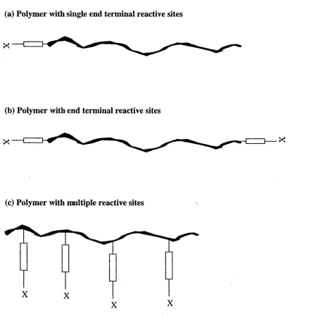
Figure 3.1 Illustration of (a) monovalent, (b) bivalent and (c) multivalent reactive polymers
Figure 3.2 Scheme to show aminolysis of HPMA copolymer precursor by MLT primary amine groups
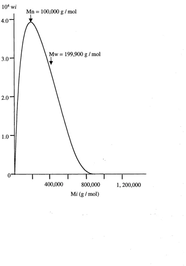
Figure 3.3 A typical polymeric molar mass distribution curve (From Young & Lovell, 1991)
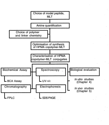
Figure 3.4 Flow chart summarising synthesis and characterisation of HPMA copolymer-MLT conjugates
Figure 3.5 Illustration of nucleophilic attack on the ONp ester of HPMA copolymer precursor
Figure 3.6 Summary of HPMA copolymer-MLT conjugation methods
Figure 3.7 Calibration of FPLC column with gel filtration standards (5 mg/ml)
Figure 3.8 UV-vis spectrum of a HPMA copolymer-MLT reaction (a) in aqueous solvent (RM 1) and (b) in DMSO (RM 6.3)
Figure 3.9 MLT UV-vis spectrum
Figure 3.10 UV-vis spectrum of ultrafiltrate of HPMA copolymer-MLT (RM 5.3) reaction mixture showing purificiation of MLT conjugate with
time
and (b) 0.3 to 5 mg/ml
Figure 3.12 FPLC elution of aminolysed HPMA copolymers (5 mg/ml)
Figure 3.13 FPLC elution of (a) unpurified HPMA copolymer-MLT (RM 3.14;
GG spacer and RM 7.1; GFLG spacer) and (b) purified HPMA
copolymer-GG-MLT (RM 3.14)
Figure 3.14 SDS PAGE (14 % Tris-HCl) gel to show; Lane (1) MLT conjugate
(RM 3), (2) Free MLT, (3) Aminolysed HPMA copolymer
precursor, (4) MWM
Figure 3.15 Calibration curve for the BCA protein assay using either free MLT
or aminolysed HPMA copolymer precursor standards. Data
represents mean ± SD (n = 4)
Figure 3.16 Illustration of the theoretical structures of HPMA copolymer-MLT
Figure 4.1 Structures o f (a) PEI and (b) dextran
Figure 4.2 Haemolytic activity o f (a) MLT and (b) HPMA copolymer-MLT
(RM 3.2) after 1 h incubation. Data represents mean ± SE (n = 48
and n = 12 respectively)
Figure 4.3 Effect of MLT content of HPMA copolymer-MLT conjugates on
haemolytic activity (10 min). Data represents mean ± SE (n = 12)
Figure 4.4 Effect of pH on haemolytic activity of MLT after 1 h incubation.
Data represents mean ± SE (n = 4)
Figure 4.5 SEM pictures of RBCs exposed to (A) control, (B) aminolysed
HPMA copolymer, (C) HPMA copolymer-MLT (RM 5.3), (D)
HPMA copolymer- MLT (RM 3.13), (E) HPMA copolymer-MLT
(RM 6.3) and (F) free MLT at their IC50 values for 10 min (X
4,000)
Figure 4.6 Cytotoxicity of HPMA copolymer-MLT (RM 3.2) against B16F10
cells after 72 h incubation. Data represents mean ± SE (n = 12)
Figure 4.7 Effect of MLT content of HPMA copolymer-MLT conjugates on
cytotoxicity against B16F10 cells after 72 h incubation. Data
represents mean ± SE (n = 18), except RM 3.2 (n = 12)
Figure 4.8 SEM pictures of B16F10 cells exposed to (A) control, (B)
aminolysed HPMA copolymer, (C) HPMA copolymer-MLT (RM
5.3), (D) HPMA copolymer-MLT (RM 3.13), (E) HPMA
copolymer-MLT (RM 6.3) and (F) free MLT at their I C 5 0 values
multidose i.p. and (c) multidose i.v.
Figure 5.2 Paper electrophoresis of (a) Bolton and Hunter diiodo reagent (b) the ^^^I-labelled MLT reaction mixture and purified product and (c)
the ‘^^I-labelled HPMA copolymer-MLT (RM 3.4) reaction
mixture and purified product
Figure 5.3 Biodistribution of (a) ^^^I-labelled MLT and (b) ^^^I-labelled HPMA copolymer-MLT (RM 3.4) administered as a single dose
(i.p.). Data represents mean ± SE (n=3)
Figure 5.4 Radioactivity in the blood after administration of ^^^I-labelled MLT or HPMA copolymer-MLT (RM 3.4). Data represents mean
± SEM (n=3). p value *** < 0.001; = not significant
Figure 5.5 Radioactivity in the kidneys after administration of ‘^^I-labelled MLT or HPMA copolymer-MLT (RM 3.4). Data represents mean
± SEM (n=3). p value ** < 0.005; = not significant
Figure 5.6 Radioactivity in the heart after administration of '^^I-labelled MLT or HPMA copolymer-MLT (RM 3.4). Data represents mean ± SE
(n=3). p value *** < 0.001; ** < 0.005; = not significant
Figure 5.7 Radioactivity in the liver after administration of '^I-labelled MLT or HPMA copolymer-MLT (RM 3.4). Data represents mean ± SE
(n=3). p value * < 0.05; = not significant
Figure 5.8 Radioactivity in the lungs after administration of ‘^^I-labelled MLT or HPMA copolymer-MLT (RM 3.4). Data represents mean ± SE
(n=3). p value * < 0.05; = not significant
Figure 5.9 Radioactivity in the spleen after administration of ‘^^I-labelled MLT or HPMA copolymer-MLT (RM 3.4). Data represents mean
± SE (n=3). p value * < 0.05; = not significant
Figure 5.10 Radioactivity in the thyroid after administration of '^^I-labelled MLT or HPMA copolymer-MLT (RM 3.4). Data represents mean
± SE (n=3). p value = not significant
Figure 5.11 Radioactivity in the tumour after administration of ‘^^I-labelled MLT or HPMA copolymer-MLT (RM 3.4). Data represents mean
± SE (n=3). p value = not significant
administration. Data represents mean ± SE (n = 3)
Figure 5.13 Biodistribution of *^^I-labelled MLT and HPMA copolymer-MLT 4 h following administration (i.p.) of 3 mg/kg MLT-equi valent.
Data represents mean ± SE (n = 3). p value * < 0.05; ** < 0.005;
= not significant
Figure 5.14 Effect of (a) MLT and (b) HPMA copolymer-MLT (RM 3.4) on animal weight after single dose i.p. (n = 2)
Figure 5.15 Effect of a single dose (i.p.) of HPMA copolymer-MLT (RM 3.7)
(10 mg/kg) and MLT (2.5 mg/kg) on s.c. B16F10 tumour size.
Data represents mean ± SD (n = 5 unless otherwise stated), p value
* < 0.05
Figure 5.16 Effect of MLT (2.5 mg/kg); (b) HPMA copolymer-MLT (RM 3.7) (10 mg/kg) and (c) control (i.p.) on animal weight. In each case the
weight change of each mouse in the group is shown
Figure 5.17 Effect of multiple doses (i.p.) of (a) MLT or (b) HPMA copolymer-MLT (RM 3.9) administered on days 0, 2 and 4 on s.c.
B16F10 tumour size. Data represents mean ± SE (n = 5)
Figure 5.18 Effect of MLT (a) 0.1 mg/kg; (b) 0.5 mg/kg or (c) 1 mg/kg (i.p.) on animal weight. In each case the weight change of each mouse
in the group is shown
Figure 5.19 Effect of HPMA copolymer-MLT (RM 3.9) (a) 0.5 mg/kg; (b) I mg/kg or (c) 2.5 mg/kg (i.p.) on animal weight. In each case the
weight change of each mouse in the group is shown
Figure 5.20 Effect of control (i.p.) on animal weight. In each case the weight change of each mouse in the group is shown
Figure 5.21 Effect of multiple doses (i.v.) of HPMA copolymer-MLT (RM 3.14) administered on days 0 and 2 on s.c. B16F10 tumour size.
Data represents mean ± SE (n = 5 unless otherwise stated)
Figure 5.22 Effect of HPMA copolymer-MLT (a) 0 .1 mg/kg; (b) 0.5 mg/kg; (c) 1 mg/kg or (d) 2 mg/kg (i.v.) on animal weight. In each case
the weight change of each mouse in the group is shown
Figure 5.23 Effect of control (i.v) on animal weight. In each case the weight change of each mouse in the group is shown
Figure 5.24 Schematic illustration of dose-response relationships of a drug (Adapted from Gloff & Benet, 1990)
List of Tables
Table 1.1
Table 1.2
Table 1.3
Table 1.4 Table 2.1 Table 2.2 Table 2.3 Table 3.1 Table 3.2 Table 3.3 Table 3.4 Table 3.5 Table 3.6 Table 3.7 Table 4.1 Table 4.2
Advantages and disadvantages of DDS used for passive tumour-
targeting by the EPR effect
Lysosomal enzymes (From Barrett & Heath, 1977 and Duncan,
1986)
Representation of examples of soluble polymeric vehicles for
parenteral drug delivery in cancer therapy
Representative cytolytic peptides (From Dathe & Wieprecht,
1999)
Batch analysis of HPMA copolymer precursors (Provided by
Polymer Laboratories Ltd, UK)
ÀKTA FPLC System (Amersham Pharmacia Biotech) used to
characterise and purify HPMA copolymer-MLT conjugates
Data to show elution profile of MLT in a PD-10 column
Reactive amino acids and their sub-classifications (Boxed amino
acids are specific to MLT)
Examples of polymer-peptide and polymer-protein conjugates that
have been studied as therapeutics
Summary of optimisation of HPMA copolymer-GG-MLT (5 mol
%) conjugation
Estimation of MLT molecular weight after FPLC elution
Maximum theoretical loading of MLT onto HPMA copolymer
intermediates
Summary of the characteristics of all HPMA copolymer-MLT
conjugates synthesised
Summary of the characteristics of all HPMA copolymer-MLT
conjugates that were subsequently used in biological experiments
Haemolysis caused by MLT and HPMA copolymer-M LT
conjugates of different MLT content. The data represents mean ±
SD (n = 18) except RM 3.2 (n = 12)
Cytotoxicity of MLT and HPMA copolymer-MLT conjugates
against B16F10 cells. The data represents mean ± SD (n = 18)
Table 4.3
Table 5.1
Table 5.2
Table 5.3
Table 5.4
Table 5.5
Table 5.6
Summary of effect of MLT and HPMA copolymer-MLT on
haemolysis of RBC after incubation for 10 min or 1 h and
cytotoxicity against B16F10 cells after 72 h
Summary of the protocols and characteristics of test compounds
used in the biodistribution studies
Total radioactivity recovery following administration (i.p.) of
labelled MLT and ’^^I-labelled HPMA copolymer-MLT (RM 3.4)
to tumour bearing mice (n = 3). Data represents mean ± SE
Total radioactivity recovery at 4 h following administration (i.p.)
of labelled MLT and ^^T-HPMA copolymer-MLT (RM 3.4
“spiked” with RM 3.9) to tumour-bearing mice (n = 3). Data
represents mean ± SE
Antitumour activity of MLT and HPMA copolymer-MLT (RM
3,9) against s.c. B16F10 after administration by single dose (i.p.).
p value = not significant
Antitumour activity of MLT and HPMA copolymer-MLT (RM
3.7) against s.c. B16F10 after administration by multiple doses
(i.p.). p value = not significant
Antitumour activity of MLT and HPMA copolymer-MLT (RM
3.14) against s.c. B16F10 after administration by multiple doses
(i.v.). p value = not significant
Acquired Immunodeficiency Syndrome AIDS
Alpha ot
Antibody Directed Enzyme Prodrug Therapy ADEPT
Bicinchoninic Acid BCA
Bovine Serum Albumin BSA
Becquerel Bq
Beta P
Carmustine BCNU
Circular Dichroism CD
Counts Per Minute CPM
Curie Ci
Daltons Da
Deoxyribonucleic Acid DNA
3-(4,5-Dimethylthiazol-2-yl)-2,5-diphenyltetrazolium bromide MTT
Divinylether-Maleic Anhydride Copolymer DIVEMA
Dimethyl Formamide DMF
Dimethyl Sulphoxide DMSO
Doxorubicin DOX
Drug Delivery System DDS
Elution Volume Vg
Enhanced Permeability and Retention Effect EPR
Epidermal Growth Factor EOF
Epsilon e
Ethylenedi amine te traacetic acid EDTA
Fast Performance Liquid Chromatography FPLC
Foetal Bovine Serum FBS
Food and Drug Administration FDA
Fourier Transform Infrared Spectroscopy FTIR
Forward Scatter FSC
Galactosylated V-(2-Hydroxypropyl)methacrylamide copolymer- PK2
Doxorubicin
Gamma Y
Gene Directed Enzyme Prodrug Therapy GDEPT
Haemoglobin Hb
50 % Haemolysis Concentration Hbgo
Hanks Balanced Salt Solution HBSS
Honey Bee Venom HBV
Human Immunodeficiency Virus HIV
Human Serum Albumin HSA
V-(2-Hydroxypropyl)methacrylamide HPMA
V-(2-Hydroxypropyl)methacrylamide Copolymer-Doxorubicin PK l
V-Hydroxy succinimidyl NHS
50 % Inhibitory Concentration 1^50
Interferon IFN
Interleukin IL
Intraperitoneal i.p.
Intravenous i.v.
Maximum Tolerated Dose MTD
Molar Mass M
Melanocyte Stimulating Hormone MSH
Melittin MLT
Minimum Inhibitory Concentration MIC
Mononuclear Phagocyte System MPS
Multiple Drug Resistance MDR
p-Nitroanilide NAp
p-Nitrophenyl ONp
Not available NA
Not determined ND
Nuclear Magentic Resonance NMR
Number No.
Number Average Molar Mass Mn
Oriented Circular Dichroism OCD
Phosphate Buffered Saline PBS
Phosphatidylseiine PS
Poly(ethylene glycol) PEG
P-glycoprotein PGP
Poly(ethylenimine) PEI
Red Blood Cells
Reticulo-Endothelial System
Reverse Phase High Performance Liquid Chromatography
Room Temperature
Scanning Electron Microscopy
Sephadex G25
Size Exclusion Chromatography
Sodium Chloride
Sodium Dodecyl Sulphate
Sodium Dodecyl Sulphate Polyacrylamide Gel Electrophoresis
Sodium Hydroxide
Standard Deviation of The Mean
Standard Error of The Mean
Subcutaneous
N-Succinimidyl 3-(4-hydroxy-5-'^^I-di-iodophenyl) propionate
Sum
Superoxide Dismutase
N,N,N’ ,N’ -Tetramethylethylenediamine
Tris(hydroxymethyl)aminomethane
United Kingdom Co-ordinating Committee on Cancer Research
UV-visible
Void Volume
Volume for Volume
Viral Directed Enzyme Prodrug Therapy
Weight Average Molar Mass
Weight for Weight
RBC RES RP-HPLC RT SEM PD-10 SEC NaCl SDS SDS PAGE NaOH SD SE s.c. Bolton and Hunter reagent S SOD TEMED Tris UKCCCR UV-vis Vo v/v VDEPT Mw w/w
Amino Acid Abbreviations
Glycine Gly G
Histidine His H
Isoleucine He I
Lysine Lys K
Leucine Leu L
Methionine Met M
Asparagine Asn N
Proline Pro P
Glutamine Gin Q
Arginine Arg R
Serine Ser S
Threonine Thr T
Valine Val V
Tryptophan Trp W
Tyrosine Tyr Y
Chapter One
Chapter 1 General Introduction
1.1 Cancer and its Treatment
Cancer is a broad term which encompasses a vast range of diseases whereby
uncontrolled cell proliferation is the common factor (King, 1996). Cancer is the prime
cause of mortality in the UK (Mason, 1998). Globally, at present, 10 million people are
diagnosed with cancer each year and it is predicted this figure will double by the year
2020 (Sikora, 1999). It is hoped that the determination of the human genome sequence
will provide a greater understanding of health and disease, including the molecular
basis of cancer, and thus enable the development of appropriate medicines and improve
prognosis.
With the currently available treatments, on average an approximately 35%
“cure” (defined as long term disease-free survival (reviewed by Frei, 1985)) rate is
achieved (Purves, 1996). The means of combating neoplasia include (1) surgical
resection of tumours, (2) cytotoxic drug treatment, (3) biological response modifiers
(immunotherapy), (4) hormonal therapy and (5) irradiation. These treatments are often
given alone or in combination, but even so, are poorly effective. The major draw back
of successful treatment by chemotherapy is the selection of tumour cells which possess
an acquired or intrinsic resistance to anti-cancer drugs (King, 1996 and Hoffmann et al,
1997). The non-selective ubiquitous distribution of cytotoxic compounds equivocally
damages rapidly proliferating normal cells as well as neoplasms. This results in
haematological side effects e.g. myelosuppresion and consequent neutropenia and
thrombocytopenia. Non-haematological side effects include damage of the gastro
intestinal epithelium and hair follicles resulting in emesis and alopecia respectively
(Rang et at, 1995).
Consequently, a lot of anticancer research has progressed towards identifying
new targets, designing novel chemical entities or improvement of existing therapeutics
by the development of drug delivery systems (DDS). In the case of DDS, their design
seeks to achieve manipulation of the biodistribution of the active drug in order to
achieve selective tumour cell kill. The concept of selective drug delivery is the basis of
this thesis.
Polymer therapeutics are a class of DDS which have been developed over the
last two decades (section 1.5). At present, 8 polymer-drug conjugates are in Phase 1/11
intralysosomally to release the active drug (section 1.4.1). The primary aim of this
study was to develop (synthesise, physico-chemically and biologically characterise)
(Chapters 3, 4 and 5) the first generation of polymer-peptide (melittin, MLT)
conjugates which are active at the level of the tumour cell membrane. The secondary
aim of this thesis was to improve the understanding of the mode of action of the
N-(2-hydroxypropyl)methacrylamide (HPMA) copolymer-doxorubicin construct (PKl, FCE
28068) (Chapter 6) which is presently in Phase II clinical trials. In this introduction, the
general concepts of tumour targeting, polymer therapeutics and membrane active
agents will be reviewed.
1.2 Tumour Targeting
Drug targeting is defined as the selective delivery of active moieties to desired
pharmacological sites. Targeted drug delivery to the tumour site offers potential
advantages over conventional therapy. These include:
• Increased bioavailabilty of active agent due to prevention of premature metabolism
and excretion
• Supressed accumulation in normal tissues and thus reduction in side effects which
improves quality of life of the patient
• Decreased frequency of administration and thus improved patient compliance
• Change of route of cellular uptake, hence by-passing cell resistance mechanisms.
In principle, drug targeting may be achieved by 2 mechanisms: site-specific activity
and site-specific delivery (summarised in Figure 1.1). These will be discussed below.
Site-specific activity of the agent only at the target site is enabled by local
administration. This includes depot preparations such as polyanhydride matrix
incorporating carmustine (BCNU) (Gliadel® wafer) for glioma (Brem et al, 1995) and
direct administration e.g. intra-hepatic poly(styrene-co-maleic anhydride)-modified
neocarzinostatin (SMANCS) with lipiodol for hepatoma (Kanematsu et al, 1989 and
Konno, 1992).
Site-specific delivery of the active agent to the target site may be classified as
active and passive targeting (reviewed by Tomlinson, 1987 and Langer, 1998).
Examples of delivery systems which capitalise on selectivity following systemic
meso-Chapter 1 General Introduction
F ig u re l.l Summary of tumour targeting strategies
Site-specific activity i.e. agent active at target site
Tumour targeting
Systemic delivery Local delivery
chlorin e6 (Peterson et al, 1996), pulsatile drug delivery from polymer matrices by an
external magnetic field, ultrasound, electric current or pH (reviewed by Langer, 1998)
and prodrug therapy e.g. antibody directed enzyme prodrug therapy (ADEPT)
(Bagshawe, 1989 and Knox & Connors, 1995), gene directed enzyme prodrug therapy
(GDEPT) and viral directed enzyme prodrug therapy (VDEPT) (Ram, 1999).
Active Targeting
Active targeting (Ehrlich's “magic bullet”) (Ehrlich, 1906) involves recognition
and localisation of a ligand at the surface of a desired cell and may be defined at three
levels; first order (organ-specific), second order (cell-specific) and third order
(organelle-specific) (Duncan, 1992; Cassidy et al, 1993; Davis & Ilium, 1994; Vyas et
al, 2001). Targeting moieties explored so far for site-specific delivery of anticancer
drugs include:
1) Proteins and peptides, such as:
1) Antibodies e.g. the anti-CD33 calicheamicin immunoconjugate, gemtuzumab
ozogamicin (Mylotarg®) for CD33^ acute myeloid leukaemia (Sievers et al,
1999); the recently FDA-approved engineered (humanised) cancer therapeutic
monoclonal antibodies e.g. anti-HER2 trastuzumab (Herceptin®) for HER2^
breast cancer (Molina et al, 2001)
ii) Hormones e.g. melanocyte stimulating hormone (MSH)-targeted HPMA
copolymer-doxorubicin designed for the treatment of melanoma (O’Hare et al,
1993)
iii) Growth factors e.g. epidermal growth factor (EGF)-targeted dextran conjugates
(Andersson et al, 1991) for certain gliomas, melanomas and squamous
carcinomas
iv) Bioconjugate receptor-mediated uptake via serum proteins e.g. transferrin,
lipoproteins (reviewed by Kratz & Beyer, 1998)
2 ) Carbohydrates e.g. galactosylated HPMA copolymer-doxorubicin (PK2, FCE
28069) (Duncan et al, 1983a; Kerr et al, 1998; Julyan et al, 1999) for hepatoma via
asialoglycoprotein receptors (Poznansky & Julyan, 1984).
In practice, however, efficient and specific receptor-mediated targeting of therapeutic
concentrations of anticancer agent to the tumour site still proves to be a challenge due
Chapter 1 General Introduction
receptor, poor homogeneity of receptor within a tumour and level of expression of the
receptor in normal tissue (reviewed by Rihova, 1998 and Chari, 1998).
Passive targeting
The limitations of active targeting outlined above stimulated an interest in an
alternative methodology of tumour targeting. Colloidal and macromolecular carrier
systems have been utilised to target passively low molecular weight anticancer agents
to tumours. Passive targeting is therefore fundamental to the concept of polymer
therapeutics (defined in section 1.5). The mechanisms that cause passive targeting are
generally either: (1) selective extravasation of particles through hyperpermeable
capillaries (Matsumura & Maeda, 1986) or (2) reticulo-endothelial / mononuclear
phagocyte system (RES / MPS) selective uptake (Davis & Ilium, 1994). In contrast to
active targeting, passive targeting results from a combination of the physicochemical
characteristics of the carrier and the pathophysiological properties of the body, or more
specifically of the diseased tissue e.g. the increased permeability of tumour vasculature
which causes accumulation of the the carrier (agent) at a specific target site (reviewed
by Yokoyama & Okano, 1996 and Maeda et al, 2000).
The carrier properties which allow avoidance of RES uptake and thus provide
the prolonged plasma half-life necessary for tumour targeting include small particle
size. Particles of 100 - 200 nm diameter often escape uptake by the liver and spleen
(reviewed by Brannon-Peppas, 1995), whereas microparticles > Ip m in diameter are
rapidly filtered out from the lung capillary bed (Davis & Ilium, 1986). Particles < 7/xm
in diameter are usually captured by the RES (Bellanti, 1985). Those polymers which
are not captured by the RES but have a molecular weight < 40 kDa (the approximate
renal threshold) (Seymour et al, 1995) usually display renal clearance by glomerular
filtration. The hydrophobicity and charge of water-soluble polymers also influence
circulation time and RES localisation (reviewed by Takakura & Hashida, 1996 and
Monfardi & Veronese, 1998).
Longer circulating polymer-anticancer conjugates exploit the hyperpermeability
of solid tumour vasculature to allow tumour-specific targeting. This process has been
termed as the Enhanced Permeability and Retention (EPR) effect (Matsumura &
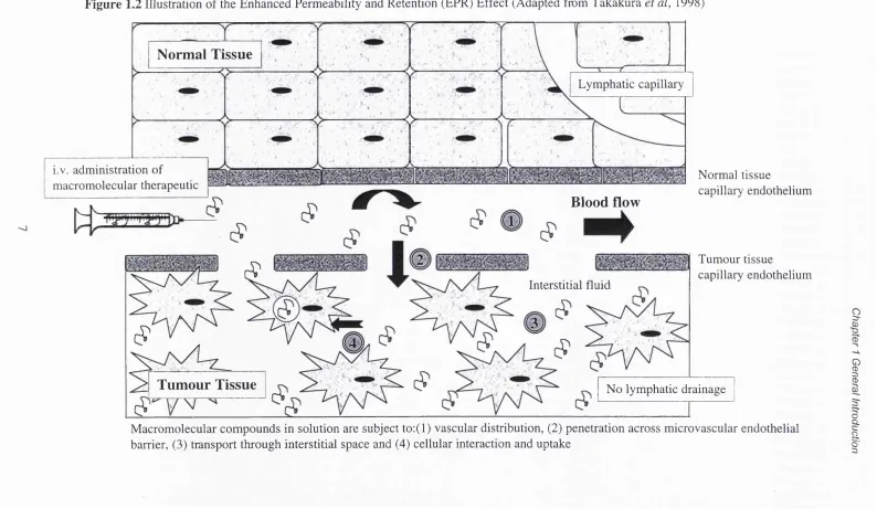
i.v. administration of macromolecular therapeutic
N orm al T issu e
Lymphatic capillary
B lood flow
Interstitial fluid
T u m o u r T issu e No lymphatic drainage j
Normal tissue
capillary endothelium
Tumour tissue capillary endothelium
Macromolecular compounds in solution are subject to:(l) vascular distribution, (2) penetration across microvascular endothelial barrier, (3) transport through interstitial space and (4) cellular interaction and uptake
I
CDChapter 1 General Introduction
1.3 The EPR Effect for P assive Tumour Targeting
As a tumour develops, it initially obtains its nutrients and oxygen from adjacent
blood vessels. However, as growth proceeds, the tumour can not grow beyond ~ 1 - 2
mm in diameter without the need to develop new vasculature to provide oxygen and
nutrients and to remove metabolic byproducts (Kerbel, 1990). Characteristically, a high
density of angiogenic vessels develop from pre-existing vasculature (reviewed by
Hanahan & Folkman, 1996 and Carmeliet & Jain, 2000). Antiangiogenic compounds
such as combrestatin (Chaplin et al, 1999) are already under clinical investigation and
they provide the advantage that in theory, the destruction of one endothelial cell allows
the destruction of - 100 tumour cells (Chaplin et at, 1999). Angiogenic tumour vessels
are highly irregular and highly permeable due to their discontinuous endothelium (Jain,
1987a). This flawed architecture allows tumour-selective extravasation of long
circulating macromolecules which pass into the tumour interstitial space (reviewed by
Takakura et al, 1998).
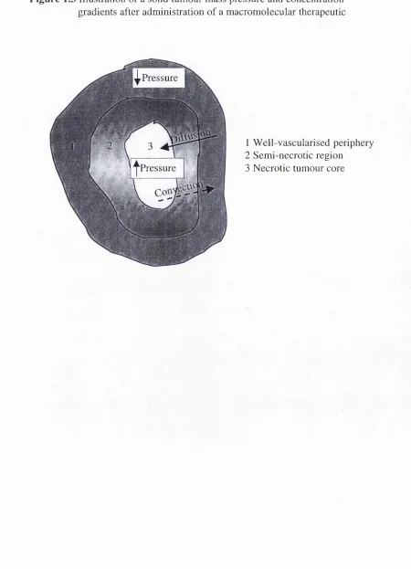
The rate of macromolecular transport in and out of the tumour is regulated by
local physiological conditions. Trans vascular transport occurs mainly by diffusion
(along a concentration gradient), convection (along a pressure gradient) or both
(reviewed by Joyner & Kern, 1990). The average vascular surface area per unit tissue
weight decreases with tumour growth. It therefore follows that reduced transvascular
exchange (extravasation) occurs in large tumours, in comparison with small tumours. In
larger solid tumours, the tumour interstitium is characterised by a large interstitial fluid
volume and a uniformly high fluid pressure at the tumour core (up to 30 mm Hg)
relative to normal tissues (reviewed by Jain, 2001) due to its confined space and absent
lymphatic drainage (reviewed by Jain, 1987b). This pressure abruptly drops at the
tumour periphery ( 5 - 1 5 mm Hg) (Jain & Baxter, 1988 and Baxter & Jain, 1989) and
allows adequate extravasation of macromolecules into the tumour site, which then
slowly diffuse towards the tumour core. The high intratumoural volume at the core
promotes a higher diffusion rate and thus facilitates initial polymer-drug concentration
in tumour tissue. On the other hand, high tumoural interstitial pressure may limit
convective extravasation of macromolecules and thus compromise tumoural delivery of
macromolecules (Jain, 1997; Figure 1.3).
The balance of these events, in combination with absent or ineffectual tumoural
lymphatic drainage aid tumoural localisation of macromolecules (Maeda & Matsumura,
1989; Jain, 1994). The phenomenon of EPR may thus be used for targeting
Figure 1.3 Illustration of a solid tumour mass pressure and concentration gradients after administration of a macromolecular therapeutic
X Pressure
Pressure
Chapter 1 General Introduction
macromolecular anti-cancer drugs (Maeda, 1991). Likewise, macromolecular anti
inflammatory drugs may be targeted to inflammatory sites (Murakami et al, 1996)
which due to local structural changes, chemical composition (e.g. histamine &
bradykinin) and contraction of endothelial cells also display EPR (Maeda et al, 1988
and Seymour, 1992).
Since the early observations of Matsumura & Maeda (1986), a number of
studies using in vivo models have been used to better understand macromolecular
targeting to tumours (reviewed by Duncan, 1999). In particular, studies have sought to
relate tumour targeting to parameters such as tumour location within the body
(Fukumura et al, 1997); vascular density (Smith et al, 1988); microvessel permeability
(reviewed by Jain, 1987a) and extracellular volume fraction (Graff et al, 2000).
Using HPMA copolymers of narrow polydispersity (10 - 800 kDa), Seymour et
al (1995) and Noguchi et al (1998) showed tumour targeting in both B16F10 and
Sarcoma 180 models respectively did not show molecular weight dependency in early
phase ( - 1 0 min) and long term (72 h) tumoural accumulation. Higher molecular
weight copolymers (> 40 kDa) of size above the renal threshold were retained in the
circulation. Recent in vivo studies by Sat & Duncan (1998) indicate that tumour size
and not tumour type or properties of macromolecular DDS examined (albumin-evans
blue complex, PK l, dendrimer-DOX conjugate and liposomal DOX) will determine the
amount of tumoural localisation following i.v. administration. Using a s.c. B16F10
murine tumour model, PKl and albumin-evans blue complex showed size-dependent
uptake in small tumours with 13.8 ± 2.5 % dose/g tumour and 12.6 ± 4.9 % dose/g
tumour respectively as compared to large tumours 1.6 ± 1.1 % dose/g tumour (PKl)
and 2.7 % dose/g tumour (albumin-evans blue complex). Similarly, Harrington et al
(1998) found better uptake of "^In-DTPA-labelled stealth liposomes in small tumours
(< 0.1 g) 15.1 ± 10.8 % dose/g tumour in comparison to large tumours (> Ig) with 3 ±
1.3 % dose/g tumour. Using a panel of human tumour xenografts and murine tumour
models, tumour type was found not to be of major significance in determining EPR-
mediated tumour uptake (Sat, 1999).
1.3.1 Vectors that Utilise EPR for Tumour Targeting
Several types of carrier have been used as DDS for tumour targeting by the EPR
effect. These include: particulate delivery vehicles e.g. liposomes (Gabizon et al, 1982;
Torchilin et al, 1988; Gregoriadis 1988; Allen et al, 1995); serum proteins such as
human serum albumin (HSA) (reviewed by Kratz & Beyer, 1998), cells e.g
erythrocytes (Bax et al, 1999); DNA (Deprez-de Campeneere & Trouet, 1980);
nanoparticles (10 - 1000 nm) (reviewed by Brannon-Peppas, 1995; Monfardi &
Veronese, 1998; Soppimath et al, 2001); block copolymers which in aqueous
conditions form polymeric micelles (Yokoyama et al, 1992 and Kataoka & Kwon
1995); soluble delivery vehicles e.g. natural biodegradable polymers such as the
polysaccharides e.g. dextrans (reviewed by Mehvar, 2000); soluble synthetic polymers
e.g. PEG (reviewed by Zalipsky, 1995; Greenwald et al, 2000; Harris et al, 2001);
bioresponsive polymers for endosomolytic delivery (Richardson et al, 1999 and Murthy
et al, 1999) and dendrimers (Malik et al, 1999). Some advantages and disadvantages of
these carriers are listed in Table 1.1.
The liposomal systems were the first to progress into the market. They do,
however, display some limitations. For example, palmar-plantar erythrodysesthesia,
commonly refererred to as "hand-foot syndrome", characterised by ulcers in the hands
and feet displayed by liposomal DOX (Doxil® in USA or Caelyx® in the EU) is due to
prolonged circulation time leading to enhanced extravasation through microvessels and
subsequent toxicity of DOX on kératinocytes (Gordon et al, 1995). Appearance of
"hand-foot syndrome" was the dose limiting toxicity that led to the dosing schedule of
50 mg/m^ every 4 weeks for Caelyx. In contrast, free DOX is administered at a dose of
60 - 75 mg/m^ every 3 weeks (Martindale, 2002) when used to treat AIDS-related
Kaposis’s sarcoma.
Disadvantages of the current design of polymer-drug conjugates are discussed in
section 1.5.1.
Prior to defining polymer therapeutics, the mechanism of endocytic uptake of
macromolecules passively accumulated in the tumour interstitium will be described in
detail.
1.4 Endocytic Capture of Polymer Therapeutics
Eukaryotic cells use a system of intracellular organelles which are functionally
interconnected by a series of carrier vesicles to remain in constant communication with
Chapter 1 General Introduction
Table 1.1 Advantages and disadvantages of DDS used for passive tumour-targeting by
the EPR effect
Advantages (Reference) Disadvantages (Reference)
Liposomes^
No degradation of entrapped drug (Monfardi L im ited drug loading capacity
& Veronese, 1998) (Langer, 1998)
Altered pharmacokinetics (no RES uptake) Possible impairment of macrophages
by PEGylation (Allen et al, 1995) involved in tumour control (Gabizon
etal, 1982)
Protection of drugs from degradation Leakage of drug in blood (Soppimath
(Knight, 1981) et al, 2001)
Decreased toxicity of parent compound Poor storage stability (Soppimath et
(Knight, 1981) al, 2001)
HSA^
Readily available (Kratz & Beyer, 1998) Polyfunctionality thus heterogeneous
products (Peters, 1985)
Good biological stability (Yuan et al, 1995) Structure and properties can not be
altered (Drobnik, 1989)
Biodegradable (Kratz & Beyer, 1998)
Non-toxic & non-immunogenic (Kratz & Beyer, 1998)
DNA
Decreased toxicity of parent compound Instability of conjugates in vivo
(non-(Deprez-de Campeneere & Trouet, 1980) covalent) (Trouet & Jolies, 1984)
Ease of preparation of complexes (Trouet et Conjugates do not overcome MDR
al, 1980) (Atassi et al, 1974)
Nanoparticles
Good stability (Soppimath et al, 2001) Matrix pore size governs size of drug
to be used (Monfardi & Veronese, 1998)
Controlled release properties (depot) H ydrolytically labile compounds
(Soppimath gr aZ, 2001) damaged by diffusion of water into
matrix (Monfardi & Veronese, 1998)
Oral administration of peptides or proteins O rganic so lv en ts may cau se
(Soppimath et al, 2001) dénaturation of protein / peptide drugs
(Monfardi & Veronese, 1998)
Advantages (Reference) Disadvantages (Reference) Polymeric micelles^
No renal clearance based on molecular Control of stability and drug release
weight (Kwon & Kataoka, 1995) may be difficult
Good water solubility irrespective of drug Industrial scale-up may be difficult
loading (Yokoyama et al, 1990)
Functionality based on distinctive character In d u stria l c h a ra c te ris a tio n o f
of constituent polymers (Yokoyama et al, entrapped and free drug may be
1990) difficult
Naturally Occurring Polymers
Easily available (Monfardi & Veronese, C om m ercial upscale is lim ited
1998) (Duncan & Seymour, 1989)
Biocompatible (M onfardi & Veronese, Chemical modification may confer
1998) immunogenicity, pyrogenicity and
hinder degradation (Drobnik, 1989)
Biodegradable (Brocchini, 2000) Lack structural uniformity (Brocchini,
2000) Synthetic Polymers^
Biocompatible (Vasey et al, 1999) Drug must possess chemical group for
polymer derivatisation (Monfardi & Veronese, 1998)
Decreased metabolism of conjugated drug Chemical modification of drug may
e.g. proteins (Nucci gr aZ, 1991) reduce activity of parent compound
(Monfardi & Veronese, 1998)
Commercial scale production with control of Internalisation of conjugate necessary
molecular weight characteristics (Godwin et for activation (Rejmanova et al 1985)
al, 2001)
Versatile chemistry (Brocchini, 2000) Not biodegradable (Brocchini, 2000)
Bioresponsive Polymers
pH-responsive, thus controlled delivery R e la tiv e ly low tr a n s f e c tio n
(Ferruti et al, 2000) (Richardson et al, 2001)
Non-immunogenic & biocompatible viral-
Chapter 1 General Introduction
Advantages (Reference) Disadvantages (Reference) Dendrimers
Precisely controlled architecture (Tomalia et Sm all size may lim it tum our
al, 1985 and Hawker & Fréchet, 1990) localisation (Malik et al, 1999)
Monodisperse (Liu & Fréchet, 1999) Biocompatibility not fully understood
(Roberts et al, 1996 and Malik et al.
1997)
Large number of chemical functionalities (Liu & Fréchet, 1999)
^ In the market
* In Phase I/II clinical trials
import and export molecules by a process that has been termed endocytosis, a general
term that encompasses pinocytosis (cell drinking) and phagocytosis (cell eating)
(Silverstein et al, 1977). The potential value of the endocytic route in drug delivery (so
called lysosomotropic delivery) was first proposed by de Duve and co-workers (de
Duve et al, 1974).
The pinocytic capture of macromolecules and indeed of small water soluble
polymer therapeutics (< 0.2 pm diameter vesicles) occurs by either fluid-phase
pinocytosis (non-clathrin-mediated internalisation and caveolae) involving the uptake
of molecules in solution (i.e. solutes) or alternatively by adsorptive pinocytosis
whereby the molecules are internalised in association with cell surface receptors via
clathrin-coated vesicles. The latter can be either non-specific receptor-mediated
pinocytosis or specific receptor-mediated uptake. Subsequent migration of the newly
formed intercellular vesicles (pH 6 - 6.8) (Mellman et al, 1986) along microtubules in
the cell cytoplasm and fusion with late endosomes in the peri-nuclear region follows to
form prelysosomes. These exist at pH 5 - 5.5 as maintained by an intramembranal
proton pump (Geisow, 1982) which then mature into or fuse with pre-existing
lysosomes.
Lysosomes usually have pH in the range of 4 - 5 and they contain a cocktail of
hydrolytic enzymes. Lysosomal enzymes act either in concert or in sequence to liberate
low molecular weight degradation products e.g. amino acids and sugars which usually
traverse the lysosomal membrane and are used by the cell in synthetic metabolic
pathways. After fusion of endosomal and lysosomal vesicles the membrane is often
recycled back to the cell membrane (reviewed by Mellman, 1996).
Lysosomal enzymes consist of a variety of hydrolytic enzymes including endo-
and exopeptidases (Table 1.2), with diverse functions broadly ranging from the
production of metastatic peptides to protein turnover (Bogyo et al, 2000). These
intracellular proteases are found mainly within lysosomes, but not exclusively. They
can also be within the proximity of cells effecting lysosomal enzyme secretion
(Duncan, 1986).
Cysteine proteases of the papain superfamily have been found to display a
sub-Chapter 1 General Introduction
Table 1.2 Lysosomal enzymes (From Barrett & Heath, 1977 and Duncan, 1986)
Enzyme Activity Optimal
pH Endopeptidases (hydrolases which cleave away from ends of polypeptides)
Acrosin Cleaves arginyl and lysyl bonds in low
MW substrates
8.0
Lysosomal elastase Cleaves valyl bonds in low MW substrates 8.5
Cathepsin G Cleaves phenylalanyl and other
chymotrypsin bonds
7.5
Cathepsin B Papain-like specificity 3.5 - 6.0
Cathepsins H & L Little action on Cathepsin B synthetic
substrate
5.0
Cathepsin D Proteins & peptides of at least 5 residues;
preferably bonds between hydrophobic amino acyl residues
2 .8 -5 .0
Cathepsin E Similar to Cathepsin D & pepsin
Exopeptidases (hydrolases which cleave near ends of polypeptides)
Lysosomal aminopeptidases Cleave N-terminal amino acyl residues various
Lysosomal
carboxypeptidase A
Cleave C-terminal residues; broad specificity (excluding Arg & Lys)
5.0
(Cathepsin A & I)
Lysosomal
carboxypeptidase B
Cleave C-terminal residues; broad specificity (including Arg & Lys)
6.2
Lysosomal
carboxypeptidase C
Cleaves C-terminal when subterminal residue is Pro
5.5
Tyrosine acid carboxypeptidase
Cleaves C-terminal residues; preferably hydrophobic
NA
Lysosomal dipeptidase Cleaves dipeptides; broad specificity 5.5
Dipeptidylpeptidase I (Cathepsin C)
Cleave N-terminal dipeptides from amide, arylamide, ester & peptide substrates; broad specificity; except if terminal
residue is Arg or Lys & 2"*^ or 3"*^ residue is Pro
5 .0 -6 .0
Dipeptidylpeptidase II Cleave N-terminal dipeptides from amide,
arylamine and peptide substrates; preferably Arg or Lys terminal
4 .5 -5 .5
classification of these enzymes is defined by the nature o f their active site. The
presence of a chemical group that exhibits the reactivity to catalyse a reaction and
specificity to a substrate which binds sufficiently tightly at the active site for a reaction
to occur (Duncan, 1986 and Y an et al, 1998). Thus Cathepsin B which is believed to be
responsible for the conversion of high molecular weight polymer-drug conjugates to
release the active moiety by cleavage of tailor-made peptidyl sequences (Duncan et al,
1983b and Duncan, 1986) is a cysteine protease in that it has cysteine in its active site
(which confers its nucleophilicity) and a pH optimum which favours acidic conditions
and a substrate preference for peptidyl amide bonds.
The selective endocytic uptake and subsequent release of cytotoxic agents
within lysosomes o f target cells by coupling these agents to a suitable carrier have been
investigated and exploited in the fields of cancer and infectious diseases (de Duve et al,
1961 and reviewed by Duncan, 1999). Low molecular weight compounds are randomly
taken up into the cell by diffusion. Their subsequent conjugation to macromolecular
carriers restricts their uptake to the pinocytic route, thus conveying tumour tissue
specificity to this new chemical entity (Duncan et al, 1983b). The notion of the
synthesis of drug conjugates with lysosomally labile linkers ("spacer"), cleaved either
by hydrolytic enzymes or the intralysosomal acidic pH which would allow subsequent
drug release into the cytosol, where it would exert its pharmacological effect, was first
proposed by Trouet and co-workers (Trouet et al, 1972 and Trouet, 1978). In vitro
incubation of a serum albumin-tetrapeptide-daunorubicin conjugate in purified rat
lysosomal fractions achieved ~ 80 % drug release (Trouet et al, 1982). This conjugate
was also found to remain stable in the blood stream in vivo (Trouet & Jolies, 1984), a
prerequisite for directed delivery.
The limitation of lysosomal delivery is the restriction to active agents which are
themselves not inactivated in the lysosomal conditions. In addition, in the case of
enzymatic cleavage, the successful release of the active agent is reliant upon the
sufficiency o f indigenous enzyme required. P K l, currently in Phase II clinical trials in
the UK for breast, colon and non small cell lung cancer is such a lysosomotropic agent
(Vasey et al, 1999). As lysosomes are present in all mammalian cells types, the tumour-
selective uptake of such lysosomotropic compounds by the EPR effect is therefore
Chapter 1 General Introduction
1.5 Polymer Therapeutics
Although biomedical polymers (contact lenses, prostheses, implants, kidney
dialysis membranes, etc.) have a long history, the use of water soluble polymers as
therapeutics is relatively new.
The term polymer therapeutics, coined by Duncan (1992), may be defined as a
family of prodrugs which incorporate polymers as the core constituent, schematically
illustrated in Figure 1.4. These comprise of polymer-drug conjugates, polymer-protein
or polymer-peptide conjugates, polymeric micelles and and non-viral gene delivery
constructs (reviewed by Brocchini & Duncan, 1999 and Duncan, 2000).
It should be noted that polymeric drugs are distinguished from polymer prodrug
carriers as they themselves exert a pharmacological effect in their own right. Polymeric
carriers, which transport the pharmacophore covalently via pendant side chains or to
terminal groups of the polymer to the target site, do not convey this inherent activity.
Polym eric drugs with intrinsic activity include glatiramer acetate injection
(Copaxone®) which has been marketed for multiple sclerosis (Gilbert, 1998),
divenylether-maleic anhydride copolymer (DIVEMA) (Gros et al, 1981) and dextrin-2-
sulphate which is presently under clinical evaluation as an anti-AIDS therapy (Shaunak
etal, 1998).
Polymer-Drug Conjugates
Several soluble polymers have been used in macromolecular systems in cancer
(reviewed by Duncan, 1992; Duncan et al, 1996; Brocchini, 2000) and other diseases
e.g leishmaniasis (Nan et al, 2001), dyslipidaemias and artherosclerosis (Lovrek et al,
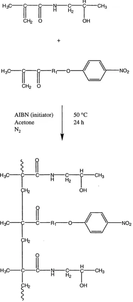
2000). Ringsdorf pioneered this model in 1975 (Figure 1.5). He proposed the covalent
attachment of drugs to a water soluble polymer backbone by means of a biodegradable
spacer with the possible incorporation of a targeting residue e.g. antibodies and sugars,
(see section 1.2) to convey receptor-mediated pinocytosis. In limiting the cellular
uptake of high molecular weight species to endocytosis (Duncan et al, 1981), polymer-
drug conjugates provide the opportunity to direct the drug to the particular cell type
where drug action is demanded by means of the EPR effect. A wide range of polymers
(both biodegradable and non-biodegradable) have been employed in polymeric
prodrugs to improve the targeting of anticancer compounds (Table 1.3).
Figure 1.4 Illustration of examples of polymer therapeutics: (A) polymeric drug with intrinsic activity; (B) polymer-drug conjugate; (C) polymer-protein conjugate and (D) polymeric micelle (Adapted from Brocchini & Duncan, 1999)
B
drug
C D
Figure 1.5 Illustration depicting the “Ringsdorf model”
Synthetic hydrophilic repeating unit
Targeting moiety
Table 1.3 Representation of examples of soluble polymeric vehicles for parenteral drug delivery in cancer therapy
Polymer Conjugated Cytotoxic
Compound
A ntitum our Activity Reference
Non-Degradable Synthetic Polymers
PEG Podophyllotoxin Similar or improved activity in vivo of various analogues
compared to free drug
Greenwald et al, 1999
Taxol Improved solubility and reduction of toxicity, but no
improved antitumour activity in vivo
Greenwald et al, 1996
Doxorubicin Reduction on toxicity compared to free drug and
increased tumour uptake in vivo
StntQv et al, 1995
HPMA Doxorubicin (PKl) Phase II Clinical Trials (CRC / Pharmacia) Vasey etal, 1999
Doxorubicin & Galactose (PK2) Phase I / I I Clinical Trials (CRC / Pharmacia) Ferry et al, 1999
Platinate (AP5280) Phase I Clinical Trials (Access Pharmaceuticals) Gianasi et al, 1999
Paclitaxel (PNU 166945) Phase I Clinical Trials (Pharmacia) Huniunk et al, 1998
Melphalan Improved activity in vivo compared to free drug Duncan et al, 1991
Dendrimers e.g. Poly (amidoamine)
Cisplatin Improved activity and reduced toxicity in vivo compared
to free drug
Malik et al, 1999
Degradable N atural or Synthetic Polymers
Naturally occuring
Serum proteins e.g.
Albumin Methotrexate Phase I Clinical Trials (German Association for Medical
Oncology)
Hartung et al, 1997; Burger
et al, 2001
9
I
I
to
Com pound
Degradable N atural or Synthetic Polymers (continued)
Polysaccharides e.g, Dextran
Mitomycin
Daunomycin
RES uptake in vivo
Improved activity in vivo
Takakura et al, 1987;
Bernstein et al, 1978
Alginate Daunomycin In vitro release of free drug in acidic pH only; antitumour
activity in vivo
Al-Shamkhani & Duncan, 1995
N atural derivatives Poly(aminoacids)
e.g. Poly(L-Iysine) Methotrexate Antitumour activity and over-riding of resistance in vitro,
but polymer related toxicity observed in vivo
Ryser & Shen, 1978; Chu et
al, 1981
Poly(L-glutaniic acid)
Doxorubicin Antitumour activity in vivo, but immunogenic van Heeswijk et al, 1984;
Hoes et al, 1993
Camptothecin (CPT) Increased water solubility and antitumour activity in vivo de Vries et al, 2000
Paclitaxel Phase I Clinical Trials (company) Li et al, 1998
Poly[A^-(2- hydroxyethyl)-L- glutamine] (PHEG)
Mitomycin C (MMC) Biocompatibility in vivo with reduction in toxicity and
improved antitumour activity
Soyez et al, 1997; de Marre
et al, 1995
Daunorubicin Hepatotoxic in vivo; not suitable for clinical development Hrdina et al, 1991
g
I
I
I
3*
Polymer Conjugated Cytotoxic Compound
A ntitum our Activity Reference
Degradable N atural or Synthetic Polymers (continued)
Polyesters e.g.
Poly(a-malic acid)
Polymeric micelles e.g.
5-Fluorouracil Slight improvement in antitumour actitivty in vivo Ouchi et al, 1990
PEG-Aspartate Doxorubicin Improved antitumour activity in solid tumours in vivo Yokoyama et al, 1990; 1991
Polymers with Inherent Activity
Divenyl ether - maleic anhydride copolymer
(DIVEMA) Cyclophosphamide
Methotrexate
Doxorubicin
No difference in in vivo activity compared to free drug
Improved antitumour activity in vivo compared to free
drug and adjuvant effect of polymer observed
Improved antitumour activity in vivo compared to free
drug
Hirano et al, 1980
Przybylski etal, 1978
Pratesi et al, 1990
Polym er-Protein Conjugates
PEG Poly(styrene-co- maleic acid) (SMA) Interleukin-2 L-Asparaginase Neocarzinostatin
Phase I / I I Clinical Trials (Enzon)
In Clinical Use for non-Hodgkin’s Lymphoma
In Clinical Use (Japan) for Hepatocarcinoma; Co administration with Lipiodol
Meyers et al, 1991
Park etal, 1981
Matsumura & Maeda, 1986; Konno, 1987
î
I
Macromolecular polymeric prodrugs enable the attainment of key factors in the
principle of drug delivery: (1) the solubility of hydrophobic drugs is enhanced; (2) the
pharmacokinetic profile of the pharmacophore is improved and (3) systemic toxicities
are minimal as the payload is protected and cytotoxic action is observed after release of
the active moiety specifically in the target cells, i.e. pharmacological activity of the
active agent remains intact. In addition, the improved therapeutic index of the active
agent bound allows a higher dose to be administered. For example, the dose for PK l in
Phase II clinical trials is 280 mg/m^ (Vasey et al, 1999) ~ 4 - 5 times greater than
doxorubicin (DOX) which is typically administered at 60 - 80 mg/m^ (Martindale,
2002).
The ideal characteristics for a polymer used as a drug carrier have been
previously reported (reviewed by Duncan et al, 1996). These include:
1) Non-immunogenic
2) Possess suitable chemical functionality for covalent linkage to drug
3) Release of drug in required site of action (stability en route to target site)
4) Hydrophilic i.e. water soluble in physiological conditions
5) Biodegradable or excreted
6) Commercially available.
In respect of the polymer-drug conjugate, stability of the payload en route to the desired
site of action is paramount for desired non-systemic toxicity.
Design of HPMA Copolymer for Tumour Targeting
HPMA copolymer, in the field of polymer-drug conjugates, has been by far the
most systematically designed and extensively studied, and has delivered several
conjugates into pre-clinical and clinical development for treatment of solid tumours
(Vasey et al, 1999; Gianasi et al, 1999; reviewed by Duncan, 2000).
Neutral, hydrophilic water soluble linear polymers such as HPMA copolymer display a
loose, random flexible coil which is in constant dynamic motion. HPMA homopolymer
was originally developed in the Czech Republic as a plasma expander and is non-
immunogenic and has been shown to be non-toxic at doses of up to 30 g/kg (Duncan,
1992). Following extensive evaluation of biodistribution (circulating half-life, renal
clearance, organ and tumour uptake) of HPMA copolymer (Seymour et al, 1987; 1995),