ABSTRACT
SIMMONS, ALICIA NICOLE. The Role of TGFß-Activated Kinase 1 Signaling in Reactive Oxygen Species and Intestinal Homeostasis. (Under the direction of Dr. Jun Ninomiya-Tsuji).
Proper function of intestinal epithelial cells is essential for overall homeostasis of the host in that if intestinal epithelial cells are unable to carry out their functions, structural disruptions may occur leading to leaky barriers, invasion of bacteria, and cell death. In this study we investigated the role of TGF-β activated kinase 1 (TAK1) in intestinal homeostasis. TAK is a member of the mitogen activated protein kinase (MAPK) kinase kinase
(MAPKKK) family and is activated by various chemical and physical stressors and inflammatory cytokines to promote cell survival through activation of MAPKs and a transcription factor, NF-κB. We used a mouse model harboring an inducible intestinal epithelial specific deletion of the Tak1 gene.
We initially found that when Tak1 is deleted, cell death, upregulation of reactive oxygen species (ROS), and loss of Paneth cells, which play a major role in gut microbe regulation, are induced in the intestine. We hypothesize that TAK1 regulation of the
antioxidant pathway prevents microbial-induced ROS and is integral for Paneth cell integrity. To test the involvement of bacteria, we depleted commensal bacteria using an antibiotic cocktail. We found that bacteria depletion greatly reduced ROS in Tak1-deficient intestinal epithelium. Furthermore, intestinal epithelial-specific deletion of Myd88, a common adaptor protein of several Toll-like receptors, also reduced the accumulation of ROS. These
deletion. These results suggest that Paneth cell depletion is independent of ROS
accumulation. We later found that deletion of receptor interacting kinase 3 (Ripk3), which is a mediator of one type of cell death, necroptosis, prevented Paneth cell loss. These results demonstrate that TAK1 regulates ROS and Paneth cells, both of which are critical for intestinal homeostasis. Chronic intestinal inflammatory diseases such as Crohn’s disease are associated with oxidative stress and loss of Paneth cells. Our results provide a better
understanding of the regulatory mechanisms for ROS and Paneth cells, which could lead to new approaches to control these diseases.
In a subsequent study we analyzed the role of TAK1 in the regulation of the antioxidant transcription factor Nrf2. Nrf2 is a leucine-zipper transcription factor that regulates a number of antioxidant enzymes thereby playing a major role in the cellular antioxidant system. We found that a protein kinase, TAK1 physically interacts with Nrf2 through Keap1 and upregulates Nrf2 protein stability, suggesting that TAK1 regulates ROS through Nrf2.
The Role of TGFß-Activated Kinase 1 Signaling in Reactive Oxygen Species and Intestinal Homeostasis
by
Alicia Nicole Simmons
A dissertation submitted to the Graduate Faculty of North Carolina State University
in partial fulfillment of the requirements for the degree of
Doctor of Philosophy
Cellular and Molecular Toxicology
Raleigh, North Carolina 2015
APPROVED BY:
_______________________________ _______________________________
Jun Ninomiya-Tsuji James Bonner
Committee Chair
_______________________________ _______________________________
DEDICATION
BIOGRAPHY
Alicia Nicole Simmons was born on October 26, 1985 to Lucius and Christine Simmons Jr, in Winston-Salem, NC. She is the oldest of two and has a younger brother, Lucius Simmons, III. Alicia attended elementary school at Forest Park Elementary followed by middle school at Atkins Middle, both which are located in Winston-Salem, NC. In 2004, Alicia graduated high school form East Forsyth High School and was admitted to and enrolled at Johnson C. Smith University in Charlotte, North Carolina. She later graduated Cum Laude from Johnson C. Smith University in May 2008 with a Bachelor of Science degree in Biology.
After graduating from JCSU, Alicia moved to Chapel Hill, NC and worked as a post baccalaureate student for two years at the University of North Carolina at Chapel Hill (UNC). While there Alicia worked under the mentorship of Dr. Shawn Ahmed, PhD in the department of Genetics where she studied germ cell immortality using the nematode roundworm Caenorhabditis elegans (C. elegans). Additionally, Alicia learned and accomplished a lot while at UNC and attended and presented at her first International Conference at UCLA in the summer of 2009.
her written preliminary examination and successfully defended her oral preliminary
examination of October of the same year. In the summer of 2015 Alicia plans to finish up her work towards her PhD and then begin her postdoctoral fellowship position at the NIEHS in the Signal Transduction Research group of Dr. Xiaoling Li, PhD.
ACKNOWLEDGMENTS
First I would like to thank God for allowing me to pursue my dreams and making it possible for me to complete what I set out to do many years ago. I have known since I was in elementary school what my passion was and I am so grateful that my dream is now my reality. I next would like to thank my mentor & advisor Dr. Jun Ninomiya-Tsuji for her dedication, support, and training. She went above and beyond the duties of a mentor and I am forever appreciative for her teachings and mentorship. She pushed me like no other mentor ever has. I also want to thank my committee members Yoshi Tsuji, Robert Smart, James Bonner, and Anthony Blikslager for their valued support, mentorship, and availability whenever I needed them. I appreciate their project advice and questions that they asked that encouraged me to think outside of the box. I would like to also acknowledge the former and current Tsuji lab members: Sho Morioka, September Mihaly, Yuka Ikeda, Yosuke
Sakamachi, and Kazu Sai. Thank you for your experimental advices and suggestions as well as your expertise in how to carry out good, clean science.
I also want to acknowledge my younger brother who I love so much. Lucius Simmons III, thank you for being the best brother a girl could ask for. I know growing up with me as a big sister has not been an easy task but you are the best and I wouldn’t trade you for anything in the world. I next would like to thank not just my immediate family members but also my extended family, grandparents, aunts, uncles, cousins, and friends for always keeping me uplifted and checking on me to see how this PhD journey was going. I appreciate you all and will never forget any of you. You all had a hand in the woman I am today.
have always been supportive of anything and everything that I have wanted to do. I knew that when I embarked on this journey it would be a long and tough road but you always reiterated
that I could do this and that it was destiny. Thank you for making me take study breaks, cooking me food whenever I just worked so hard that I would only eat junk food because it
was easier and quicker. You took me on walks, told me jokes, and will forever be the best vacation partner a girl could ask for. I appreciate you and love you! You are definitely one of
TABLE OF CONTENTS
LIST OF FIGURES ... x
GENERAL INTRODUCTION ... 1
1. The Intestine ... 1
2. Inflammatory Bowel Disease (IBD) ... 2
3. Pathogenesis of IBD ... 3
4. Oxidative Stress ... 5
5. Oxidative Stress and IBD ... 6
6. Gut Microbiota Involvement ... 7
7. Paneth Cells & IBD ... 8
8. TAK1 ... 8
9. The role of TAK1in vivo ... 9
10. The mechanism of cell death ... 11
11. Cell death in Tak1 deficient cells and tissues ... 13
12. Pathogenic phenotype of Tak1 deficient intestinal epithelium ... 14
13. TAK1 Regulation of Oxidative Stress ... 15
14. Keap1/Nrf2 Signaling ... 15
15. Nrf2 and Inflammatory Disorders ... 16
HYPOTHESES ... 17
MANUSCRIPT I: TAK1-dependent Paneth cell maintenance is required for prevention of intestinal oxidative stress ... 18
1. ABSTRACT ... 19
3. RESULTS ... 21
4. DISCUSSION ... 28
5. MATERIALS & METHODS ... 30
6. ACKNOWLEDGEMENTS ... 34
7. FIGURE LEGENDS ... 35
8. SUPPLEMENTARY FIGURE LEGENDS ... 39
9. FIGURES ... 40
10. MANUSCRIPT I REFERENCES ... 49
MANUSCRIPT II: TAK1-dependent and redox-independent regulation of Nrf2-Keap1 is required for prevention of oxidative tissue injury ... 54
1. ABSTRACT ... 55
2. INTRODUCTION ... 66
3. RESULTS & DISCUSSION ... 59
4. MATERIALS & METHODS ... 64
5. ACKNOWLEDGEMENTS ... 66
6. FIGURE LEGENDS ... 67
7. FIGURES ... 70
8. MANUSCRIPT II REFERENCES ... 75
GENERAL DISCUSSION ... 80
1. Role of Paneth cells in Host Defense and Intestinal Integrity ... 80
2. Paneth cells are regulated by Necroptosis ... 81
3. TAK1 is a critical regulator of Paneth cells ... 83
5. Why are Paneth cells sensitive to necroptosis? ... 84
6. How does Tak1 deletion cause necroptosis? ... 85
7. Role of TAK1 in cellular antioxidant system ... 86
LIST OF FIGURES GENERAL INTRODUCTION
Figure 1. Generating a TAK1 intestinal epithelium conditional knockout mouse … ... 10
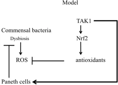
Figure 2. Model ... 17
MANUSCRIPT I: TAK1-dependent Paneth cell maintenance is required for prevention of intestinal oxidative stress Figure 1. Tak1 deletion depletes Paneth cells ... 40
Figure 2. Antibiotics treatment reduces ROS and apoptosis ... 41
Figure 3. Myd88 deletion reduces ROS and apoptosis ... 42
Figure 4. Antibiotic treatment of Myd88 deletion does not block Paneth cell loss ... 43
Figure 5. Ripk3 deletion partially rescues Paneth cell loss and ROS accumulation ... 44
Figure S1. Heterozygous deletion of Tak1 and inducible Cre expression does not cause Paneth cell loss ... 45
Figure S2. Tak1-deficient colonic crypts are relatively intact ... 46
Figure S3. Intestinal injury is slightly improved by antibiotic treatment or Myd88 deletion in Tak1-deficient intestinal epithelium ... 47
MANUSCRIPT II: TAK1-dependent and redox-independent regulation of Nrf2-Keap1 is required for prevention of oxidative tissue injury ... 56
Figure 1. TAK1 upregulates Nrf2, and binds to Nrf2 through Keap1 ... 69
Figure 2. TAK1 stabilizes Nrf2 ... 70
Figure 3. Ablation of TAK1 upregulates Keap1 ... 71
Figure 5. Electrophilic activation of Nrf2 blocks ROS accumulation in Tak1-deficient intestinal epithelium ... 73 GENERAL DISCUSSION ... 79
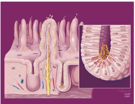
Figure 1. Paneth cells at the base of the crypts discharging antimicrobial peptides
GENERAL INTRODUCTION
Chronic inflammation is associated with a diverse set of human diseases not limited to the well known inflammatory diseases but also many cancers, and metabolic diseases. Therefore, better understanding of the mechanisms leading to chronic inflammation is much anticipated. Among the many diseases associated with inflammatory conditions, I focus on inflammatory bowel disease (IBD), which is known to be associated with oxidative stress induced tissue injury. I fortunately found a very effective mouse model system to investigate intestinal chronic inflammation, which was previously developed by our research group. These mice harbor gene deletion of a protein kinase, Tak1 that exhibits profound IBD-like oxidative injury in the intestine. In this dissertation project, I explore the mechanism by which chronic oxidative injury is developed. This introduction serves to provide background information regarding intestine physiology, IBD, oxidative stress, and TAK1, which will support my hypothesis for this project described at the end of the introduction.
The Intestine
The intestine consists of a small and large intestine. The small intestine contains the
duodenum, jejunum, and the ileum, which is where most of the nutrient and water absorption occurs whereas the large intestine also known as the colon or large bowel is the last part of the digestive system where waste is stored as feces before defecation. A layer of microvilli, which contain enzymes needed for digestion as well as nutrient transporters, covers the intestinal epithelial cells. Two major regions of the intestinal epithelium are the villus and the crypt of Lieberkuhn. The villus lining contains a single layer of epithelial cells that is
from the crypt stem cell. The stem cells continuously proliferate to support the constant turnover of all of the differentiated epithelial cells. These differentiated cells include the absorptive enterocytes, goblet cells, and enteroendocrine cells. Additionally, in the small intestine, the specialized epithelial Paneth cells are also generated from the stem cells where they reside at the crypt base, and their role is to secrete antimicrobial peptides, defensins, and lysozymes to aid in intestinal homeostasis (Ayabe et al., 2000). We note that Paneth cells are not present in the large intestine.
Inflammatory Bowel Disease (IBD)
Inflammatory bowel disease (IBD) is a chronic disease of the colon and small intestine that primarily consists of ulcerative colitis (UC) and Crohn’s disease (CD) and is one of the major health problems in the US. This chronic disease involves interaction of immune cells,
density in the large intestine (Geboes, 2008). Whereas, features that favor CD include inflammation, tissue damage of the ileum (ileitis), discontinuous crypts, mucin preservation, discontinuous inflammation, Paneth cells loss, and epitheloid granulomas (Geboes, 2008). The presence or absence of these features is dependent upon duration and activity of the disease as well as medical treatment (Kleer and Appelman, 1998).
Although the pathogenesis of IBD has been extensively studied there is still a large gap in our understanding at the molecular level of how phenotypes associated with IBD as well as how genetics, and exogenous factors impact our ability to handle the circumstances of IBD. My goal of the study is to provide a molecular mechanism of development of IBD-like disorders that will potentially lead to a new approach to combat the major problems associated with IBD including Paneth cell loss by investigating our mouse model that resembles IBD pathologies.
Pathogenesis of IBD
Both genetic and environmental factors have been associated with the pathogenesis of IBD. These factors can lead to the destruction of the intestinal barrier, translocation of microbial products, and chronic inflammation.
It is widely accepted that the pathogenesis of IBD results when there are overly aggressive acquired immune responses towards commensal bacteria in susceptible
in patients with IBD as well as experimental colitis mouse models (Atreya et al., 2000; Kai et al., 2005). Additionally, in experimental ulcerative colitis mouse models (Sugimoto et al., 2008), dendritic cells and neutrophils produce increased levels of IL-22, which is a member of the anti-inflammatory IL-10 cytokine family (Dumoutier et al., 2000a; Dumoutier et al., 2000b), that induces the production of antibacterial proteins and chemokines.
Several genetic mutations and polymorphisms are closely associated with IBD pathogenesis. Some include mutated genes associated with autophagy (ATG16) (Hampe et al., 2007), with the anti-inflammatory cytokine IL-10 (Franke et al., 2008), and
polymorphisms in NOD2 leading to increased risk for Crohn’s disease (Hugot et al., 2001; Ogura et al., 2001). ATG16L1 (T100A) gene mutation is associated with Paneth cell granule abnormalities (Rioux et al., 2007). Autophagy is a cellular homeostasis process by which a double-membraned structure, the autophagosome, surrounds the cytoplasm to degrade captured proteins and cytoplasmic organelles (Cadwell et al., 2008). In patients with healthy ileums, Paneth cells in the crypts of the ileum have granules and secrete peptides such as antimicrobial peptides and lysozyme to control the gut microbiota. However, loss of autophagy caused by the ATG16L1 mutation in patients with Crohn's disease or through a decrease in the gene product in Atg16L1HM mice results in fewer granules or diffused granule contents in the cells (Cadwell et al., 2008; Deuring et al., 2014). NOD2 is a intracellular pattern recognition receptor that plays a role in immune response by recognizing bacteria moiety, muramyl dipeptide, and activating NF-κB. About 50 different variations in the
NOD2 gene is associated with an increase risk of CD (Fritz et al., 2011). NOD2 mutation is known to increase NF-κB activation thereby potentiating inflammation (Maeda et al., 2005).
production in Paneth cells (Wehkamp et al., 2004). Thus, the combination of alterations in gut environment such as changes in gut bacteria or nonpathogenic/pathogenic virus infection with autophagy genes or NOD2 mutations may promote CD pathogenesis as suggested by a previous study conducted by (Cadwell et al., 2010).
Oxidative Stress
Oxidative stress is caused by an imbalance between the production of reactive oxygen species (ROS), including superoxides and peroxides, and the availability of antioxidant defenses (Du et al., 2009). ROS are generated and produced naturally as a product of cellular metabolism. During oxidative phosphorylation electrons are passed to electron carriers (cytochrome c and ubiquinone). The final electron acceptor is an oxygen molecule and typically this reaction will combine with hydrogen to form water. However, small amounts of peroxide are also produced, which is one of the most common reactive oxygen species. Intracellular ROS that can cause damage to lipids, proteins, and DNA, which could disrupt plasma and organelle membrane integrity and protein function and/or inactivating some enzymes by oxidation of cofactors (Cederbaum et al., 2009). The cells in the body can naturally produce antioxidants and enzymes to reduce ROS. There are three main pathways that are involved in the removal of ROS that involve reduced glutathione, thioredoxin, and catalase. One of the most common antioxidants is glutathione, which provides electrons to the oxidants so that ROS are reduced (Du et al., 2009). Both glutathione and thioredoxin pathways rely heavily on NADPH production for sustaining their activity, whereas catalase
Oxidative stress has been linked to several diseases and pathologies such as increased inflammation, cancers, and atherosclerosis (Le Lay et al., 2014). Additionally, oxidative damage in the epithelium is causally associated with chronic inflammatory diseases such as inflammatory bowel disease (IBD) as discussed below.
Oxidative Stress & IBD
It is widely accepted that under normal homeostatic conditions, the primary role of ROS is to defend against invading microorganisms. In the intestine, as generally discussed above, dietary antioxidants such as vitamin E, or vitamin C as well as glutathione, thioredoxin, and enzymatic antioxidants like superoxide dismutase and catalase protect tissues from the damaging effects of ROS (Puertollano et al., 2011; Gough and Cotter, 2011). However, an excess amount of ROS and impairment in production or maintenance of the antioxidants has been shown to lead to intestinal oxidative damage. Such aberrant ROS accumulation can be induced through several mechanisms. One prominent cause of ROS in the intestine is
leukocyte infiltration, where these cells produce large amounts of ROS and reactive nitrogen species (RNS) (Espey, 2013). However, ROS are generated not only by immune cells but also in intestinal epithelial cells, which will be described in a later section. Nonetheless, ROS accumulation is closely associated with pathology of IBD. Indeed, it has been well
Gut microbiota Involvement
Bacterial phyla Firmicutes and Bacteroidetes account for over 90% of the intestinal
microbiota (Eckburg et al., 2005). Several studies have documented that there are changes in the gut microbiota (dysbiosis) in patients with IBD and dysbiosis is emerging as the cause of IBD (Dalal and Chang, 2014). Dysbiosis may be at least in part due to the increasing
consumption of the western type diets (high fat, low fiber), which are high in processed sugars, protein, and overall calories. For example, many people who consume a high fat, low fiber diet generally have an increase in Bacteroidetes and Actinobacteria, while the bacteria Firmicutes and Proteobacteria are associated with low fat, high fiber diets (Wu et al., 2011). The mechanism by which dysbiosis in IBD is induced is not yet clear. Recently, it was reported that bacteria short chain fatty acid (SCFA) production is decreased in IBD patients, which is necessary for intestinal mucosal homeostasis. Therefore, this could lead to an
exacerbated pro-inflammatory phenotype in these hosts (Smith et al., 2013). Dysbiosis is also clearly associated with increased oxidative stress (Henao-Mejia et al., 2012).
Paneth cells & IBD
defensins, lysozyme and other antimicrobial peptides is usually indicative of IBD pathogenesis where microbes are able to invade the mucosa and cause inflammation. Specifically, CD patients display a reduction in defensins (Wehkamp et al., 2005). In contrast, UC exhibits mainly colon abnormalities and goblet cells have a defective and/or reduced ability to secrete mucin, which disturbs the mucosal barrier leading to colonic inflammation and UC (McCormick et al., 1990; Pullan et al., 1994), and this is consistent with the fact that Paneth cells are present only in the small intestine.
TAK1
Transforming growth factor-β-activated kinase 1 (TAK1) is a member of the mitogen-activated protein kinase kinase kinase (MAPKKK) family and is mitogen-activated by various cytokines, such as interleukin-1 (IL-1) and tumor necrosis factor (TNF), and ligands of Toll-like receptors and NOD-Toll-like receptors, which recognize bacterial and viral moieties (Kim et al., 2008; Sato et al., 2005; Takaesu et al., 2001; Ninomiya-Tsuji et al., 1999). In response to these stimuli, TAK1 is activated through interaction with TAK1 binding proteins: TAK1 binding protein 1 (TAB1), TAK1 binding protein 2 (TAB2), and TAK1 binding protein 3 (TAB3) (Mihaly et al., 2014a; Scholz et al., 2010; Inagaki et al., 2008; Kishimoto et al., 2000; Shibuya et al., 1996). It is known that TAK1 regulates mitogen-activated protein kinase (MAPK) pathways leading to activation of c-Jun N-terminal kinase (JNK) and p38, and also IKK signaling pathways to activate NF-κB (Ikeda et al., 2014; Omori et al., 2012; Ishitani et al., 2003; Takaesu et al., 2000). Although MAPK and NF-κB are extensively
studied, TAK1 can activate several other downstream targets including Ski-related novel
2004), which could include other unidentified targets.
The role of TAK1 in vivo
The role of TAK1 during development is not completely elucidated but it is known that mice deficient in Tak1 die in utero at around E10.5 (Shim et al., 2005b; Jadrich et al., 2006). These mice have morphological defects such as neural tube defects and reduced vessel architecture. Because the early lethality prevents detailed analysis of the roles of TAK1, our group has been using tissue specific and/or inducible gene knock out mice in order to fully understand the role of TAK1 in vivo. We used the LoxP-Cre system where exon 2 of the Tak1 gene is flanked by two loxP sites so that when Cre recombinase is activated, a truncated form of TAK1, TAK1Δ, lacking the ATP binding domain is expressed (Sato et al., 2005). TAK1Δ is
unfunctional and often unstable in cells. The phenotypes of our Tak1 exon 2 deletion mice are identical to total deletion of Tak1 gene expression (Sato et al., 2005; Shim et al., 2005a; Jadrich et al., 2006). For the intestinal epithelial specific deletion, we used the intestinal epithelial specific promoter (villin promoter) driven inducible Cre system that contains a fusion protein of Cre recombinase and a mutant estrogen receptor (CREERT2). The CREERT2 can be activated by treatment of the synthetic estrogen tamoxifen but not endogenous estrogen (Indra et al., 1999). Generating tissue specific TAK1 inducible knock out mice show the importance of TAK1 in tissue integrity. For example, endothelial-specific Tak1 deletion causes defects in vascular formation, as well as increased cell death and vessel
TAK1 plays a diverse set of roles in different tissues, but that TAK1 is in general important for cell survival.
The mechanism of cell death
(TRAF2 and TRAF5), cellular inhibitor of apoptosis 1 and 2 (cIAP1/2), and receptor
interacting protein kinase 1 (RIPK1) are recruited to the receptor complex (TNFR1 Complex I). Under some circumstances, the TNF bound receptor complex dissociates from TNFR1 leading to molecular reorganization and formation of a cytosolic death-inducing signaling complex (DISC) including TRADD, FAS-associated protein with a death domain (FADD), RIPK1 and caspase 8, known as Complex IIa (Micheau and Tschopp, 2003b). In this complex caspase-8 dimerizes, self-cleaves, and is activated to further activate executioner caspases such as caspase-3, which leads to apoptotic cell death.
In contrast, the intrinsic pathway is mainly activated by non-receptor signaling associated with Bcl-2 family proteins (Ledgerwood and Morison, 2009). For example, DNA damage activates p53, which transcriptionally activates several pro-apoptotic Bcl-2 family proteins (i.e. Puma, Noxa). Bcl-2 regulates mitochondrial integrity, and activation of pro-apoptotic Bcl-2 causes mitochondrial membrane permeabilization (Vander Heiden et al., 1997). Cyctochrome c and other cell death factors are released by MMP, which induces the activation of caspase-9 which in turn cleaves and activates downstream caspases such as caspase-3, -6, and -7 (Zamzami et al., 1998; Boldin et al., 1996; Muzio et al., 1996; Srinivasula et al., 1996).
There are several mechanisms to prevent apoptosis. One is the NF-κB, which is
known to upregulate antiapoptotic proteins such as cellular FLICE (FADD-like
IL-1β-converting enzyme)-inhibitory protein (c-FLIP) and inhibitor of apoptosis (IAP) to inhibit the activation of caspases (Chang et al., 2006; Wang et al., 1998).
2003). Characteristics of necrosis include: cell swelling, membrane rupture, inflammation, and release on intracellular contents such as nucleotides and lipids into the extracellular space. Necrotic cells trigger inflammation, which is caused by the release of such cellular contents so-called, danger associated molecular patterns (DAMPs) or also referred to as alarmins that stimulate pattern recognition receptors on macrophages, dendritic cells, and natural killer cells (Oppenheim and Yang, 2005; Chen et al., 2007). This stimulation enables the cells the ability to activate T cells and elicit immune responses (Trinchieri and Sher, 2007).
Several new forms of cell death have been recently reported, which are considered programmed but different from apoptosis. One of such types of cell death that I want to describe here is necroptosis. Necroptosis is induced by death receptors, toll-like receptors, interferons, intracellular RNA & DNA sensors, as well as other unidentified mediators (Pasparakis and Vandenabeele, 2015). Most information regarding necroptosis comes from studies regarding TNF signaling which is a potent inducer of cell death (Vandenabeele et al., 2010). As discussed above, TNF recruits several proteins to form Complex 1, which leads to activation of NF-κB and MAPKs. Deubiquination of RIPK1 cIAP proteins promotes the
formation of Complex II leading to apoptosis. However, when cells have high expression of one protein kinase, receptor interacting protein kinase 3 (RIPK3), which is a structurally related protein kinase of RIPK1, or when caspases are inhibited, RIPK1 forms a complex with RIPK3 to initiate necroptosis (Vandenabeele et al., 2010; Micheau and Tschopp, 2003a; Wang et al., 2008). As mentioned above, Tak1 deletion leads to ROS accumulation in
Cell death in Tak1 deficient cells and tissues
Tak1 deficient cultured cells including fibroblasts and keratinocytes grow normally and do not exhibit any increase in cell death compared to wild type cells (Omori et al., 2006; Morioka et al., 2009). However, following treatment with TNF, these cells die, indicating that TNF is the cause of cell death in Tak1 deficient cells. This cell death is associated with caspase activation and also increased ROS. TNF induced early cell death (apoptosis) is blocked by inhibition of caspases (Omori et al., 2008), but Tak1 deficient cells still die with long-term incubation of TNF (Morioka et al., 2014b). Several recent reports indicate that Tak1 deletion cause necroptotic cell death following TNF treatment (Lamothe et al., 2013; Arslan and Scheidereit, 2011; Dondelinger et al., 2013). Thus, Tak1 deficiency induces apoptosis and necroptotic cell death upon TNF stimulation. In contrast to the in vitro observations, in vivo epithelial cells die without any exogenous stimulation. This led us to question of what is the endogenous activator of cell death in Tak1 deficient tissues? One prominent possibility is endogenous TNF, which is known to be constantly expressed at certain levels in many tissues and provide basal immunity to prevent invasion of
microorganisms (Pasparakis et al., 1996). Indeed, ablation of TNF signaling in the Tak1 deficient epidermis, cell death is largely reduced (Omori et al., 2006). Cell death in the Tak1 deficient endothelium is also largely rescued by compound deletion of the TNF receptor (Morioka et al., 2012). In the intestinal epithelium, TNF deletion rescued of cell death and inflammatory conditions in neonatal intestinal epithelial specific Tak1 deficient mice
and germline deletion of TNF receptor 1 (hereafter referred to as Tak1IEKO Tnfr1-/-) which is the major receptor of TNF induced cell death, develop normally until two weeks of age; however, intestinal epithelial cell death and inflammation are observed in the adults.
Pathogenic phenotype of Tak1 deficient intestinal epithelium
Our recent studies in the Tak1IEKO Tnfr1-/- intestinal epithelium have revealed several interesting features. For example, mice with Tak1IEKO Tnfr1-/- intestinal epithelium have a profound accumulation of ROS positive cells, which is correlated with caspase activation leading to cell death (Kajino-Sakamoto et al., 2010). These mice also have a loss of barrier function (Kajino-Sakamoto et al., 2010) as shown by disrupted intestinal morphology and leaky tight junctions. Most interestingly, Paneth cells are depleted in these adult Tak1IEKO Tnfr1-/- mice. These characteristics are very similar and resemble IBD pathology.
Thus, Tak1IEKO Tnfr1-/- mice are potentially a useful model for IBD, and we asked by what mechanism are Paneth cells depleted and ROS are accumulated in the Tak1 deficient intestinal epithelium?
TAK1 Regulation of Oxidative Stress
How does TAK1 regulate the level of ROS? One prominent possibility is through the regulation of the NF-κB pathway, which is downstream of TAK1 and regulates antioxidant
systems. However, our group previously demonstrated that loss of NF-κB does not increase
Keap1/Nrf2 Signaling
An important mechanism in cellular defense against oxidative stress is activation of an anti-oxidant transcription factor NF-E2 related factor-2 (Nrf2). Nrf2 is a leucine zipper DNA binding protein that, under normal conditions, is sequestered in the cytoplasm by its binding partner Keap1. Keap1 is an adaptor protein that is responsible for the degradation of Nrf2 by recruiting the ubiquitination complex, E1, E2 and E3 Cullin 3 (Kobayashi et al., 2004). Under oxidative stress or electrophilic stress, the Keap1- Cullin 3 ubiquinator system is disrupted and Nrf2 cannot be degraded. This results in accumulation of Nrf2 in the cytoplasm and Nrf2 is then translocated into the nucleus leading to transcriptional activation of target anti-oxidant genes. In the nucleus Nrf2 binds to antioxidant response element (ARE) to regulate a number of antioxidant enzymes thereby playing a major role in the cellular antioxidant system. Ablation of Nrf2 is known to be associated with increased sensitivity to oxidative stress and with inflammatory diseases, cancers, and cardiovascular disease
(Mitsuishi et al., 2012; Wu et al., 2014).
Nrf2 and Inflammatory disorders
It has been suggested that Nrf2 may contribute to maintaining intestinal homeostasis by preventing excess ROS production in the intestinal epithelium (Mariani et al., 2014). It was shown that deletion of Nrf2 causes some of the IBD pathology seen in mouse models (Khor et al., 2008). Nrf2 knockout mice show a decreased expression of antioxidant enzymes such as heme oxygenase-1 (HO-1), NAD(P)H dehydrogenase, quinone 1 (NQO-1), and
glutathione s transferase (GST) and are highly susceptible to colitis mouse models.
crypts with infiltration of inflammatory cells (Khor et al., 2008; Khor et al., 2006).
Hypotheses (see Fig. 2 Model)
Hypothesis 1: Commensal bacteria induce ROS in the Tak1 deficient intestinal epithelium and TAK1 regulates Paneth cell maintenance directly or through ROS.
Hypothesis 2: TAK1 regulates ROS through Nrf2.
MANUSCRIPT I
TAK1-dependent Paneth cell maintenance is required for
prevention of intestinal oxidative stress
Alicia N. Simmons1, Rie Kajino-Sakamoto1,2, and Jun Ninomiya-Tsuji1,3
1Department of Biological Sciences, North Carolina State University, Raleigh, NC
27695-7633 USA
2Current address: Division of Molecular Pathology, Aichi Cancer Center Research Institute, Nagoya, Aichi 464-8681 JAPAN
3To whom correspondence should be addressed: Jun Ninomiya-Tsuji
E-mail: Jun_Tsuji@ncsu.edu
ABSTRACT
INTRODUCTION
TAK1 (MAP3K7) is a member of mitogen-activated protein kinase kinase kinase (MAP3K), and an indispensable signaling intermediate of proinflammatory cytokine and TLR/NLR signaling pathways leading to activation of transcription factors, NF-κB and AP-1 (reviewed in (Mihaly et al., 2014)). NF-κB and AP-1 induce expression of a number of proinflammatory and cell survival genes including several antioxidant genes (Sen and Packer, 1996). TAK1 was also found to regulate another redox transcription factor, Nrf2 (Kajino-Sakamoto et al., 2010). Through these transcription factors, TAK1 participates in the maintenance of the cellular antioxidant system. Deletion of Tak1 impairs the cellular redox balance resulting in ROS accumulation in cultured cells (Morioka et al., 2009; Omori et al., 2010; Omori et al., 2008). Tak1 deficiency causes cell death primarily through apoptosis (Morioka et al., 2014) but also induces a regulated type of necrosis so-called necroptosis (Arslan and Scheidereit, 2011; Lamothe et al., 2013; Vanlangenakker et al., 2011; Vucur et al., 2013). Increased ROS are causally associated with apoptosis in Tak1-deficient cells (Morioka et al., 2009; Omori et al., 2011; Omori et al., 2008), whereas the mechanism by which Tak1 deficiency induces necroptosis is not yet clear.
Furthermore, we found that one specific cell type of the intestinal epithelium, Paneth cells, was depleted in the Tak1-deficient small intestine (shown in the current study). Paneth cells reside at the base of the crypts in the small intestine and are specialized to secret antimicrobial enzymes and peptides such as lysozyme C and α-defensins, which control commensal microbiota (Bevins and Salzman, 2011). Paneth cells are a unique cell type among the specialized intestinal epithelial cells, which have a very long life span of around 6-8 weeks, while other cells are constantly renewed about every 3-6 days in the mouse intestinal epithelium (Ireland et al., 2005; Sancho et al., 2003). Inflammatory bowel disease (IBD) is a group of chronic inflammatory diseases in the intestine, which is characterized by oxidative damage in the intestinal epithelium and are sometime associated with degradation of Paneth cells (Kaser et al., 2010; Xavier and Podolsky, 2007). One type of IBD, Crohn’s disease is specifically characterized by ileitis and dysfunction of Paneth cells, which resemble the Tak1-deficient intestinal epithelium. In the current study, we sought to determine the mechanism by which Tak1 deficiency causes IBD-like pathology i.e. increased ROS and loss of Paneth cells. We postulated two scenarios: one is that Tak1 deficiency causes oxidative stress due to an impaired cellular redox system, which is the cause of Paneth cell loss: the other is that Tak1 deficiency causes Paneth cell death, which results in disruption of normal gut microbiota leading to increased ROS. Better understanding of the relationship between two major IBD disorders: oxidative stress and Paneth cell loss could shed new insights into IBD pathogenesis, which is still largely undetermined.
RESULTS
intraperitoneal injection of tamoxifen for 3 consecutive days (day 3), intestinal epithelium TAK1 protein was diminished and Tak1 deletion was afterward maintained without additional tamoxifen treatment (Kajino-Sakamoto et al., 2010) (supplementary Fig. S1A). We found that the number of Paneth cells (granulated cells in the base of crypts) was gradually decreased starting at day 4 and depleted around day 7 (Fig. 1C). We note here that heterozygous deletion of Tak1, Villin.CreERT2 Tak1flox/+ Tnfr1-/-, did not exhibit any abnormality with tamoxifen treatment (supplementary Fig. S1B), demonstrating that the observed phenotypes are dependent on Tak1 deletion but not on artifacts from inducible Cre expression. While the small intestine exhibited Paneth cell loss, the colon was found to be relatively intact in Tak1IE-IKO Tnfr1-/- mice even after 2 months (supplementary Fig. S2). Paneth cell loss was observed around day 7, which is much shorter than the lifespan of Paneth cells. Thus, the cause of Paneth cell depletion should not be due to impairment in the renewal processes but should be due to premature removal (cell death) of pre-existing Paneth cells. Indeed, we observed morphologically disrupted Paneth cells in Tak1IE-IKO Tnfr1-/- crypt at day 4 (Fig. 1C, top right panel, arrows). These results suggest that Tak1 deletion induces Paneth cell depletion, which is likely to be caused by Paneth cell death.
Gut bacteria are the cause of accumulation of ROS in the Tak1-deficient intestinal epithelium.
CM-H2DCFDH to a fluorescent product that is trapped inside of the cells (Halliwell and Whiteman, 2004). Earlier studies have shown CM-H2DCFDH staining using tissue sections in the intestine (Kajino-Sakamoto et al., 2010) and in the endothelium (Liu et al., 2013). We used unfixed freshly frozen sections and ROS positive signals were validated by treatment of a ROS scavenger, butylated hydroxyanisole (BHA) as shown previously (Kajino-Sakamoto et al., 2010). Bacterial moieties are major inducers of ROS in the intestinal epithelium (reviewed by (Lambeth and Neish, 2014)). Thus, we postulated that depletion of gut bacteria could reduce ROS in the Tak1-deficient intestinal epithelium. We treated mice with an antibiotic cocktail, ampicillin (1 g/l), vancomycin (1 g/l), neomycin sulfate (1 g/l), and metronidazole (1 g/l), which is commonly used for depletion of commensal bacteria (Rakoff-Nahoum et al., 2004), for 4 weeks and subsequently treated with tamoxifen to delete Tak1. In the absence of antibiotic treatment, ROS were highly increased by Tak1 gene deletion and ROS accumulation was predominantly observed in the lower part of the crypts at day 7-10 (Fig. 2A), which is consistent with our previous results at day 3 (Kajino-Sakamoto et al., 2010). The level of ROS was greatly reduced with the pre-treatment of antibiotics (Fig. 2A and 2B). Apoptotic cells were also observed in the lower part of crypts (Fig. 2C), and antibiotic treatment greatly reduced the number of apoptotic cells in the Tak1IE-IKO Tnfr1 -/-intestinal epithelium (Fig. 2C and 2D). These results suggest that commensal bacteria are the cause of ROS accumulation and apoptotic cell death in the Tak1-deficient intestinal epithelium.
TLR-MyD88 pathway mediates ROS accumulation.
mitochondrial ROS production (Laroux et al., 2005; Lipinski et al., 2009; West et al., 2011). We asked whether TLR signaling is responsible for ROS accumulation in the Tak1-deficient intestinal epithelium. TLR signaling pathways are mediated through two key adaptor proteins i.e. MyD88 and TIR-domain-containing adapter-inducing interferon-β (TRIF) (Kawai and Akira, 2010). Among them, TLR-MyD88 pathway is implicated in activation of ROS production (Laroux et al., 2005; Lipinski et al., 2009). To test the involvement of TLR-MyD88 signaling in Tak1 deficiency-induced ROS, we utilized the inducible Myd88-deficient system (Hou et al., 2008). We generated mice harboring compound inducible deletion of Tak1 and Myd88 on a background of Tnfr1-/- (Tak1IE-IKO, Myd88IE-IKO Tnfr1-/-). We examined ROS levels in Tak1IE-IKO, Myd88IE-KO Tnfr1-/- and Myd88 heterozygous inducible deletion littermate (Tak1IE-IKO, Myd88Het Tnfr1-/-) mice. Myd88 heterozygous intestinal epithelium exhibited increased ROS similar to Tak1 single-deficient intestinal epithelium (Fig. 3B), but homozygous deletion of Myd88 alleviated accumulation of ROS (Fig. 3A and 3B). Apoptotic cells were also decreased in Tak1IE-IKO, Myd88IE-KO Tnfr1 -/-mice (Fig. 3C and 3D). Thus, Myd88 deletion resembles the antibiotic treatment, suggesting that commensal bacteria-induced TLR-MyD88 signaling is the primary pathway to induce excess ROS accumulation in the Tak1-deficient intestinal epithelium. We note here that this partial prevention of ROS accumulation by the antibiotic treatment or Myd88 deletion slightly improved intestinal injury (supplementary Fig. S3).
Paneth cell loss is independent on gut bacteria or MyD88
eosin (H&E) staining revealed that granulated cells in the base of crypt were still not observed in the antibiotic treated Tak1IE-IKO Tnfr1-/- intestinal epithelium (Fig. 4A), and only a few crypt base cells was detected as positive with a Paneth cell marker, lysozyme (Fig. 4B and 4C). Similarly, Paneth cells were not increased in Tak1IE-IKO Myd88IE-KO Tnfr1 -/-intestinal crypts compared to control mice (Fig. 4D-F). Thus, ROS are not the cause of loss of Paneth cells in the Tak1-deficient intestinal epithelium. These results suggest that commensal bacteria are causally involved in increased ROS in the Tak1-deficient intestinal epithelium, whereas Paneth cells are depleted through a bacteria-ROS-independent mechanism.
RIPK3-dependent cell death is involved in Paneth cell loss and is the cause of ROS accumulation.
DISCUSSION
pathways leading to NF-κB and AP-1, which is activated by a variety of inflammatory stimuli including TNF, IL-1 and Toll-like receptor ligands. In these signaling pathways, another protein kinase RIPK1 is ubiquitilyated, which serves as a scaffold of signaling molecules including TAK1 (Ea et al., 2006). Both RIPK1 and TAK1 are essential molecules in these inflammatory signal transduction pathways. Recently, intestinal epithelial specific deletion of Ripk1 is reported to deplete Paneth cells (Dannappel et al., 2014; Takahashi et al., 2014). Thus, deletion of either Tak1 or Ripk1 results in Paneth cell loss. This raises the possibility that impairment of inflammatory signaling causes Paneth cell loss. Given the fact that intestinal epithelium is constantly exposed to gut bacteria and immune cell-derived cytokines, it may not be surprising that proper inflammatory signaling from bacteria and cytokines is involved in the maintenance of Paneth cells. Homeostatic intestinal inflammatory signaling may be one of the key factors to maintain Paneth cells through preventing RIPK3-dependent necroptosis.
tissue damage and neonatal lethality when Tak1 is deleted (Kajino-Sakamoto et al., 2008). This suggests that additional mechanisms are involved in the oxidative damage by Tak1 deletion. Tak1 deletion has been shown to reduce the capacity of cellular antioxidant system through downregulation of antioxidant transcription factors such as NF-κB, AP-1 and Nrf2 (Kajino-Sakamoto et al., 2010; Omori et al., 2008). Thus, the impaired antioxidant system may contribute to the high accumulation of ROS in the Tak1-deficient intestinal epithelium. These collectively suggest that homeostatic inflammatory signaling, which activates TAK1 in the intestine, is important not only for Paneth cell maintenance but also for integrity of cellular antioxidant system. Our results reveal the potential importance of homeostatic inflammatory signaling in the intestinal epithelium.
Our results clearly demonstrate that removal of bacterial-induced ROS did not block Paneth cell loss, whereas blockade of Paneth cell loss reduces ROS accumulation. Thus, Paneth cell necroptosis is not the result of excess ROS accumulation but rather the cause of dysbiosis-induced ROS. Paneth cells are emerging as the critical modulation system of intestinal homeostasis (Adolph et al., 2013; Clevers and Bevins, 2013). Our current study further illustrates the importance of Paneth cells in the regulation of oxidative stress.
MATERIALS AND METHODS
Mice
Histology, ROS and immunofluorescent staining
For hematoxylin and eosin (H&E) staining, a part of ileum was fixed in 4% paraformaldehyde and embedded into paraffin, and cross sections were stained by H&E. Sections are scored in a blinded fashion on the scale described previously (Kajino-Sakamoto et al., 2008). Remaining ileums were embedded into optimum cutting temperature (OCT) compound and frozen immediately. Cryosections (8 µm) were incubated with the ROS staining dye (CM-H2DCFDA, Life Technologies) for 30 min at room temperature. To detect apoptotic cells, cryosections were fixed with 4% paraformaldehyde and cleaved caspase 3 was detected using a polyclonal antibody against cleaved caspase 3 (1:200, Cell Signaling). Bound antibodies were visualized by the Alexa Fluor 488 fluorescence dye-conjugated secondary antibody (1:1000, Life Technologies). Nuclei were counterstained with DAPI. Images were visualized using a fluorescent microscope (BX41; Olympus) controlled by the CellSens imaging software (Olympus). Random portions of the intestine were selected and images were visualized and photographed using the same exposure times. More than 100 crypts per mouse were counted and representative pictures are shown.
Analysis of Paneth Cells
selected and photographed using the same exposure times. More than 100 crypts per mouse were counted.
Immunoblot analysis of intestinal epithelial cells
The small intestine was harvested and flushed with phosphate buffer saline (PBS). One end of the intestine was tied off, filled with Hanks’ Balanced Salt Solution (HBSS, Sigma) containing 10 mM EDTA and incubated in a PBS bath at 37oC for 5-10 min. The contents (intestinal epithelial cells) were collected and lysed in a cell extraction buffer containing 20 mM HEPES (pH 7.4), 150 mM NaCl, 12.5 mM β-glycerophosphate, 1.5 mM MgCl2, 2 mM EGTA, 10mM NaF, 2 mM DTT, 1 mM Na3VO4, 1 mM phenylmethylsulfonyl fluoride, 20 µM aprotinin, and 0.5% Triton X-100. Proteins were electrophoresed on SDS-PAGE and transferred to Hypond-P membranes (GE Healthcare). The membranes were immunoblotted with anti-TAK1 (Ninomiya-Tsuji et al., 1999) and β-actin (AC-15, Sigma), the bound antibodies were visualized with horseradish peroxidase-conjugated antibodies against rabbit or mouse IgG using the ECL Western blotting system (GE Healthcare).
Statistical Analysis
ACKNOWLEDGMENTS
We thank Dr. Akira for Tak1-floxed mice, Dr. Robine for Villin.CreERT2 transgenic mice and Dr. Dixit for Ripk3-/- mice. We thank for North Carolina State University Histology Laboratory for preparation of paraffin sections and Biological Resources Facility for animal care. This work was supported by National Institutes of Health Grant GM068812 (to J. N-T.).
Author contributions statement
AS and RK performed experiments. AS, RK and JNT designed the experiments, analyzed data and wrote the manuscript.
Conflict-of-interest disclosure
FIGURE LEGENDS Figure 1
Tak1 deletion depletes Paneth cells.
(A) H&E staining (upper panels) and immunofluorescent staining of Paneth cell marker, lysozyme (red) (lower panels) of control and the non-inducible version of intestinal epithelial-specific Tak1-deficient ileum on a Tnfr1-/- background at postnatal day 17. Scale bars, 20 µm.
(B) H&E staining of the crypts of ileum (3 months old). Scale bars; upper panels, 20 µm; lower panels (high magnifications), 10 µm.
(C) H&E staining of control and the inducible version of intestinal epithelial-specific Tak1-deficient crypts of ileum on a Tnfr1-/- background. Tamoxifen was treated for 3 consecutive days and analyzed at 4, 7 or 2 months after the initial tamoxifen treatment. Black arrows indicate structurally disrupted Paneth cells. Black scale bars, 20 µm: red scale bars, 10 µm.
Figure 2
Antibiotic treatment reduces ROS and apoptosis.
(A) ROS were determined by ROS sensitive dye (CM-H2DCFDA) staining using fresh unfixed cryosections at day 7-10.
(C) Immunofluorescent staining of cleaved caspase 3. Scale bars, 50 µm.
(D) Cells with cleaved caspase 3-positive staining were counted (more than 100 crypts per mouse). 3 mice per treatment group were analyzed. The plots show medians (line) and 10th and 90th percentiles (whiskers). **, p < 0.01 (two-tailed unpaired Student’s t test).
Figure 3
Myd88 deletion reduces ROS and apoptosis.
(A) ROS sensitive dye staining of Myd88 heterozygous or Myd88 homozygous deletion Tak1IE-IKO Tnfr1-/- ileum crypts at day 10-12. Scale bars, 50 µm.
(B) ROS puncta in each crypt of samples in (A) was counted (more than 100 crypts per mouse). Myd88 heterozygous deletion Tak1IE-IKO Tnfr1-/- mice; n = 7; Myd88 homozygous deletion Tak1IE-IKO Tnfr1-/- mice; n = 8. The box plots show medians (line), lower and upper quartiles (boxes), 10th and 90th percentiles (whiskers) and an outlier. **, p < 0.01 (two-tailed unpaired Student’s t test).
(C) Immunofluorescent staining of cleaved caspase 3. Scale bars, 50 µm.
(D) Quantification of cleaved caspase 3-positive staining (more than 100 crypts per mouse). Myd88 heterozygous deletion Tak1IE-IKO Tnfr1-/- mice; n = 3; Myd88 homozygous deletion Tak1IE-IKO Tnfr1-/- mice; n = 4. The box plots show medians (line), lower and upper quartiles (boxes), and 10th and 90th percentiles (whiskers). **, p < 0.01 (two-tailed unpaired Student’s t test).
Figure 4
(A) H&E staining and (B) immunofluorescent staining of lysozyme in the crypts of ileum with and without antibiotic treatment at day 7-10. Scale bars, (A), 20 µm; (B), 40 µm.
(C) Quantification of lysozyme staining-positive cells. More than 100 crypts per mouse were counted. Control Tnfr1-/- mice; without (n = 5) and with (n =5) antibiotics. Tak1IE-IKO Tnfr1-/- mice; without (n = 4) and with (n = 7) antibiotics. The box plots show medians (line), lower and upper quartiles (boxes), and 10th and 90th percentiles (whiskers). NS, not significant (one-way ANOVA).
(D) H&E staining and (E) immunofluorescent staining of lysozyme of Myd88 heterozygous or Myd88 homozygous deletion Tak1IE-IKO Tnfr1-/- ileum crypts at day 10-12. Scale bars, (D), 20 µm; (E), 50 µm.
(F) Quantification of (E). Myd88 heterozygous deletion Tak1IE-IKO Tnfr1-/- mice; n = 6; Myd88 homozygous deletion Tak1IE-IKO Tnfr1-/- mice; n = 7. The box plots show medians (line), lower and upper quartiles (boxes), and 10th and 90th percentiles (whiskers). NS, not significant (one-way ANOVA).
Figure 5
Ripk3 deletion partially rescues Paneth cells loss and ROS accumulation.
(A) H&E staining and (B) immunofluorescent staining of lysozyme of the indicated mouse genotype ileum crypts at day 14. Scale bars, (A), 20 µm; (B), 50 µm.
(C) Quantification of (B). Ripk3-/-, n = 5; Tak1IE-IKO Ripk3+/-; n = 3; Tak1IE-IKO Ripk3-/-; n = 5. The box plots show medians (line), lower and upper quartiles (boxes), and 10th and 90th percentiles (whiskers). *, p < 0.05 (one-way ANOVA).
SUPPLEMENTARY FIGURE LEGENDS Figure S1
(A) Intestinal epithelial cells were collected at day 12 and TAK1 protein was analyzed by immunoblotting. β-actin is shown as a loading control. Asterisk indicates a non-specific band. In this Tak1-floxed system (Sato et al., 2005), a truncated version of TAK1 (TAK1Δ) lacking the ATP binding site was produced by the gene deletion but it was expressed at a lower level compared to intake TAK1 presumably due to instability of a mutant protein. (B) Heterozygous deletion of Tak1 and inducible Cre expression does not cause Paneth cell loss. H&E staining of the ileum crypts at day 8. Scale bars, 20 µm.
Figure S2
Tak1-deficient colonic crypts are relatively intact. H&E staining of the colonic crypts at day 4, day 7 and 2 months after the initiation of Tak1 gene deletion. Arrows indicate cells morphologically showing apoptotic features. Scale bars, 20 µm.
Figure S3
Intestinal injury is slightly improved by antibiotic treatment or Myd88 deletion in Tak1-deficient intestinal epithelium.
MANUSCRIPT I REFERENCES
Adolph, T. E., Tomczak, M. F., Niederreiter, L., Ko, H. J., Bock, J., Martinez-Naves, E., Glickman, J. N., Tschurtschenthaler, M., Hartwig, J., Hosomi, S. et al. (2013). Paneth cells as a site of origin for intestinal inflammation. Nature 503, 272-276.
Arslan, S. C. and Scheidereit, C. (2011). The prevalence of TNFalpha-induced necrosis over apoptosis is determined by TAK1-RIP1 interplay. PLoS One 6, e26069.
Bevins, C. L. and Salzman, N. H. (2011). Paneth cells, antimicrobial peptides and maintenance of intestinal homeostasis. Nat Rev Microbiol 9, 356-368.
Bry, L., Falk, P., Huttner, K., Ouellette, A., Midtvedt, T. and Gordon, J. I. (1994). Paneth cell differentiation in the developing intestine of normal and transgenic mice. Proc Natl Acad Sci U S A 91, 10335-10339.
Clevers, H. C. and Bevins, C. L. (2013). Paneth cells: maestros of the small intestinal crypts. Annu Rev Physiol 75, 289-311.
Dannappel, M., Vlantis, K., Kumari, S., Polykratis, A., Kim, C., Wachsmuth, L., Eftychi, C., Lin, J., Corona, T., Hermance, N. et al. (2014). RIPK1 maintains epithelial homeostasis by inhibiting apoptosis and necroptosis. Nature 513, 90-94.
Ea, C. K., Deng, L., Xia, Z. P., Pineda, G. and Chen, Z. J. (2006). Activation of IKK by TNFalpha requires site-specific ubiquitination of RIP1 and polyubiquitin binding by NEMO. Mol Cell 22, 245-257.
el Marjou, F., Janssen, K. P., Chang, B. H., Li, M., Hindie, V., Chan, L., Louvard, D.,
Chambon, P., Metzger, D. and Robine, S. (2004). Tissue-specific and inducible Cre-mediated recombination in the gut epithelium. Genesis 39, 186-193.
Gunther, C., Martini, E., Wittkopf, N., Amann, K., Weigmann, B., Neumann, H., Waldner, M. J., Hedrick, S. M., Tenzer, S., Neurath, M. F. et al. (2011). Caspase-8 regulates TNF-alpha-induced epithelial necroptosis and terminal ileitis. Nature 477, 335-339.
Hou, B., Reizis, B. and DeFranco, A. L. (2008). Toll-like receptors activate innate and adaptive immunity by using dendritic cell-intrinsic and -extrinsic mechanisms. Immunity 29, 272-282.
Ireland, H., Houghton, C., Howard, L. and Winton, D. J. (2005). Cellular inheritance of a Cre-activated reporter gene to determine Paneth cell longevity in the murine small intestine. Dev Dyn 233, 1332-1336.
Kajino-Sakamoto, R., Inagaki, M., Lippert, E., Akira, S., Robine, S., Matsumoto, K., Jobin, C. and Ninomiya-Tsuji, J. (2008). Enterocyte-derived TAK1 signaling prevents epithelium apoptosis and the development of ileitis and colitis. J Immunol 181, 1143-1152.
Kajino-Sakamoto, R., Omori, E., Nighot, P. K., Blikslager, A. T., Matsumoto, K. and Ninomiya-Tsuji, J. (2010). TGF-β-activated kinase 1 signaling maintains intestinal integrity by preventing accumulation of reactive oxygen species in the intestinal epithelium. J. Immunol 185, 4729-4737.
Kaser, A., Zeissig, S. and Blumberg, R. S. (2010). Inflammatory bowel disease. Annu Rev Immunol 28, 573-621.
Kawai, T. and Akira, S. (2010). The role of pattern-recognition receptors in innate immunity: update on Toll-like receptors. Nat Immunol 11, 373-384.
Lambeth, J. D. and Neish, A. S. (2014). Nox enzymes and new thinking on reactive oxygen: a double-edged sword revisited. Annu Rev Pathol 9, 119-145.
Lamothe, B., Lai, Y., Xie, M., Schneider, M. D. and Darnay, B. G. (2013). TAK1 is essential for osteoclast differentiation and is an important modulator of cell death by apoptosis and necroptosis. Mol Cell Biol 33, 582-595.
Laroux, F. S., Romero, X., Wetzler, L., Engel, P. and Terhorst, C. (2005). Cutting edge: MyD88 controls phagocyte NADPH oxidase function and killing of gram-negative bacteria. J Immunol 175, 5596-5600.
Liu, Y., Collins, C., Kiosses, W. B., Murray, A. M., Joshi, M., Shepherd, T. R., Fuentes, E. J. and Tzima, E. (2013). A novel pathway spatiotemporally activates Rac1 and redox signaling in response to fluid shear stress. J Cell Biol 201, 863-873.
Madison, B. B., Dunbar, L., Qiao, X. T., Braunstein, K., Braunstein, E. and Gumucio, D. L. (2002). Cis elements of the villin gene control expression in restricted domains of the vertical (crypt) and horizontal (duodenum, cecum) axes of the intestine. J Biol Chem 277, 33275-33283.
Mihaly, S. R., Ninomiya-Tsuji, J. and Morioka, S. (2014). TAK1 control of cell death. Cell Death Differ 21, 1667-1676.
Morgan, X. C., Tickle, T. L., Sokol, H., Gevers, D., Devaney, K. L., Ward, D. V., Reyes, J. A., Shah, S. A., LeLeiko, N., Snapper, S. B. et al. (2012). Dysfunction of the intestinal microbiome in inflammatory bowel disease and treatment. Genome Biol 13, R79.
Morioka, S., Broglie, P., Omori, E., Ikeda, Y., Takaesu, G., Matsumoto, K. and Ninomiya-Tsuji, J. (2014). TAK1 kinase switches cell fate from apoptosis to necrosis following TNF stimulation. J. Cell Biol. 204, 607-623.
Morioka, S., Omori, E., Kajino, T., Kajino-Sakamoto, R., Matsumoto, K. and Ninomiya-Tsuji, J. (2009). TAK1 kinase determines TRAIL sensitivity by modulating reactive oxygen species and cIAP. Oncogene 28, 2257-2265.
Newton, K., Sun, X. and Dixit, V. M. (2004). Kinase RIP3 is dispensable for normal NF-κBs, signaling by the B-cell and T-cell receptors, tumor necrosis factor receptor 1, and Toll-like receptors 2 and 4. Mol Cell Biol 24, 1464-1469.
Ninomiya-Tsuji, J., Kishimoto, K., Hiyama, A., Inoue, J., Cao, Z. and Matsumoto, K. (1999). The kinase TAK1 can activate the NIK-IκB as well as the MAP kinase cascade in the IL-1 signalling pathway. Nature 398, 252-256.
Omori, E., Matsumoto, K. and Ninomiya-Tsuji, J. (2011). Non-canonical β-catenin
degradation mediates reactive oxygen species-induced epidermal cell death. Oncogene 30, 3336-3344.
Omori, E., Morioka, S., Matsumoto, K. and Ninomiya-Tsuji, J. (2008). TAK1 regulates reactive oxygen species and cell death in keratinocytes, which is essential for skin integrity. J Biol Chem 283, 26161-26168.
Pfeffer, K., Matsuyama, T., Kundig, T. M., Wakeham, A., Kishihara, K., Shahinian, A., Wiegmann, K., Ohashi, P. S., Kronke, M. and Mak, T. W. (1993). Mice deficient for the 55 kd tumor necrosis factor receptor are resistant to endotoxic shock, yet succumb to L.
monocytogenes infection. Cell 73, 457-467.
Rakoff-Nahoum, S., Paglino, J., Eslami-Varzaneh, F., Edberg, S. and Medzhitov, R. (2004). Recognition of commensal microflora by toll-like receptors is required for intestinal
homeostasis. Cell 118, 229-241.
Sancho, E., Batlle, E. and Clevers, H. (2003). Live and let die in the intestinal epithelium. Curr Opin Cell Biol 15, 763-770.
Sato, S., Sanjo, H., Takeda, K., Ninomiya-Tsuji, J., Yamamoto, M., Kawai, T., Matsumoto, K., Takeuchi, O. and Akira, S. (2005). Essential function for the kinase TAK1 in innate and adaptive immune responses. Nat Immunol 6, 1087-1095.
Sen, C. K. and Packer, L. (1996). Antioxidant and redox regulation of gene transcription. FASEB J 10, 709-720.
Takahashi, N., Vereecke, L., Bertrand, M. J., Duprez, L., Berger, S. B., Divert, T., Goncalves, A., Sze, M., Gilbert, B., Kourula, S. et al. (2014). RIPK1 ensures intestinal homeostasis by protecting the epithelium against apoptosis. Nature 513, 95-99.
Vandenabeele, P., Galluzzi, L., Vanden Berghe, T. and Kroemer, G. (2010). Molecular mechanisms of necroptosis: an ordered cellular explosion. Nat Rev Mol Cell Biol 11, 700-714.
Vanlangenakker, N., Vanden Berghe, T., Bogaert, P., Laukens, B., Zobel, K., Deshayes, K., Vucic, D., Fulda, S., Vandenabeele, P. and Bertrand, M. J. (2011). cIAP1 and TAK1 protect cells from TNF-induced necrosis by preventing RIP1/RIP3-dependent reactive oxygen species production. Cell Death Differ 18, 656-665.
Inflammatory Hepatocarcinogenesis but Promotes Cholestasis by Controlling Caspase-8- and JNK-Dependent Compensatory Cell Proliferation. Cell Rep 4, 776-790.
Welz, P. S., Wullaert, A., Vlantis, K., Kondylis, V., Fernandez-Majada, V., Ermolaeva, M., Kirsch, P., Sterner-Kock, A., van Loo, G. and Pasparakis, M. (2011). FADD prevents RIP3-mediated epithelial cell necrosis and chronic intestinal inflammation. Nature 477, 330-334.
West, A. P., Brodsky, I. E., Rahner, C., Woo, D. K., Erdjument-Bromage, H., Tempst, P., Walsh, M. C., Choi, Y., Shadel, G. S. and Ghosh, S. (2011). TLR signalling augments macrophage bactericidal activity through mitochondrial ROS. Nature 472, 476-480.
MANUSCRIPT II:
TAK1-dependent and redox-independent regulation of Nrf2-Keap1 is
required for prevention of oxidative tissue injury
Alicia N. Simmons1, Kazunori Hashimoto1, Rie Kajino-Sakamoto1,2, Yoshiaki Tsuji1 and Jun Ninomiya-Tsuji1,3
1Department of Biological Sciences, North Carolina State University, Raleigh, NC 27695-7633 USA
2Current address: Division of Molecular Pathology, Aichi Cancer Center Research Institute, Nagoya, Aichi 464-8681 JAPAN
3To whom correspondence should be addressed: Jun Ninomiya-Tsuji
E-mail: Jun_Tsuji@ncsu.edu
ABSTRACT
Nuclear factor erythroid 2 (NF-E2)-related factor 2 (Nrf2) and its binding partner Kelch-like ECH associated protein 1 (Keap1) are the master regulators of cellular redox homeostasis, which control antioxidant gene expression by sensing the cellular redox levels. Here we report that a protein kinase TGF-β activated kinase 1 (TAK1) is a previously uncharacterized modulator of Nrf2-Keap1. We found that TAK1 physically interacts with Nrf2 through Keap1 and upregulates Nrf2 protein stability. Ablation of TAK1 upregulated Keap1 and reduced Nrf2 levels under steady-state conditions. Tak1 deficiency did not prevent redox-dependent increase of Nrf2. In the in vivo setting, intestinal-epithelial specific Tak1 deletion downregulated Nrf2 and profoundly increased reactive oxygen species (ROS) without any exogenous oxidative stressors, while enforced activation of Nrf2 through the redox pathway restored the level of Nrf2 and reduced accumulation of ROS in the intestine. These results demonstrate that TAK1 regulation of Keap1-Nrf2 is required for a steady-state antioxidant protection, which is critical to prevent unattended increase of ROS in the in vivo
INTRODUCTION
Nrf2 is a member of the Cap’n’ Collar/basic leucine zipper (CNC)/(bZIP) families of proteins and transcriptionally upregulates a number of antioxidant genes (Jaramillo and Zhang, 2013; Kensler et al., 2007; Kobayashi et al., 2006). Cytoplasmic Nrf2 is constantly associated with Kelch-like ECH associated protein 1 (Keap1) (Itoh et al., 2010). Keap1 acts as an adaptor molecule for the Cul3 E3 ubiquitin ligase, which targets Nrf2 for degradation via the proteasome pathway (Kang et al., 2004). Conformational changes by covalent
modifications on Keap1 block Cul3 ubiquitylation of Nrf2. Consequently, Keap1 is occupied by Nrf2 and newly synthesized Nrf2 stays as a Keap1-free form, which translocates into the nucleus (Baird et al., 2013; Itoh et al., 2003). Nuclear Nrf2 binds to a specific DNA element so-called antioxidant responsive element (ARE) and upregulates antioxidant genes including glutathione S-transferase (GST), NADPH quinone oxidoreductase-1 (NQO1), and heme oxygenase-1 (HO1) (Taguchi et al., 2011).
Nrf2-Keap1 complex is primarily regulated by the cellular redox status through modification of specific cysteine residues including Cys151, Cys273 and Cys288 in mouse Keap1
(Dinkova-Kostova et al., 2002; Eggler et al., 2007; Yamamoto et al., 2008; Zhang and Hannink, 2003). Increased cellular ROS oxidize thiols to disulfides of these cysteine residues, or electrophilic oxidative stressors; sulforaphane and quinone compounds such as tert-butylhydroquinone (tBHQ), directly modify these cysteine residues, which cause conformational changes of Keap1 and stabilize Nrf2 (Abiko et al., 2011; Li et al., 2005; Takaya et al., 2012). In addition, Nrf2-Keap1 is modulated during tumorigenesis and
with tumorigenesis (Taguchi et al., 2011). Keap1 expression level is also regulated epigenetically through promoter methylation (Wang et al., 2008). These alterations downregulate Keap1 and upregulate Nrf2, which facilitates tumorigenesis by increasing antioxidants (Jaramillo and Zhang, 2013). Tumor suppressor Trp53 stabilizes Nrf2 through sequestration of Keap1 by its target gene products, p21 and sestrins (Bae et al., 2013; Chen et al., 2009). Nrf2 stability is also modulated by changes in a cellular catabolic process,
autophagy, in which a scaffold protein p62/SQSTM1 binds to and recruits Keap1 into autophagosomes (Copple et al., 2010; Jain et al., 2010; Komatsu et al., 2010b; Lau et al., 2010). Accordingly, the level of Keap1 is downregulated and Nrf2 is upregulated by accumulation of p62/SQSTM1, which occurs when the autophagic catabolic process is disrupted and is generally associated with increased cell proliferation (Bjorkoy et al., 2009). Collectively, Nrf2 is regulated by acute oxidative stress through direct modifications on Keap1, which provides effective means to reduce excessive ROS: and Nrf2 is also
chronically upregulated by elevated anabolism (inhibition of catabolic process) in the highly proliferative cells, which helps cell proliferation by removing hazardous ROS.
TGF-β activated kinase 1 (TAK1) is an intermediate of inflammatory signal
its binding proteins TAK1 binding protein 1 (TAB1) and TAK1 binding protein 2 (TAB2), and TAK1 phosphorylates and activates downstream protein kinases (Mihaly et al., 2014). Genetic ablation of TAK1 in mouse models have revealed that TAK1 is required for