HIGHLIGHTED ARTICLE
| INVESTIGATION
A Pathway Analysis of Melanin Patterning in a
Hemimetabolous Insect
Jin Liu,* Thomas R. Lemonds,* James H. Marden,†and Aleksandar Popadi´c*,1 *Biological Sciences Department, Wayne State University, Detroit, Michigan 48202, and†Department of Biology, Pennsylvania State University, University Park, Pennsylvania 16802
ABSTRACTDiversity in insect pigmentation, encompassing a wide range of colors and spatial patterns, is among the most noticeable features distinguishing species, individuals, and body regions within individuals. In holometabolous species, a significant portion of such diversity can be attributed to the melanin synthesis genes, but this has not been formally assessed in more basal insect lineages. Here we provide a comprehensive analysis of how a set of melanin genes (ebony,black,aaNAT,yellow, andtan) contributes to the pigmentation pattern in a hemipteran,Oncopeltus fasciatus. For allfive genes, RNA interference depletion caused alteration of black patterning in a region-specific fashion. Furthermore, the presence of distinct nonblack regions in forewings and hindwings coincides with the expression ofebonyandaaNATin these appendages. Thesefindings suggest that the region-specific phenotypes arise from regional employment of various combinations of the melanin genes. Based on this insight, we suggest that melanin genes are used in two distinct ways: a“painting”mode, using predominantly melanin-promoting factors in areas that generally lack black coloration, and, alternatively, an “erasing” mode, using mainly melanin-suppressing factors in regions where black is the dominant pigment. Different combinations of these strategies may account for the vast diversity of melanin patterns observed in insects.
KEYWORDSOncopeltus fasciatus; insect pigmentation; melanin patterning; melanin-promoting factors; melanin suppressors
P
IGMENT patterns are among the most striking and vari-able features of insect morphology. An extraordinary di-versity in coloration distinguishes species, populations within species, individuals within populations, and different body regions (Wittkopp and Beldade 2009). Most insights into the mechanisms underlying such diversity have come from stud-ies on melanization in Drosophila (Wittkopp et al. 2003; Wittkopp and Beldade 2009). Melanization is the pigmenta-tion process wherein precursors (catecholamines) are con-verted into pigment molecules that are incorporated into the cuticle (Wittkopp and Beldade 2009). These studies have helped to identify a network of melanin genes and their roles in body color patterning (Wright 1987; Wittkoppet al.2003; Wittkopp and Beldade 2009). The core part of this proposed pathway is shown in Figure 1. The pathway begins with theconversion of tyrosine to dihydroxyphenylalanine (DOPA). DOPA can then be used in two different manners: to produce DOPA melanin (black) or to be converted to dopamine, an-other precursor of black melanin. In the conversions from DOPA/dopamine to black melanin, theyellowgene is thought to play an essential role in promoting these processes. How-ever, it is still unclear whetheryellowplays a role in producing DOPA melanin, dopamine melanin, or both (question marks in Figure 1). Alternatively, production of dopamine melanin can be suppressed by converting dopamine toN-b-alanyldopamine (NBAD) orN‐acetyldopamine (NADA). The NBAD branch con-tains three main genes:ebony,black, andtan. Black catalyzes the production of b-alanine, which binds to dopamine by the activity of Ebony, thus forming NBAD, the precursor of yellow sclerotin. It is also possible to convert NBAD back to dopamine by the activity of Tan. The dopamine produced under such circumstance is then converted to dopamine melanin, thus promoting dark coloration (Wittkopp et al.
2002b; Trueet al.2005). InHeliconius, Tan also has been recognized as an additional factor that promotes dark melanization (Fergusonet al.2011). The NADA branch re-lies on the function of arylalkylamine-N-acetyltransferase
Copyright © 2016 by the Genetics Society of America doi: 10.1534/genetics.115.186684
Manuscript received December 30, 2015; accepted for publication March 2, 2016; published Early Online March 16, 2016.
Supplemental material is available online atwww.genetics.org/lookup/suppl/doi:10. 1534/genetics.115.186684/-/DC1.
1Corresponding author: Biological Sciences Department, Wayne State University, 5047
(AANAT), which converts dopamine to NADA, the precursor of colorless sclerotin. These five genes, including three melanin-suppressing factors (ebony, black, and aaNAT) and two melanin-promoting factors (tan and yellow), are thought to be the main genes in the mechanism responsible for producing different melanin patterns (Wright 1987; Witt-koppet al.2003; Wittkopp and Beldade 2009).
Assays inDrosophilahave provided functional evidence as-sociatingebony,yellow, andtanwith body and wing pigmenta-tion (Walteret al.1996; Wittkoppet al.2002b; Gompelet al.
2005; Trueet al.2005; Jeonget al.2008). In addition, functional studies inTribolium have also reported the essential involve-ment ofebony,black, andyellow(Arakaneet al.2009; Tomoyasu
et al.2009; Arakaneet al.2010), whereas the pigmented phe-notype ofaaNATwas reported only inBombyx(Zhanet al.2010; Osanai-Futahashiet al.2012). A systematic functional profile of these melanin genes is lacking in any of the more basal hemi-metabolous insect lineages. In this mode of development, the embryo hatches as a miniature adult that undergoes a succes-sion of molts, with melanization occurring at each stage. Thus, as a complement to the studies already performed in holome-tabolous insects, it is important to begin evaluating the roles of melanization genes in the pigmentation of hemimetabolous species. This also establishes a foundation for understanding the evolution of the melanin pathway in insects.
Accordingly, we performed thefirst systematic functional analysis of ebony, black,tan,yellow, and aaNAT in a hemi-metabolous species, the milkweed bugO. fasciatus. This in-sect is well suited for the study of pigmentation because it features striking aposematic black/orange warning colora-tions and color patterns that are distinct in different body regions. The black coloration has been confirmed recently to be produced via the melanin pathway, whereas most of the orange coloration comes from another source (Liuet al.
2014). The RNA interference (RNAi) approach inOncopeltus
is also highly efficient and generates phenotypes displaying
systemic responses (Liu and Kaufman 2004a, b, 2005; Angelini and Kaufman 2005). This allows for the functional analysis of pigmentation genes at a whole-body scale, as was highlighted by a previous study (Liuet al.2014). The depletion of master synthesis genes (tyrosine hydroxylaseanddopa decarboxylase, which initialize the melanin pathway) (Figure 1) caused a great reduction in black coloration throughout the whole body (Liuet al.2014). In the present study, RNAi knockdown of the five melanin genes caused different regional pigment phenotypes. More specifically, the phenotypes observed in the abdomen and hindwings of RNAi adults were different from those in the head, thorax, and forewings. These observations are unexpected because RNAi depletion of ebony, black, or
aaNAT in holometabolous species (Tribolium and Bombyx) caused alterations in color patterns over the entire body (Tomoyasu et al. 2009; Arakane et al. 2010; Zhan et al.
2010; Osanai-Futahashi et al.2012). Results presented here fromOncopeltussuggest that different melanin genes are used to form black patterns in distinct body regions. Thisfinding provides novel insights regarding how pigment diversity is created in different body regions by mechanisms that could be general to other insects with complex color patterns, in-cluding other aposematic species whose color patterns play critical roles in interspecies communication.
Materials and Methods
Cloning and sequence analysis of complementary DNA (cDNA) fragments
Extraction of total RNA from embryos ofO. fasciatus, as well as the follow-up cDNA generation, nested RT-PCR, and cloning were carried out according to Liu et al. (2014) and Li and Popadi´c (2004). cDNA fragments of ebonyand aaNAT were obtained fromPeriplaneta americanaembryos (another hemi-metabolous species used here for comparison and confi rma-tion) following an established protocol from Chesebro et al.
(2009). Multiple reactions of nested PCR were performed to amplify the gene fragments. The primers used for PCR cloning, as well as the lengths of the cDNA fragments obtained, are listed in Supplemental Material,Table S1. All these cDNA fragments cover most of the coding region of each gene. The orthologies of theseOncopeltusgenes were confirmed by phylogenetic analy-sis (Figure S1andFigure S2). In the case ofOncopeltus Ubx, a previously described clone was used (Medvedet al.2015).
RNA interference (RNAi)
For all genes in this assay, double-stranded RNA (dsRNA) was generated using the entire cDNA fragment to ensure the specificity of RNAi depletion. The prepared dsRNA was then injected into the abdomens ofOncopeltusnymphs following established protocols (Chesebroet al.2009; Liuet al.2014). Approximately 2ml of dsRNA was injected at a concentration of 2–3mg/ml into each individual. Injections were performed around the beginning of the last nymphal stage (fifth nymphs).UbxRNAi was performed following an established protocol (Medvedet al., 2015). ForebonyRNAi, 20 nymphs
were injected, and 11 successfully molted into adults. Fortan
RNAi, 45 nymphs were injected, and 37 survived to adult stage. For black RNAi, 32 nymphs were injected, and 20 molted to adults. ForyellowRNAi, 45 nymphs were injected, and 38 molted to adults. ForaaNATRNAi, 25 nymphs were injected, and 17 molted to adults. All images were chosen to show the representative phenotypes. For Ubx RNAi, 17 nymphs were injected, all of which were dissected at thefifth nymphal stage forin situhybridization.
The RNAi analysis inPeriplanetawas carried out following Chesebro et al. (2009). dsRNA was prepared and injected into the ventral abdomens of late nymphs of Periplaneta. Approximately 3ml of dsRNA was injected at a concentration of 3–4mg/ml into each nymph. Of the 10 nymphs injected with ebony, six successfully molted into adults. For aaNAT
RNAi, of the 10 nymphs injected, seven successfully molted into adults. All images were chosen to show the representa-tive phenotypes.
RT-PCR analysis
To determine the efficiency of our RNAi approach, we per-formed independent RT-PCR analyses onebony,black,tan,
yellow, aaNAT, andUbx as controls (shown in Figure S3). Total RNA was extracted from the whole bodies (with inter-nal organs removed). Two individuals were used for RNA extraction in each group. The procedures of total RNA ex-traction and cDNA synthesis were carried out following Liu
et al.(2014). Primers used for RT-PCR are listed inTable S1. Complementary RT-PCR analyses onebonyandaaNAT
were performed inP. americana(Figure S4).
In addition, to determine whether the RNAi depletion was consistent throughout the body, we performed RT-PCR analyses on isolated body regions ofblackRNAi individuals that showed the greatest contrast in pigmented phenotypes between the abdomen and anterior body regions (Figure S5). Two wild-type orblackRNAififth nymphs (day 8 of development) were used. The epidermis of head, thorax, forewing, hindwing, and abdomen were dissected and sep-arated. Each regional sample was used for total RNA ex-traction and cDNA synthesis following Liuet al.(2014).
Image processing
Microscopic images were obtained using a SZX16 Microscope (Olympus) and DP72 Camera (Olympus). To minimize the possible variation in captured images, the background and all the microscope and camera settings were standardized: the images under a particular magnification were taken under the same light conditions, aperture, exposure time, and white balance.
In addition, to quantify the changes in black intensity observed in yellow and tanRNAi adults, we measured the mean grayscales (at a scale of 0–255) of the black subregions using ImageJ in separate microscopic images of the head, thorax, forewing, hindwing, and ventral abdomen. The mea-surements were carried from 10 individuals of wild-type,
yellow RNAi, or tanRNAi Oncopeltusadults. The grayscale
values were converted to brightness level as grayscale value/2553100. The converted brightness levels from these three groups were then compared to each other using group
t-tests (the level of significance was 0.05). As a control, the same measurements and analyses were also applied to the orange subregions in the dorsal abdomens.
In situ hybridization
The digoxigenin-labeled antisense RNA probes ofebonyand
aaNAT were synthesized as described in Li and Popadi´c (2004). For each gene, both sense and antisense probes were used. The formation of wing tissue is completed at day 5 of thefifth nymphal stage, after which the cuticle starts forming, which, in turn, restricts the use of postembryonicin situ hy-bridization analysis. Before day 5, the wing tissue is not com-pletely formed and cannot befixed intact. After day 6, the cuticle prevents the riboprobes from penetrating into the wing tissue. Therefore, in this assay, the wings were dissected from wild-type andUbxRNAififth nymphs that were exactly 5–6 days old. In each group, 16–22 wings were analyzed, and the images shown represent the consensus expression pat-terns. Thein situhybridization was modified from Tomoyasu
et al.(2009). Details will be provided on request.
Data availability
The authors state that all data necessary for confirming the conclusions presented in this article are represented fully within the article. The accession numbers of theO. fasciatus
sequences from GenBank are as follows: KX023894 (ebony), KX023895 (black), KX023896 (yellow), KX023897 (tan), and KX023898 (aaNAT).
Results
Functions of melanin genes on black patterns of body pigmentation in Oncopeltus
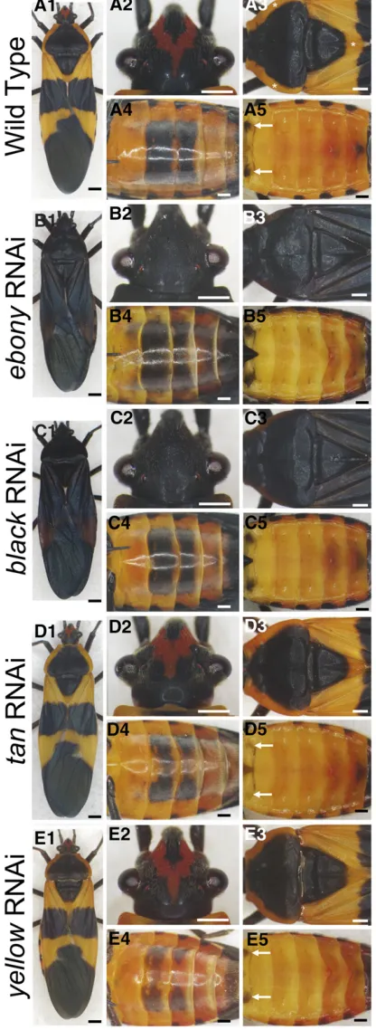
TheOncopeltusbody has warning coloration consisting of alter-nating black and orange subregions and patches (Figure 2A1). The dorsal head features a V-shaped orange stripeflanked by black pigmentation covering the rest of the head (Figure 2A2). Dorsal plates of the thoracic segments are primarily black, with the exception of orange at the lateral edges on the prothorax (T1) and posterior tip of the mesothorax (T2; asterisks, Figure 2A3). In contrast, most of the abdomen is orange, with the exception of two rectangular black patches on the ventral side of A3–A4 segments (Figure 2A4) and two small spots on the dorsal A1 segment (white arrows, Figure 2A5).
To examine the role of the melanin pathway inOncopeltus
color patterning, we clonedfive putative core melanin genes:
ebony,black,tan,yellow, andaaNAT. Phylogenetic analyses confirmed that the clones obtained are indeed Drosophila
consistent with previous RNAi analyses inOncopeltus show-ing changes in coloration encompassshow-ing the entire body (Liu et al. 2014). These findings, combined with an addi-tional RT-PCR result showing a significant reduction in the level ofblacktranscripts in all body regions (Figure S5), sup-port the conclusion that the observed phenotypes resulted from a systemic response.
Studies in Drosophila and Tribolium have shown that
ebonyplays the central role in suppressing black pigmenta-tion (Figure 1). On loss of funcpigmenta-tion ofebony, a global darken-ing of body pigmentation was observed in both species (Wittkopp et al. 2002b; Takahashi et al. 2007; Tomoyasu
et al.2009). Based on these observations, we expectedebony
RNAi Oncopeltus adults to exhibit black coloration through-out the body. Among 11 ebony RNAi adults, all but one exhibited a significant expansion of black melanization in the anterior body regions. As illustrated in Figure 2, B1–B3, all nonblack anterior subregions, including the orange V-shaped stripe on the head, the lateral edges on T1, and the posterior tip on T2, became black. However, in these 10 individuals, the expansion of black pigmentation was only moderate in posterior body regions. The black rectangle on the third abdominal segment (A3) expanded anteriorly into the A2 segment, whereas the A4 rectangle expanded poste-riorly into A5 and A6 segments (Figure 2B4). The remainder of the ventral abdomen and the non-black-pigmented dorsal abdomen were generally unaltered (Figure 2B5). Therefore, the different outcomes observed between anterior and pos-terior body regions suggested that whileebonywas critical for nonblack patches in the head and thorax, it played only a minor role in the majority of nonblack subregions in the abdomen.
Ebony is the core enzyme in the conversion of dopamine to NBAD, catalyzing the binding ofb-alanine to dopamine (Fig-ure 1). Therefore, the synthesis ofb-alanine, which is cata-lyzed by the product of theblackgene, is also critical for the NBAD branch (Wright 1987; Arakane et al.2009). Among 20blackRNAi adults, 18 showed an expansion of black col-oration in the head and thorax that covered the original orange pigmentation (Figure 2, C1–C3). In the ventral abdo-men, the effects were highly consistent among these 18 indi-viduals, showing little of the melanin expansion that was observed in ebonyknockdowns (compare Figure 2C4 with Figure 2B4). The dorsal abdomen and most of the nonblack
Figure 2 Functions ofebony,black,tan, and yellowin different body regions ofOncopeltusadults. (A1–A5) Wild-typeOncopeltusshows alter-nating black-orange patterning. Black pigmentation is present in the head (A2), thorax (A3), ventral abdomen (A4), and dorsal abdomen (A5). (B1–
B5) TheebonyRNAi adult phenotypes showed the expansion of black pigment in the head (B2) and thorax (B3), whereas such an expansion was only moderate in the ventral abdomen (B4) and barely noticeable in dorsal abdomen (B5). (C1–C5) TheblackRNAi adults showed a
portions of the ventral abdomen also remained unchanged (Figure 2, C4 and C5). These observations show thatblack
was required for the nonblack patches in the anterior body regions but not for the abdomen. Combined withebonyRNAi insights, these results establish that suppression of melanin by the NBAD branch is used differently between the anterior and posterior body regions ofOncopeltus.
While these results focus on the mechanism that creates nonblack subregions, it is equally important to understand the complementary process—the generation of black areas across the insect body. Studies inDrosophilahave shown that other than tyrosine hydroxylase (TH) and dopa decarboxy-lase (DDC), the enzymes required for the production of DOPA and dopamine, additional promoting enzymes are required for melanization (Wright 1987; Wittkoppet al.2002b; True
et al.2005; Jeonget al.2008). One of the essential enzymes is Tan, which counteracts Ebony in the NBAD branch (Figure 1), thus promoting melanin production (True et al. 2005; Jeong et al.2008). Functional analyses on tan have been reported only inDrosophila, which showed that its loss causes a global reduction of melanin patterns (True et al. 2005; Jeong et al. 2008). Of 37 tan RNAi Oncopeltus adults, 34 exhibited a consistent phenotype (Figure 2D1). Black patterns in the head and thorax were generally unaltered (Figure 2, D2 and D3). In contrast, the melanin patterns showed noticeable changes in the abdomen (Figure 2, D4 and D5). In particular, the two black spots on the dorsal A1 segment were significantly reduced (arrows in Figure 2D5). The same trend also was observed in the ventral A3 and A4 segments, exhibiting a reduction in the middle portion of each black rectangle (Figure 2D4). In three individuals, the degree of reduction was more pronounced, with either the left or right half of the A4 rectangle disappearing completely. It is worth noting that the intensity of black coloration within the remaining A3 and A4 rectangles was not significantly different from wild type (Figure S6,P.0.05). These obser-vations suggest thattanis required for proper patterning of black pigmentation in theOncopeltusabdomen but not for its intensity. Such a role fortanin abdominal melanin patterning is consistent with previous studies ofDrosophilaspecies (True
et al.2005; Jeonget al.2008).
As shown earlier, the depletions ofebony,black, andtandid not affect the majority of the nonblack subregions in the abdomen. These observations suggest that the NBAD branch of the melanin pathway is not involved in the generation of these subregions. Another possible candidate is arylalkyl-amine-N-acetyltransferase (aaNAT), which is the core gene in the NADA branch of the melanin pathway (Figure 1). Studies inDrosophilahave shown thataaNATis responsible for converting dopamine to NADA as a way of depleting mel-anin and creating colorless sclerotin (Wright 1987; Hinter-mannet al.1995; Brodbecket al.1998). Consistent with this, the depletion ofaaNAT causes an increase in melanization across the body in Bombyx(Zhanet al., 2010). In order to determine whether this mechanism also can explain the non-black subregions in the abdomen ofOncopeltus, we depleted
aaNAT infifth instar nymphs. The consequent adults, how-ever, showed no effect in color patterns in the head, thorax, or abdomen (Figure S7). This observation indicates that the NADA branch of the melanin pathway is not required for black patterns in these body regions.
In addition to the preceding four genes within the NBAD and NADA branches, we also tested the function ofyellow, another important gene that promotes melanin production (Wright 1987; Wittkoppet al.2002a, b; Jeonget al.2008; Tomoyasu et al.2009; Arakaneet al.2010). Although this gene is not within the NBAD branch, its depletion in Drosoph-ila causes a severe reduction in melanization across the whole body (Wittkopp et al. 2002b; Jeong et al. 2008). Among the 38yellowRNAi adults, 32 displayed a significant reduction in the intensity of black pigments (Figure S6,P,
0.05) in ventral A3 and A4 rectangles (Figure 2, E4 and E5). In addition, in 30 of 32 individuals, a reduction in the middle portions of the A3 and A4 rectangles also was observed (Fig-ure 2E4). These pigmented phenotypes in the abdomen are similar to those reported in Drosophila melanogaster
(Wittkoppet al.2002b; Jeonget al.2008). While abdomens showed an increase in average brightness of 51%, the reduc-tion in black intensity in the head and thorax was more mod-erate (Figure 2, E1 and E3), with an increase in average brightness of 28 and 15%, respectively (Figure S6). These
findings indicate thatyellowwas involved in regulating both the extent of melanin patterns and their intensity in the ab-domen but had much less effect in the thorax and abab-domen. Based onyellowandtanRNAi results, we speculate that these melanin-promoting factors may be critical for the melanin patterns in the posterior body regions.
Distinct black patterns between the forewing and hindwing are generated by different branches of the melanin pathway in Oncopeltus
The forewing and hindwing inOncopeltusalso have distinct melanin patterns. The forewing has an alternating black and orange pattern (Figure 3A1), whereas the hindwing is color-less at the proximal end and black throughout the distal re-gion (Figure 3A2). To determine whether the NBAD branch regulates the melanin patterns in both pairs of wings, we examined the wings ofebonyandblackRNAi adults. In both instances, black pigmentation greatly expanded into the or-ange subregions on the forewing (Figure 3, B1 and C1). How-ever, the depletion of either gene generated no noticeable changes in color patterns in the hindwing (Figure 3, B2 and C2). These observations indicate that the NBAD branch was required for repressing melanization in the nonmelanized areas on the forewing, whereas similar nonmelanized regions on the hindwing used a different mechanism.
Candidate genes responsible for suppressing melanization in the proximal hindwing include the NADA branch of the melanin pathway (Figure 1). BecauseaaNAT, the core gene within this branch, is reported to be responsible for convert-ing dopamine to colorless NADA sclerotin in Drosophila
it is possible that such a mechanism also can generate the color-less pattern in the hindwing ofOncopeltus. To test this hypoth-esis, we observed the hindwing coloration in aaNAT RNAi adults. In all 17 resulting RNAi individuals, the anal lobe region of the hindwing became melanized (Figure 3D2), whereas the black pigmentation of the forewing was not affected (Figure 3D1). These observations indicate thataaNATwas involved in suppression of melanin formation in the colorless anal lobe re-gion of the hindwing. However, there was no indication that this role was required for proper pigmentation of the forewing. In summary, theOncopeltusforewing and hindwing seem to use distinct mechanisms to generate nonblack subregions: the NBAD branch is applied to suppress melanization in the orange areas of the forewing, whereas the hindwing employs the NADA branch to generate the colorless anal lobe.
In addition to the melanin-suppressing factorsebony,black, andaaNAT, we tested the function of the melanin-promoting factorsyellowandtanin the black subregions ofOncopeltus
wings. InyellowRNAi adults, there was a significant reduc-tion in black intensity in both pairs of wings (Figure 3, E1 and E2, and Figure S6,P,0.05 in forewing andP 0.05 in hindwing). This effect was much greater in the hindwing (average brightness increased by 89%) than the forewing (19%) (Figure S6). In contrast, the depletion oftandid not generate any noticeable effect on the black patterns of either the forewing or hindwing (Figure 3, F1 and F2) nor signifi -cant reduction in their black intensity (Figure S6,P.0.05), indicating that this gene was not essential for wing melanin patterns. These observations suggest that the roles in wing melanization are distinct between different melanin-promot-ing factors:yellowis required for the proper intensity of black melanin in the wings, especially the hindwing, whereas
tan may not be involved at all in wing pigmentation in
Oncopeltus.
Differential involvement of pigmentation genes between forewing and hindwing correlates with their expression patterns
Previous studies in the wings of Drosophilaand Heliconius
have shown strong correlations between melanin patterns and the expression patterns of melanin genes (Wittkopp
et al.2002b; Gompelet al.2005; Fergusonet al.2011; Hines
et al.2012). In particular, such correlations have been seen in the D. melanogasterabdomen (Rebeizet al. 2009; Camino
et al. 2015) and forewing (Gompelet al. 2005) and occur in diverseDrosophilaspecies with divergent patterns of me-lanic pigmentation (Werneret al.2010; Arnoultet al.2013; Ordwayet al.2014; Caminoet al.2015), indicating that these correlations are functionally meaningful. Thus, we hypothe-sized that the difference in mechanisms regulating nonblack patterns between forewing (NBAD branch) and hindwing (NADA branch) comprise differential expressions of the rel-evant core genes. To test this hypothesis, we used in situ
hybridization to detect in developing wing pads ofOncopeltus
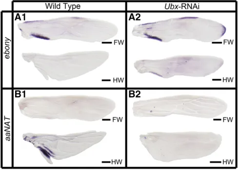
the expression patterns ofebonyandaaNAT, the two essential genes in the NBAD and NADA branches. Note that because formation of the cuticle precludes the expression analysis later in development (see Materials and Methods), we can only study early stages during which initial patterns are be-ginning to be laid out and do not fully correspond to thefinal patterns. As shown in Figure 4A1,ebonyexpression was ob-served in three distinct patches on the forewing: two are located on the anterior and posterior edges of the proximal portion and the other in the middle of the anterior edge. These locations correspond with the anterior and posterior margins of orange subregions on the forewing (Figure 3A1). The expression ofebonyon the hindwing, however, was not detectable (Figure 4A1), consistent with the fact thatebony
RNAi displayed no phenotype. However, aaNATexpression was not observed in the forewing but was present in the anal lobe region of the hindwing (Figure 4B1). This expression pattern ofaaNATwas strongly correlated with its RNAi phe-notype, where the colorless anal lobe region of the hindwing became melanized (Figure 3A2). Hence, the expression
patterns ofebonyandaaNATcorrelated with their functions in generating the nonblack patches on the forewing and hindwing.
These differences in RNAi phenotypes between forewing and hindwing pigmentation may be explained by the differ-ential activations of ebony and aaNAT. If so, this regional regulation of melanin genes would require specific selector genes (Wittkoppet al.2003; Wittkopp and Beldade 2009). In the butterfly Junonia coenia, the Hox gene Ultrabithorax
(Ubx) plays such a role by differential regulation of hindwing and forewing color patterns (Weatherbee et al.1999). Re-cently,Ubxwas shown to also control the identity of hindwing inOncopeltus(Medvedet al.2015), allowing us to test the generality of the selector gene’s role in regional regulation. Therefore, on depletion ofUbx, we would expectebonyto be expressed in the developing hindwing, whereas aaNAT
should be absent. As shown in Figure 4A2, the hindwing expresses ebonyat the proximal margins, which resembles the patterns observed in the wild-type forewing. However, the expression of aaNATis lost in the hindwing, even in a moderate phenotype showing an intermediate shape trans-formation (Figure 4B2). These findings indicate that Ubx
governs the differential activation of melanin genes between the wings. This further suggests that the different pigmenta-tion roles of melanin genes across differentOncopeltusbody regions, as shown by the present RNAi analyses, may be due to the functions of other region-specific regulatory genes.
The expression patterns ofblack,yellow, andtan, however, could not be detected usingin situhybridization. Note that the wing tissue is amenable to this procedure only during the middle part of the fifth nymphal stage (see Materials and Methods) when these three genes may not be active. To test this possibility, we examined the expression ofblack,yellow,
andtanduring the entirefifth nymphal stage (Figure S8). As predicted, expression of these genes was noticeable only dur-ing the latter half of the stage (Figure S8). These observations suggest that during the development of Oncopeltuswings, enzymes such as Ebony and AANAT, which use dopamine as direct substrates (Figure 1), perform their patterning roles at early-middlefifth nymphal stage. In contrast, Black, Yel-low, and Tan are turned on at a later stage.
Functions of melanin-suppressing factors in Periplaneta
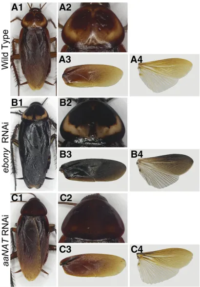
The preceding sections have shown that in Oncopeltus, melanin-suppressing factors (ebonyandaaNAT) play critical roles in the appearance of light patches of color in body regions that are mostly dark in coloration (e.g., the head, thorax, and wings). To determine whether such a mechanism is applicable to nonaposematic hemimetabolous insects, we examined the American cockroach, P. americana (Figure 5A1), whose uniform brown body coloration is also gener-ated by the melanin pathway (Lemonds 2015). In the mid-dle of the dorsal T1 plate, however, there are paired oval regions created by clear sclerotin (Figure 5A2). ThePeriplaneta
forewing is dark brown (Figure 5A3), whereas the hindwing features a brown anterior distal half and clear sclerotin in the posterior proximal half (Figure 5, A3 and A4). To de-termine how the melanin-suppressing factors can generate the color patterns inPeriplaneta, we performed RNAi deple-tion of ebony and aaNAT using previously reported gene fragments (Bembenek et al. 2005; Blenau and Baumann 2005).
Adults fromebonyRNAi treatments showed a significant increase in overall melanization, with black pigment covering most of their bodies (Figure 5B1). However, the clear sclero-tin on the dorsal T1 plate remained (Figure 5B2). The wings also showed a substantial increase in pigmentation (Figure 5, B3 and B4), especially the forewing, which became black in color (Figure 5B3). These observations indicate that in
Periplaneta,ebonyplays an essential role in maintaining the proper intensity of melanization in areas that are already melanized, but it has no effect on clear sclerotin regions. In other words, ebonycan regulate the darkness levels of the already-established melanin pattern, but it cannot alter the pattern. Thisfinding is similar to that of a previous report in
Tribolium, where the colorless patch on the hindwing was not altered byebonyRNAi (Tomoyasuet al.2009).
The aaNAT RNAi adults retained wild-type brown pig-mentation (Figure 5C1). However, the clear sclerotin areas in the T1 plate became melanized (Figure 5C2), with the overall coloration in the forewing and hindwing remaining similar to wild type (Figure 5, C3 and C4). These results reveal that aaNAT is responsible for creating some (T1 plate), but not all, colorless regions (posterior half of the hindwing). Alternatively, it is possible that these latter re-gions of clear sclerotin are produced by putative aaNAT
paralogs (Mehere et al. 2011; Barbera et al. 2013; Long
et al.2015). Thesefindings suggest that in bothOncopeltus
andPeriplaneta, some aspects of melanin patterning may be
conserved (e.g., creating clear sclerotin by using AANAT in hindwing inOncopeltusand in T1 inPeriplaneta).
Discussion
Pigmentation functions of melanin genes in Oncopeltus
In this study, we performed a comprehensive functional anal-ysis of the putative core melanin genes using RNAi. Although there has been limited success in RNAi silencing by direct body cavity injection inDrosophila, this approach has proven to be highly effective in other insects (Liet al.2015). This is espe-cially the case in Oncopeltus, which is characterized by a strong systemic gene depletion following RNAi treatment (Liu and Kaufman, 2004a, b, 2005; Angelini and Kaufman 2005). The recent RNAi analysis of the essential enzymes for
the production of black melanin in this species also showed a systemic reduction in coloration encompassing the entire body (Liu et al.2014). In this study, the whole-body RNAi response is documented by RT-PCR results showing similar reduction in the amount of transcript across different body regions in black RNAi individuals (Figure S5). Hence, the observed region-specific effects of RNAi against melanin genes are likely true phenotypes resulting from systemic sponses rather than low-penetrance phenotypes. These re-sults suggest distinct regional utilization of melanin genes.
The NBAD branch of the melanin pathway has been re-ported to be essential for melanin patterning inDrosophila
(Wright 1987; Wittkoppet al.2002b). In this pathway, both
blackandebonycan suppress melanin formation via the pro-duction of b-alanine and NBAD, respectively (Figure 1). In
Oncopeltus, RNAi knockdown of these two genes caused the nonblack subregions to become black in the head (Figure 2, B2 and C2), thorax (Figure 2, B3 and C3), and forewing (Figure 3, B1 and C1), whereas most of the nonblack patches in the hindwing (Figure 3, B2 and C2) and abdomen (Figure 2, B4 and B5 and C4 and C5) were not affected. Thesefi nd-ings suggest that the melanin-suppressing role ofebonyand
black is crucial in the head, thorax, and forewing but not necessary for proper coloration in the hindwing and abdomen. This is in contrast with findings inDrosophilaandTribolium, where loss of function of either ebony or blackresults in an overall darkening of body pigmentation (Wright 1987; Wittkopp
et al.2002b; Arakaneet al.2009; Tomoyasuet al.2009). Another gene of the NBAD branch, tan, counteracts the function ofebonyand promotes black pigmentation by con-verting NBAD back to dopamine (Wright 1987; True et al.
2005; Jeonget al.2008).tanRNAi phenotypes inOncopeltus
indicate that it is essential for the black patterns present in the abdomen (Figure 2, D4 and D5) but is not a significant player in forming the melanin patterning in the head (Figure 2D2), thorax (Figure 2D3), and wings (Figure 3, F1 and F2). Again, these results are different from those in Drosophila, wheretanmutants display a reduction in melanization span-ning the entire body (Trueet al.2005; Jeonget al.2008).
Despite the fact that the reactions catalyzed by tanand
ebonyappear as a circuit from the biochemical perspective (Figure 1), in Oncopeltus, they are employed differently in distinct body regions. It is possible that in hemimetabolous insects, only half of this circuit is essential for melanization in one specific body region, whereas the other half might be active but not required for pigmentation. Instead, they might be involved in other biological processes such as behavior (Wittkopp and Beldade 2009). Subsequently, in more derived groups such as dipterans, the entire tan-ebonycircuit (both reactions) became fully involved in melanization (Trueet al.
2005). Extending future studies of tanto additional hemi-metabolous species may show how the NBAD branch of the melanin pathway has changed during insect evolution.
Finally, the NADA branch also can suppress melanin pro-duction by transforming dopamine to NADA, thus creating colorless tissue (Wright 1987). The key gene in this pathway
is aaNAT (Wright 1987; Hintermannet al.1995; Brodbeck
et al.1998). Here we observed that the role ofaaNATis re-stricted to the anal lobe region of the hindwing (Figure 3D2), which correlates with its expression profile (Figure 4B1). This observation is in contrast to the depletion ofaaNATin the silkwormBombyx, which causes a global darkening of the adult body (Zhanet al.2010; Osanai-Futahashiet al.2012). Thus, Oncopeltus appears to preferentially activate aaNAT
expression in wings to create the colorless tissue in the anal lobe region. It is worth noting thataaNATmay not be the only gene used in this region because the anterior portion of the proximal half of the hindwing remains colorless whenaaNAT
is depleted by RNAi (Figure 3D2). Future studies will need to assess whether other members of the AANAT family are in-volved in generating colorless patches in this region (Mehere
et al.2011; Longet al.2015).
The pigmented phenotypes ofyellowhave been reported in bothDrosophilaandTribolium, with distinct results. Loss of
yellow causes global reduction of melanin in Drosophila
(Wittkopp et al.2002b), whereas only the hindwing is af-fected in Tribolium (Tomoyasu et al. 2009; Arakane et al.
2010). InOncopeltus,yellowRNAi adults displayed a mix of features previously observed in Drosophila and Tribolium: black coloration in the abdomen and hindwing is greatly re-duced, but other body regions were only moderately affected
(Figure S6). Therefore, the restriction ofyellowfunction
ap-pears to have changed during insect evolution. At present, we cannot rule out the possibility that in regions where yellow
RNAi was observed to have a moderate effect, other yellow
family members might play additional roles in melanization (Hanet al.2002). However, currently the functional roles of most of the yellow family genes are yet to be determined. Even for the yellow gene itself, despite the fact that it is known to be required for melanization in Drosophila and
Tribolium (Wright 1987; Wittkopp et al. 2002a, b; Jeong
et al.2008; Tomoyasuet al.2009; Arakaneet al.2010; Bray
et al. 2014), its enzymatic activity has not yet been estab-lished. In terms of the position ofyellowin the melanin path-way, two different hypotheses have been proposed (Figure 1): it may be essential for the production of either DOPA melanin (Wright 1987; Walteret al.1996) or dopamine mel-anin (Wittkoppet al.2002b, 2003). Our present results and another recent report (Liuet al.2014) show that black pig-ment in the hindwing is dopamine melanin, suggesting that
yellowmay be involved only in dopamine melanin production. Overall, our results show that the pigmentation functions of thefive genes under study are regionalized inOncopeltus. This is in contrast with previous studies inDrosophila,Tribolium, and
Bombyx, in which depletion of ebony, black, tan, aaNAT, or
yellow resulted in alteration of melanin patches throughout the body (Wright 1987; Walter et al.1996; Wittkoppet al.
2002b; Trueet al.2005; Gibertet al.2007; Jeonget al.2008; Arakaneet al.2009; Tomoyasuet al.2009; Zhanet al.2010; Osanai-Futahashiet al.2012). Hence, the insight from Onco-peltus is that the entire melanin pathway can be split into different sections that are used in different body regions.
In this study, the regional patterns of melanin genes are observed from analyses at a whole-body scale. To obtain a deeper comprehension of how complex melanin patterns are generated will require analyzing pigmentation genes at afiner morphologic scale (e.g., a subregion of a segment). Classical studies inDrosophilahave shown that selector genes involved in a general anteroposterior axis determination, such as
hedgehog (hh), engrailed (en), and optomotor-blind (omb), also regulate melanin patterning in the abdominal segments (Kopp and Duncan 1997; Koppet al.1997). It is tempting to speculate that similar mechanisms may account for the pres-ence of centrally positioned black rectangles on A3 and A4 segments in Oncopeltus. Also, other axis-determination mechanisms, especially mediolateral, may also be involved in generating the black spots located on the lateral edges of each abdominal segment (Figure 2A5). Furthermore, it is interesting to note that hh anden regulate melanin genes in a cell-autonomous manner in Drosophila (Kopp et al.
1997). In contrast, melanin genes themselves can function nonautonomously, semiautonomously, or autonomously in different body regions (Hanna 1953; Hotta and Benzer 1970; Boryczet al.2002; Wittkoppet al.2002b; Trueet al.
2005). At present, we do not know whether such region-specific cell autonomy of melanin genes also may account for production of the regional color patterns observed in
Oncopeltus. Extending future studies in this direction will be required to gain a better understanding of the regulatory mechanisms that generate species-specific melanin patterns in insects in general.
Region-specific employment of melanin genes
To better understand the regional utilization of melanin genes, we provide a summary of the general principles that
Figure 6 Three proposed modes of insect melanin patterning, as illus-trated by pigmentation inOncopeltus. Thefinal melanin patterns (right) are generated from the preliminary background (left) via different modes of melanin patterning. In the head, thorax, and forewing, where the background is fully black, the nonmelanin patches are generated by
appear to guide melanin patterning (Figure 6). In this sum-mary, we refer to a putative case as the “preliminary back-ground,” in which only the basic enzymes required for melanin production, such as TH and DDC, would be active. In other words, none of the promoting or melanin-suppressing factors function in such a background. Under the
first scenario, the“painting”mode would be used in a body region that lacks melanin patterns, such as the Oncopeltus
abdomen, which is predominantly orange. The preliminary background in this case would be nonmelanin because most of the abdomen may not be capable of producing black pig-ments. The dark melanin is“painted”onto specific areas of this background, which requires melanin-promoting factors (tanandyellow) to ensure the proper boundary and intensity of black patterns. Under a second scenario, the “erasing” mode would apply to a body region where dark melanin is the predominant pigment (as observed in the head, thorax, and forewing inOncopeltus, as well as the body and wings of
Periplaneta). In this instance, the preliminary background would be fully melanized, and the melanin suppressors such as ebony, black, and aaNAT are used for“erasing”melanin production in a specific area or for lowering the overall mel-anin intensity. In addition to situations where one of these two modes is used exclusively, there are also regions in which the melanin-promoting factors and suppressors can both play active roles, such as the hindwing inOncopeltus, where melanin and nonmelanin subregions are equally distrib-uted. Under this scenario, the preliminary background is defined as a region undergoing melanization but lacking a proper level of intensity. Finalizing the melanin patterns requires both the melanin-suppressing factor (aaNAT), which generates the nonmelanin patches, and the melanin-promoting factor (yellow), which intensifies the dark color within melanin patches.
In terms of their generality, these three scenarios can account for most of the functional results in the previously studied species. The painting mode may be evidenced in the dark-colored pterostigma on the hindwing of Tribolium. In those studies, this specific black pattern becomes lighter in
yellow RNAi individuals, whereas ebonyRNAi has no effect on theflanking nonblack subregions (Tomoyasuet al.2009; Arakaneet al.2010). The erasing mode may be evidenced in the butterflyPapilio, where the default fully melanized fore-wing was observed inebonymutant adults (Kochet al.2000). Another example is the depletion ofebonyorblackin Tribo-lium, which results in the general blackening of the whole body (Arakaneet al.2009; Tomoyasuet al.2009). In addition to the present results inOncopeltus, the mixed mode has been fully confirmed only inD. melanogaster, where functional anal-yses revealed thatebonyandyellowcontribute equally to both wing and body pigmentation (Wittkoppet al.2002b; Gompel
et al.2005). In general, our summary of the principles of mel-anin patterning can serve as a practical framework explaining the diversity in melanin coloration observed in previously re-ported insects. A broader taxonomic sampling in basal groups, from which we can infer the ancestral melanin patterning, will
be required to determine whether this framework can be ap-plied to a wider range of insect species.
Acknowledgments
We thank Victor Medved for help with cloning gene fragments in Oncopeltus. We also thank Mark VanBerkum, Patricia Wittkopp, William Branford, and two anonymous re-viewers for helpful comments that greatly improved this manuscript. This work was supported in part by National Institutes of Health grant GM-071927 to A.P. and a WSU Rumble Fellowship to J.L. J.H.M.’s participation was facili-tated by National Science Foundation grants IOS-0950416 and IOS-1354667 and a HITS grant from the Huck Institutes of the Life Sciences.
Literature Cited
Angelini, D. R., and T. C. Kaufman, 2005 Functional analyses in the milkweed bug Oncopeltus fasciatus (Hemiptera) support a role for Wnt signaling in body segmentation but not appendage development. Dev. Biol. 283: 409–423.
Arakane, Y., J. Lomakin, R. W. Beeman, S. Muthukrishnan, S. H. Gehrke et al., 2009 Molecular and functional analyses of amino acid decarboxylases involved in cuticle tanning in Tribo-lium castaneum. J. Biol. Chem. 284: 16584–16594.
Arakane, Y., N. T. Dittmer, Y. Tomoyasu, K. J. Kramer, S. Muthukrishnan et al., 2010 Identification, mRNA expression and functional analysis of several yellow family genes in Tribolium castaneum. Insect Biochem. Mol. Biol. 40: 259–266.
Arnoult, L., K. F. Su, D. Manoel, C. Minervino, J. Magrina et al., 2013 Emergence and diversification of fly pigmentation through evolution of a gene regulatory module. Science 339: 1423–1426.
Barbera, M., B. Mengual, J. M. Collantes-Alegre, T. Cortes, A. Gonzalez et al., 2013 Identification, characterization and analysis of expression of genes encoding arylalkylamine N-acetyltransferases in the pea aphid Acyrthosiphon pisum. Insect Mol. Biol. 22: 623–634.
Bembenek, J., K. Sakamoto, and M. Takeda, 2005 Molecular clon-ing of a cDNA encodclon-ing arylalkylamine N-acetyltransferase from the testicular system of Periplaneta americana: primary protein structure and expression analysis. Arch. Insect Biochem. Physiol. 59: 219–229.
Blenau, W., and A. Baumann, 2005 Molecular characterization of the ebony gene from the American cockroach, Periplaneta amer-icana. Arch. Insect Biochem. Physiol. 59: 184–195.
Borycz, J., J. A. Borycz, M. Loubani, and I. A. Meinertzhagen, 2002 tan and ebony genes regulate a novel pathway for trans-mitter metabolism at fly photoreceptor terminals. J. Neurosci. 22: 10549–10557.
Bray, M. J., T. Werner, and K. A. Dyer, 2014 Two genomic regions together cause dark abdominal pigmentation in Drosophila ten-ebrosa. Heredity 112: 454–462.
Brodbeck, D., R. Amherd, P. Callaerts, E. Hintermann, U. A. Meyer et al., 1998 Molecular and biochemical characterization of the aaNAT1 (Dat) locus in Drosophila melanogaster: differential expression of two gene products. DNA Cell Biol. 17: 621–633. Camino, E. M., J. C. Butts, A. Ordway, J. E. Vellky, M. Rebeizet al.,
Chesebro, J., S. Hrycaj, N. Mahfooz, and A. Popadic, 2009 Diverging functions of Scr between embryonic and post-embryonic de-velopment in a hemimetabolous insect, Oncopeltus fasciatus. Dev. Biol. 329: 142–151.
Ferguson, L. C., L. Maroja, and C. D. Jiggins, 2011 Convergent, modular expression of ebony and tan in the mimetic wing pat-terns of Heliconius butterflies. Dev. Genes Evol. 221: 297–308. Gibert, J. M., F. Peronnet, and C. Schlotterer, 2007 Phenotypic plasticity in Drosophila pigmentation caused by temperature sen-sitivity of a chromatin regulator network. PLoS Genet. 3: e30. Gompel, N., B. Prud’homme, P. J. Wittkopp, V. A. Kassner, and S. B.
Carroll, 2005 Chance caught on the wing: cis-regulatory evo-lution and the origin of pigment patterns in Drosophila. Nature 433: 481–487.
Han, Q., J. Fang, H. Ding, J. K. Johnson, B. M. Christensenet al., 2002 Identification of Drosophila melanogaster yellow-f and yellow-f2 proteins as dopachrome-conversion enzymes. Biochem. J. 368: 333–340.
Hanna, A., 1953 Non-autonomy ofyellowin gynandromorphs of Drosophila melanogaster. J. Exp. Zool. 123: 523–560.
Hines, H. M., R. Papa, M. Ruiz, A. Papanicolaou, C. Wang et al., 2012 Transcriptome analysis reveals novel patterning and pig-mentation genes underlying Heliconius butterfly wing pattern variation. BMC Genomics 13: 288.
Hintermann, E., P. Jeno, and U. A. Meyer, 1995 Isolation and characterization of an arylalkylamine N-acetyltransferase from Drosophila melanogaster. FEBS Lett. 375: 148–150.
Hotta, Y., and S. Benzer, 1970 Genetic dissection of the Drosoph-ila nervous system by means of mosaics. Proc. Natl. Acad. Sci. USA 67: 1156–1163.
Jeong, S., M. Rebeiz, P. Andolfatto, T. Werner, J. True et al., 2008 The evolution of gene regulation underlies a morpholog-ical difference between two Drosophila sister species. Cell 132: 783–793.
Koch, P. B., B. Behnecke, and R. H. ffrench-Constant, 2000 The molecular basis of melanism and mimicry in a swallowtail but-terfly. Curr. Biol. 10: 591–594.
Kopp, A., and I. Duncan, 1997 Control of cell fate and polarity in the adult abdominal segments of Drosophila by optomotor-blind. Development 124: 3715–3726.
Kopp, A., M. A. Muskavitch, and I. Duncan, 1997 The roles of hedgehog and engrailed in patterning adult abdominal seg-ments of Drosophila. Development 124: 3703–3714.
Lemonds, T. R., 2015 The contribution of the melanin pathway to overall body pigmentation changes during ontogenesis of Periplaneta Americana. Master of Science Thesis, Wayne State University, Detroit.
Li, H., and A. Popadic, 2004 Analysis of nubbin expression pat-terns in insects. Evol. Dev. 6: 310–324.
Li, Z., B. Zeng, L. Ling, J. Xu, L. Youet al., 2015 Enhancement of larval RNAi efficiency by over-expressing Argonaute2 in Bombyx mori. Int. J. Biol. Sci. 11: 176–185.
Liu, J., T. R. Lemonds, and A. Popadic, 2014 The genetic control of aposematic black pigmentation in hemimetabolous insects: insights from Oncopeltus fasciatus. Evol. Dev. 16: 270–277. Liu, P. Z., and T. C. Kaufman, 2004a Kruppel is a gap gene in
the intermediate germband insect Oncopeltus fasciatus and is required for development of both blastoderm and germband-derived segments. Development 131: 4567–4579.
Liu, P. Z., and T. C. Kaufman, 2004b hunchback is required for suppression of abdominal identity, and for proper germband growth and segmentation in the intermediate germband insect Oncopeltus fasciatus. Development 131: 1515–1527.
Liu, P. Z., and T. C. Kaufman, 2005 even-skipped is not a pair-rule gene but has segmental and gap-like functions in Oncopeltus
fasciatus, an intermediate germband insect. Development 132: 2081–2092.
Long, Y., J. Li, T. Zhao, G. Li, and Y. Zhu, 2015 A new arylalkyl-amine N-acetyltransferase in silkworm (Bombyx mori) affects integument pigmentation. Appl. Biochem. Biotechnol. 175: 3447–3457.
Medved, V., J. H. Marden, H. W. Fescemyer, J. P. Der, J. Liuet al., 2015 Origin and diversification of wings: Insights from a neo-pteran insect. Proc. Natl. Acad. Sci. USA 112: 15946–15951. Mehere, P., Q. Han, B. M. Christensen, and J. Li, 2011 Identification
and characterization of two arylalkylamine N-acetyltransferases in the yellow fever mosquito, Aedes aegypti. Insect Biochem. Mol. Biol. 41: 707–714.
Ordway, A. J., K. N. Hancuch, W. Johnson, T. M. Wiliams, and M. Rebeiz, 2014 The expansion of body coloration involves coordi-nated evolution in cis and trans within the pigmentation regula-tory network of Drosophila prostipennis. Dev. Biol. 392: 431–440. Osanai-Futahashi, M., T. Ohde, J. Hirata, K. Uchino, R. Futahashi et al., 2012 A visible dominant marker for insect transgenesis. Nat. Commun. 3: 1295.
Rebeiz, M., J. E. Pool, V. A. Kassner, C. F. Aquadro, and S. B. Carroll, 2009 Stepwise modification of a modular enhancer underlies adaptation in a Drosophila population. Science 326: 1663–1667.
Takahashi, A., K. Takahashi, R. Ueda, and T. Takano-Shimizu, 2007 Natural variation of ebony gene controlling thoracic pig-mentation in Drosophila melanogaster. Genetics 177: 1233–1237. Tomoyasu, Y., Y. Arakane, K. J. Kramer, and R. E. Denell, 2009 Repeated co-options of exoskeleton formation during wing-to-elytron evolution in beetles. Curr. Biol. 19: 2057–2065. True, J. R., S. D. Yeh, B. T. Hovemann, T. Kemme, I. A. Meinertzhagen
et al., 2005 Drosophila tan encodes a novel hydrolase required in pigmentation and vision. PLoS Genet. 1: e63.
Walter, M. F., L. L. Zeineh, B. C. Black, W. E. McIvor, T. R. Wright et al., 1996 Catecholamine metabolism and in vitro induction of premature cuticle melanization in wild type and pigmenta-tion mutants of Drosophila melanogaster. Arch. Insect Biochem. Physiol. 31: 219–233.
Weatherbee, S. D., H. F. Nijhout, L. W. Grunert, G. Halder, R. Galant et al., 1999 Ultrabithorax function in butterfly wings and the evolution of insect wing patterns. Curr. Biol. 9: 109–115. Werner, T., S. Koshikawa, T. M. Williams, and S. B. Carroll,
2010 Generation of a novel wing colour pattern by the Wing-less morphogen. Nature 464: 1143–1148.
Wittkopp, P. J., and P. Beldade, 2009 Development and evolution of insect pigmentation: genetic mechanisms and the potential consequences of pleiotropy. Semin. Cell Dev. Biol. 20: 65–71. Wittkopp, P. J., K. Vaccaro, and S. B. Carroll, 2002a Evolution of
yellow gene regulation and pigmentation in Drosophila. Curr. Biol. 12: 1547–1556.
Wittkopp, P. J., J. R. True, and S. B. Carroll, 2002b Reciprocal functions of the Drosophila yellow and ebony proteins in the development and evolution of pigment patterns. Development 129: 1849–1858.
Wittkopp, P. J., S. B. Carroll, and A. Kopp, 2003 Evolution in black and white: genetic control of pigment patterns in Dro-sophila. Trends Genet. 19: 495–504.
Wright, T. R., 1987 The genetics of biogenic amine metabolism, sclerotization, and melanization in Drosophila melanogaster. Adv. Genet. 24: 127–222.
Zhan, S., Q. Guo, M. Li, J. Li, X. Miaoet al., 2010 Disruption of an N-acetyltransferase gene in the silkworm reveals a novel role in pigmentation. Development 137: 4083–4090.
GENETICS
Supporting Information
www.genetics.org/lookup/suppl/doi:10.1534/genetics.115.186684/-/DC1
A Pathway Analysis of Melanin Patterning in a
Hemimetabolous Insect
Jin Liu, Thomas R. Lemonds, James H. Marden, and Aleksandar Popadic´
Fig. S1.
Phylogenetic analysis of
Drosophila melanogaster
and
Oncopeltus facsiatus
Fig. S2.
Phylogenetic analysis of
yellow
gene family.
Oncopeltus
Yellow (OfY‐Y)
is
orthologous to Yellow protein in
Drosophila melanogaster
(DmY‐Y) and
Tribolium
castaneum
(TcY‐Y).
Phylogeny was inferred using the Neighbor‐Joining method
based upon amino acid sequences. Branch support values are bootstrap percentages
from 500 replicates.
Fig. S3.
RT‐PCR analysis of
ebony
,
black
,
aaNAT, tan, yellow,
and
Ubx
mRNA in
Oncopeltus
5
thnymphs. Only trace levels of individual gene transcripts were
Fig. S4.
RT‐PCR analysis of
ebony
and
aaNAT
mRNA in
Periplaneta americana
.
Fig. S5.
RT‐PCR analysis of
black
mRNA in isolated body regions of
Oncopeltus
5
thnymphs at Day8 of development. There is a significant reduction in black transcript
in every body region in
black
RNAi individuals compared to wild type 5
thnymphs.
Fig. S6.
The comparison of black intensity in isolated body regions of wild type,
tan
RNAi and
yellow
RNAi adults. The levels of black in the melanized subregions in the
head, thorax, forewing, hindwing, and ventral abdomen are shown in average
percent brightness. The error bars are showing the 95% confidence intervals. The
depletion of
tan
did not show significant changes in the black intensity in all body
regions (P > 0.05). The black intensity in
yellow
RNAi is significantly different from
that in wild type in all body regions (P < 0.05). The amount of reduction in black is
most significant in the hindwing and ventral abdomen, whereas it is moderate in the
head, thorax, and forewing. In both
tan
RNAi and
yellow
RNAi adults, the brightness
of the orange subregions of the dorsal abdomen are not significantly different from
those in the wild type (P > 0.05). Abbreviations: FW, forewing; HW, hindwing.
Fig. S7.
Phenotype of
aaNAT
RNAi in black coloration of
Oncopeltus
body. (A1‐A5)
Wild type fully‐melanized
Oncopeltus
adults establish black pigmentation in the
head (A2), thorax (A3), ventral abdomen (A4), and dorsal abdomen (A5). (B1‐B5)
aaNAT
RNAi adults showed similar black coloration as wild type, which can be
observed in the head (B2), thorax (B3), ventral abdomen (B4) and dorsal abdomen
(B5).
Fig. S8.
RT‐PCR analyses of
black
,
yellow
, and
tan
mRNA in the developing wings of
Oncopeltus
throughout the entire 5
thnymphal stage
.
The chosen time points are:
Day2 (initialization of adult melanin cycle), Day5 (formation of wing tissue), Day8
(localization of melanin enzymes), and freshly molted adults (initialization of
melanin process). The expression levels of the targeted mRNA are dynamic among
these four time points. At Day2 and Day5, none of these three genes were detectable
in the wings. Starting by Day8, the expression of
black, yellow
and
tan
showed up
with different patterns.
black
was expressed in both Day8 and adult forewings.
However, its expression in the hindwing was only observed at Day8. The expression
of
yellow
was observed in both wings at Day8, which were significantly reduced in
the adults.
tan
expression was very low on day 8 in both wings. The expression in
the forewing later reached its peak in adult, whereas the hindwing expression was
not detected at adult stage.
Primers for PCR cloning Primers for RT‐PCR Length of
fragment Corresponding AA residues of Dm orthologs Ofebony Forward: 5’ ACGACCGCAACTCTCAAACT 3’
5’ TCACCAGCTCTTCTCGTTGA 3’ Reverse: 5’ TGCTGAGTTAGAGGGCTGGT 3’ 5’ TGCCAGTAAAGCCTCTGGAT 3’
Forward: 5’ CTTCTCCCGACCTCATTCTG 3’
Reverse: 5’ GGTCCACAGGGCATCTAAAA 3’
2333 bp 135~822 (out of 879aa)
Ofblack Forward: 5’ GGTGCAGCGTCTTCTCCTAC 3’ 5’ GGCCGGTACTGACACTCTGT 3’ Reverse: 5’ AGAAGTTTGGGTGTGCCTTG 3’ 5’ TTGTTCCTTCACGCATCATC 3’
Forward: 5’ GGCCGGTACTGACACTCTGT 3’
Reverse: 5’ TTGTTCCTTCACGCATCATC 3’
1160 bp 147~532 (out of 575aa)
Ofyellow Forward: 5’ ATTCGCCTGGAAGTATGTGG 3’ 5’ GGACAAAGGTGTCCCGAGTA 3’ Reverse: 5’ GAGAATTCTGGCAGCTGAGG 3’ 5’ ATCGGCAACCTGTGGTAGAG 3’
Forward: 5’ AGTCCAGAAACACCCCTCCT 3’
Reverse: 5’ ATCGGCAACCTGTGGTAGAG 3’
1062 bp 80~408 (out of 541aa)
Oftan Forward: 5’ CGTCCGTTCCTTACTTCTCG 3’ Reverse: 5’ CCTGGCATTGCAGTTGAATA 3’
Forward: 5’ GGACATTCTACCAGCCCAAA 3’
Reverse: 5’ CAATCGATCTCCAGCCTGAT 3’
1338 bp 6~386 (out of 387aa)
Of
aaNAT Forward: 5’ TGGGCGAAAGAGAGAATGAC 3’ 5’ CCAGGAAGACGTGGAAAGAG 3’ Reverse: 5’ GTGCAGACTTTGCAGTGTGG 3’ 5’ TCGGTCTTCCATCATTGTCA 3’
Forward: 5’ CCAGGAAGACGTGGAAAGAG 3’
Reverse: 5’ CAAGGCTGTAGATGCTGTGG 3’
554 bp 30~206 (out of 240aa)
Paebony Forward: 5’ TCAAGACGTGGGTATGCAGT 3’ 5’ GAATGGCAAGACAGACCGAC 3’ Reverse: 5’ CATCTGCATCTGGGTGAACG 3’ 5’ TGCTGTGTTAGGGGATTGGT 3’
Forward: 5’ GAATGGCAAGACAGACCGAC 3’
Reverse: 5’ TGCTGTGTTAGGGGATTGGT 3’
880 bp 538~833 (out of 879aa)
Pa
aaNAT Forward: 5’ TGCGAAGCAACGAGTGTAAC 3’ 5’ CACCGATGGAGGTCTACGTT 3’ Reverse: 5’ AGGAACCAGTGGCAAATCAC 3’ 5’ AGGCTGTCCATTCTTGCAGT 3’
Forward: 5’ CACCGATGGAGGTCTACGTT 3’
Reverse: 5’ AGGCTGTCCATTCTTGCAGT 3’
646 bp 6~221 (out of 240aa)
![Figure 1 A summary of the melanin pathway in insects. [Redrawn fromWright (1987).] Black melanin originates from either DOPA or dopamine,which are converted from tyrosine by TH and DDC](https://thumb-us.123doks.com/thumbv2/123dok_us/1530641.1187670/2.603.48.294.47.148/figure-summary-redrawn-fromwright-originates-dopamine-converted-tyrosine.webp)