Analysis of Human Knee Joint during MMH
Using Soft Computing Technique
S.Dinesh1, J.Anish Jafrin Thilak2, T.Rajeshkumar3, P.Suresh4
M.E Scholar, Department of Mechanical Engineering, Karpagam College of Engineering, Coimbatore, Tamil Nadu,
India1
Assistant Professor, Department of Mechanical Engineering, Karpagam College of Engineering, Coimbatore,
Tamil Nadu, India2&3
Professor, Department of Mechanical Engineering, Karpagam College of Engineering, Coimbatore, Tamil Nadu, India4
ABSTRACT: Bio medical image processing is an emerging field for analyzing medical images to develop models for clinical analysis and medical intervention. Recent development in biomechanics, the model developed can be used for mechanical stress and vibrational analysis of human body parts. The Segmentation of joints image model from CT scan noise images are done using Canny Edge Detection Algorithm and Sobel Edge Detection Techniques. The most difficult problem is to find the correct boundary in a noisy image of joints. A standard result can be used to find out absolute edges from noise images. Finally solution is produced by the canny edge detection algorithm as a accurate boundary in a noise images of joints.
KEYWORDS: Finite Element Modelling, Sobel Edge Detection Techniques, Canny Edge Detection Algorithm, Magnitude and Edge Length, CT scan image of Knee joint.
I. INTRODUCTION
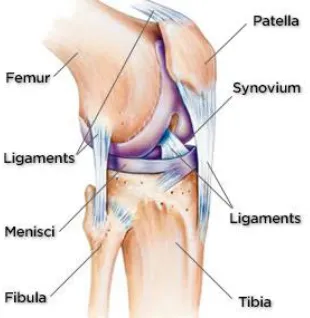
The production for knee joint is provided by composite structure. Most industrial plant carry out manual material handling operation. A stable solution along with minimization of error is produce through the variational operations. Approximately a large circle is implemented with the connection of many tiny straight lines. A more complex equation over a larger domain connects many simple element equation over many small sub domains named finite element is encompassed by FEM. During manual material handling employee get trouble on their joint. In order to avoid this trouble in joint finite element analysis should be performed. The part that plays a vital role in research is the knee joint region, which is a part of knee joint. In literature the relationship between anatomy knee region and knee joint are put together. The FEM modeling the joints is analyzed under some injuries. The explanation along with its uses in the field of orthopedics for the joint region is analyzed by finite element analysis. Edge detection technology is the fundamental tool used to elucidate proper information from the images. This provides the outline of an image. By using this tool noise get eliminated and to get better enhanced image. The boundary detection of the CT scanned image of the joints Canny Edge Detection Algorithm has been preferred which is based on the magnitude and edge length algorithm. Canny edge detection algorithm that is used to detect the boundary of the joints image from the noisy CT scanned image.
II.PROPOSED WORK SEQUENCE
The flow diagram of the proposed system of normal Knee image Edge Detection and ROI ( Region of Interest
Fig. 1. Work Sequence flow chart Normal Knee Anatomy EDGE DETECTION
CT SCAN NOISY IMAGE
AVERAGE MAGNITUDE IMAGE
DENSITY OF EDGE LENGTH INITIAL POSITION MAPPING
Fig. 2. Description of Normal Knee Anatomy
III. PRE-PROCESSING OF INITIAL POSITION OF EDGE PARAMETERS DETECTION
Step 1: To find the average magnitude
[1]
Step 2: To find the density of the edge length. The density of the edge length is find out from [2]
Where C(i,j) is the number of connected pixels at each position of pixel.
Step 3: To find the Initial position of map from summation of density of edge Length and average magnitude.
[3] Step 4: To find the thresholding of the initial position map. If
[4]
Then P(1, 2) is the initial position of the edge following. And then we obtained the initial position by setting Tmax to
92% of the maximum value. Figure 3 shows the significant results of preprocessing.
(d) (c)
Fig.3. (a). Noisy Knee image, (b). Average Magnitude Image, (c). Density of the Edge Length
(d). Final Noise removed image
IV. CANNY EDGE DETECTION ALGORITHM
Multi-stage algorithm in the canny edge detector allow to detect a wide range of edges in images. structural information from different vision objects can be extracted using step edge detection technique and reduce the amount of data to be processed. It has been employed in various computer vision systems. Canny has been proved for the requirements for the application of edge detection on various vision systems are relatively similar. An edge detection is the only solution to address these requirements and can be implemented in a wide range of situations. The general criteria for edge detection includes:
1. Detection of edge with low error rate.
2. The edge point detected from the operator should accurately localize on the center of the edge.
3. A given edge in the image should only be marked once, and there is no possibility for creation of false edges in the images.
[5]
Step 1:To find the average magnitude
[6]
Step 2:To find the density of the edge length. The density of the edge length is find out from [7]
Where C(i,j) is the number of connected pixels at each position of pixel.
Step 3:To find the Initial position of map from summation of average magnitude and density of edge Length.
[9]
Then P(1, 2) is the initial position of the edge following. And then we obtained the initial position by setting Tmax to
92% of the maximum value.
(a) (b)
(d) (c)
Fig.4. (a). Noisy Knee image (b). Density of the Edge Length (c). Initial Position map,
(d). Final Thresholding of edge map
V. SOBEL EDGE DETECTION TECHNIQUE
In this technique 2D spatial gradient measurement is performed on an image and also it emphasizes regions of high spatial frequency. Generally to find the approximate absolute gradient magnitude at each point in an input greyscale image it is used . In theory slightly, the operator consists of a pair of 3x3 convolution masks. One mask is quitly the
other rotated by 90o. It is very similar to the Roberts cross operator. These masks are designed to respond maximally to
edges running horizontally and vertically relative to the pixel grid, one mask for each of the two perpendicular locations. The masks can be utilised separately to the input image, to produce separate measurements of the gradient
component in any orientation that is Gx and Gy. These can be combined together to find the absolute magnitude of the
gradient at every each point and the orientation of that gradient. The gradient magnitude is given below in Figure 5.
Although typically, an approximate magnitude is computed using:
│G│=│Gx│+ │Gy│ [11]
Which is much faster to compute. The orientation of the angle edge (related to the pixel grid) giving rise to the spatial
gradient is given by:
α = arctan(Gy/Gx) - 3π/4 [12]
In that case, orientation 0 is taken to mean that the direction of maximum contrast from black to white runs from left to right on the image, and another angles are measured anti-clockwise from this. Often, this absolute magnitude is the only output the user look at the two components of the gradient are conveniently computed and added in a single pass over the input image using by pseudo convolution operator shown in Figure 5.
Using this mask the approximate magnitude is given by:
│G│=│(N1+2xN2+N3)–(N7+2xN8+N9)│+ (N3+2x N8+N9)–(N1+2xN4+N7)│ [13]
The Sobel operator is slower to compute than the Roberts cross operator, but its larger convolution mask smooth’s the input image to a greater extend and so makes the operator less sensitive to noise [5,6]. The problem can be avoided by using type of an image that supports pixel values with a larger range.
(a) (b)
(d) (c)
Fig.5. (a). Noisy Knee image (b). Density of the Edge Length (c). Initial Position map
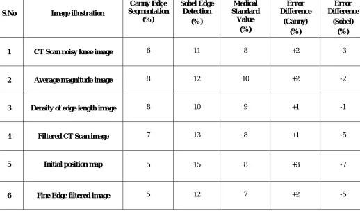
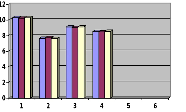
To evaluate the efficiency of the proposed method in addition to the visual inspection, The objects encompassed by the contour obtained using the five snake model and the suggested method are compared with that manually drawn by skilled doctors from the medical hospital from the above table shows the average results of probability of error in image segmentation of sobel edge detection and canny edge detection algorithm were compared with accepted medical values and also predict the error difference. From that it shows the error difference value is minimum and also negligible. So the canny edge detection algorithm produces nearer to the standard value. Fig.6 Shows the comparative analysis of canny edge detection value, Sobel Operator value and the Medical standard value which is collected from the standard Hospital.
Table 1. Average Results of Probability of Error in Image Segmentation
S.No Image illustration
Canny Edge Segmentation
(%)
Sobel Edge Detection
(%)
Medical Standard
Value (%)
Error Difference
(Canny) (%)
Error Difference
(Sobel) (%)
1 CT Scan noisy knee image 6 11 8 +2 -3
2 Average magnitude image 8 12 10 +2 -2
3 Density of edge length image 8 10 9 +1 -1
4 Filtered CT Scan image 7 13 8 +1 -5
5 Initial position map 5 15 8 +3 -7
0
2
4
6
8
10
12
1
2
3
4
5
6
Canny
Sobel
Normal
Fig 6. Comparative Analysis Graph for Canny Edge Detection value, Sobel Value
and Medical Standard value
VII. CONCLUSION
This paper provide the comparative analysis of different edge detection algorithm like Sobel edge detection and canny edge detection algorithm from that canny edge detection algorithm produce better results for noisy joints image. The proposed method can be applied for medical imaging and also be applied to any image processing problems.
REFERENCES
[1]. Bycoung-su Kim, Hyun Lee and Whoi-Yul Kim, “Rapid Eye Detection Method for Non-Glasses Type 3D Display on Portable Devices”, IEEE Transactions on consumer Electronics, 2010, Vol.56, No.5, pp.2498-2505.
[2]. C.C. Chibelushi, J.S. Mason, and F. Deravi, "Integration of acoustic and visual speech for speaker recognition," EUROSPEECH'93, 1993, pp.345-348.
[3]. Deborah Rankin, Bryan Scotney, Philip Morrow, Rod McDowell and Barbara Pierscionek, “Comparing and Improving Algorithms for Iris Recognition”, 13th International Machine Vision and Image Processing Conference, 2009, pp. 99-104.
[4]. K. Bae, S.-I. Noh, and J. Kim, “Iris feature extraction using independent component analysis,” in Proc. Conf. Audio and Video Based Biometric Person Authentication, Guildford, U.K., Jun. 2003, pp.838–844.
[5]. Natalia A.Schmid, Manasi V,Ketkar, Harshinder Singh and Bojan Cukic, “Performance Analysis of Iris-Based Identification System at the Matching Score Level”, IEEE Transactions on Information Forensics and Security, 2006, Vol.1, No.2, pp. 154-168.
[6]. Suresh P., Kesavan R., and M.Madheswaran., ‘Finite Element Modeling and Analysis of Magnetic Resonance Imaging of Lumbar Spine Using Pro/E Software’, International Journal of computational Intelligence and health Care Informatics (IJCIHCI), Vol.I, No.1 Page No: 50-56, 2008.
[7]. N. Theera-Umpon and S. Dhompongsa, ―Morphological granulometric features of nucleus in automatic bone marrow white blood cell classification, IEEE Trans. Info. Tech. in Biomed., vol. 11(3), pp. 353-359, 2007.
[8]. Suresh P. and Kesavan R., ‘Ergonomic Experimental Analysis of Eye Strain on VDT users: A strategic Prevention Perspective’, ICFAI Journal of Business Strategy, Vol.IV, No.3 Page No: 63-72, September 2007.
[9]. Antonious Rohlmann, Jorge Callisse and George Bergmann (1999), ‘Estimation of trunk muscle forces using the finite element method and in vivo loads measured by telemeterised internal spinal fixation devices’, Journal of Biomechanics, vol. 32, pp. 727 – 731.
Management and Engineering, 2009, pp. 714-717.
[10].R.M.Farouk, R.Kumar and K.A.Riad, “Iris matching using multi-dimensional artificial neural network”, IET Computer Vision, 2001, Vol.5, Issue 3, pp. 178-184.
[11].Ross and A.K. Jain, "Multimodal biometrics: an overview" Proceedings of 12th European Signal Processing Conference, 2004, pp.1121-1224.
[12].Ross, A.K. Jain, and J. Qian, "Information fusion in biometrics" Proceedings of Audio- and Video-based Biometric Person Authentication'01, June, 2001, pp. 354-359. Suresh P. and Kesavan R., ‘Ergonomic analysis of spine during lifting of loads by Finite Element Modeling – A Case Study’, Industrial Engineering Journal, Vol.1, Issue No.7, page No: 7-10, January 2009.
[13].A.K. Jain, A. Ross, and S. Prabhakar, "An introduction to biometric recognition," IEEE Trans. on Circuits and System for Video Technology Special Issue on Image- and Video-based Biometrics, 2004, vol. 14, pp. 4-20.
[14]. E.Punarselvam, P.Suresh and R.Parthasarathy, ‘Segmentation of CT Scan Lumbar Spine Image Using Median Filter and Canny Edge Detection Algorithm, International Journal of Computer Science and Engineering, Vol.5, No.9, September 2013, pp. 806-814.
[15].Agaian, S. S., Baran, T. A., & Panetta, K. A. (2003). Transform-based image compression by noise reduction and spatial modificat ion using Boolean minimization. IEEE Workshop on Statistical Signal Processing, 28 Sept.-1 Oct. pp. 226 – 229.
[16].Baker, S., & Nayar, S. K. (1996). Pattern rejection. Proceedings of IEEE Conference Computer Vision and Pattern Recognition, 544-549. [17].Canny, J. F. (1986). A computational approach to edge detection. IEEE Trans Pattern Analysis and Machine Intelligence, 8(6), 679-698. [18].Chang-Huang, C. (2002). Edge detection based on class ratio. 152, sec.3, Peishen Rd., Shenkeng, Taipei, 22202, Taiwan, R.O.C.
[19].Folorunso O., Vincent, O. R., & Dansu, B. M. (2007). Image edge detection: A knowledge management technique for visual scene analysis. Information Management and Computer Security, 15(1), 23-32.