YEASTBOOK
GENOME ORGANIZATION & INTEGRITY
Everything You Ever Wanted to Know About
Saccharomyces cerevisiae
Telomeres: Beginning
to End
Raymund J. Wellinger*,1and Virginia A. Zakian†,2
*Department of Microbiology and Infectiology, RNA-Group, Faculty of Medicine, Université de Sherbrooke, Sherbrooke, QC J1H 5N4, Canada, and †Department of Molecular Biology, Princeton University, Princeton, New Jersey 08544
ABSTRACTThe mechanisms that maintain the stability of chromosome ends have broad impact on genome integrity in all eukaryotes. Budding yeast is a premier organism for telomere studies. Many fundamental concepts of telomere and telomerase function werefirst established in yeast and then extended to other organisms. We present a comprehensive review of yeast telomere biology that covers capping, replication, recombination, and transcription. We think of it as yeast telomeres—soup to nuts.
TABLE OF CONTENTS
Abstract 1073
Introduction 1074
Sequence and Structure of Telomeric Regions 1074
Telomeric Chromatin 1076
Telomere binding proteins: direct binders
and associated proteins 1076
Telomere dedicated proteins vs.proteins doing double duty 1079
How many more genes affect telomere biology? 1079
The Capping Function 1079
Classical chromosome capping 1079
Alternative ways of capping 1080
Crosstalk between DNA damage checkpoint activation
and DNA repair 1081
Regulated resection 1081
Telomere Replication 1082
Continued
Copyright © 2012 by the Genetics Society of America doi: 10.1534/genetics.111.137851
Manuscript received December 14, 2011; accepted for publication March 29, 2012 Available freely online through the author-supported open access option.
1Present address: Department of Microbiology and Infectious Diseases, Faculty of Medicine, Université de Sherbrooke, Québec, J1E 4K8 Canada.
CONTENTS,continued
Semiconservative replication of telomeric
and subtelomeric DNA 1082
Telomere maintenance via telomerase 1083
End replication problems and the discovery of telomerase: 1083
Characteristics of components of the telomerase holoenzyme: 1085
Est1p: 1085
Est2p: 1086
Est3p: 1086
TLC1: 1086
Regulation of telomerase by the cell cycle: 1086
Regulation of telomerase by telomere length: 1087
In vitrotelomerase assays: 1089
Telomere maintenance via recombination 1090
Type I survivors: 1091
Type II survivors: 1091
Telomeric length control by telomeric rapid deletions: 1091
Transcription at Telomeres 1091
Telomere-associated RNA 1091
Telomere silencing or TPE 1092
Telomeres and Nuclear Organization 1093
Higher order chromatin structure and telomere folding 1093
Telomere organization in mitotic cells 1094
Telomeres in meiosis 1095
Outlook 1096
E
ukaryotic chromosomes are linear DNA molecules with physical ends, called telomeres. It is estimated that as many as 10,000 DNA damaging events occur each day in every cell in the human body (Loeb 2011). Perhaps the most hazardous of these events are double-stranded DNA breaks (DSBs), which create chromosome ends at internal sites on chromosomes. Thus, a central question is how cells distin-guish natural ends or telomeres from DSBs. Telomeres on one hand are essential for the stable maintenance of chro-mosomes: they must be retained—they cannot be lost by degradation or fused with other ends. Exactly the opposite applies to DSBs: they must be repaired by either homolo-gous or nonhomolohomolo-gous recombination, and this repair of-ten involves regulated degradation of the DSB. In fact, unrepaired DSBs lead to cell cycle arrest to provide time for their repair. Capping is used to describe how telomeres prevent their degradation and recombinational fusion (Muller 1938; McClintock 1939). Perhaps as a consequence of capping, the regions near telomeres are gene poor. In many organisms, telomere proximal genes are subjected to a special type of transcriptional regulation called telomere position effect (TPE), where transcription of genes near te-lomeres is metastably repressed. Another key role for telo-meres is to provide the substrate for a special mechanism of replication. Telomere replication is carried out by telomer-ase, a specialized ribonucleoprotein complex that ismecha-nistically related to reverse transcriptases (Greider and Blackburn 1987).
The biology of telomerase has broad ramifications for human health and aging. Therefore, the discovery of telomerase and studies on telomere capping by Elizabeth Blackburn, Carol Greider, and Jack Szostak, were honored with the 2009 Nobel Prize in Medicine. All three prize winners carried out research in single-cell organisms, in-cluding budding yeast. As described in this review, Saccha-romyces cerevisiae continues to be a premier organism for telomere research.
Sequence and Structure of Telomeric Regions
Like most organisms whose telomeres are maintained by telomerase, the ends ofS. cerevisiaechromosomes consist of nonprotein coding repeated DNA (Figure 1A). There are 3006 75 bp of simple repeats, typically abbreviated C1-3A/TG1-3.
from preexisting telomeric DNA (Wang and Zakian 1990; Teixeira et al.2004). When many copies of the same telo-mere are sequenced from a given colony, the exact sequence of the internal half is the same from telomere to telomere while the terminal half turns over much more rapidly (Wang and Zakian 1990). Thus, under most conditions, only the terminal half of the telomere is subject to degradation and/or telomerase lengthening. These repeats in conjunc-tion with the proteins that bind them are necessary and sufficient for telomere function.
As in most eukaryotes, the very ends ofS. cerevisiae chro-mosomes are not blunt ends. Rather the G-rich strand extends to form a 39 single strand tail or G tail (Figure 1A). Throughout most of the cell cycle, G tails are short, only 12 to 15 nucleotides (nt) (Larriveeet al.2004). How-ever, G tails are much longer, $30–100 nt in size, during a short period in late S/G2 phase when they can be detected readily by nondenaturing Southern hybridization (Wellinger
et al.1993a,b). Long G tails are not due solely to telomerase-mediated lengthening as they are seen in late S/G2 phase even in telomerase-deficient cells (Wellinger et al. 1996;
Dionne and Wellinger 1998). G tails are generated by cell-cycle–regulated C-strand degradation, which is dependent on the kinase activity ofCdk1p(Cdc28p; Franket al.2006; Vodenicharov and Wellinger 2006). This generation is oblig-atorily linked to semiconservative DNA replication, which occurs prior to C-strand degradation (Wellinger et al.
1993a; Dionne and Wellinger 1998).
Also similar to most organisms, yeast telomeric regions contain subtelomeric, middle, repetitive elements, often called TAS elements (telomere associated sequences; Figure 1A andhttp://www.nottingham.ac.uk/biology/people/louis/ telomere-data-sets.aspx).S. cerevisiaehas two classes of TAS elements, X and Y9. Y9 is found in zero to four tandem copies immediately internal to the telomeric repeats (Chan and Tye 1983a,b). About half of the telomeres in a given strain lack Y9, and the identity of Y9-less telomeres differs from strain to strain (Horowitz et al. 1984; Zakian et al.
1986). Y9comes in two sizes, Y9long (6.7 kb) and Y9short (5.2 kb) (Chan and Tye 1983a,b), which differ from each other by multiple small insertions/deletions (Louis and Haber 1992). X is present at virtually all telomeres and is much more heterogeneous in sequence and size. Although X is found on all telomeres, it is composed of a series of repeats, many of which are present on only a subset of telomeres. When telomeres contain both X and Y9, X is centromere proximal to Y9. Short tracts of telomeric DNA are sometimes found at the Y9–X and Y9–Y9 junctions (Walmsleyet al.1984; Figure 1A).
Subtelomeric regions are dynamic, undergoing frequent recombination (Horowitz et al. 1984; Louis and Haber 1990). Moreover, subtelomeric repeats diverge rapidly even among related yeast strains (Chan and Tye 1983a,b). X and Y9both contain potential replication origins or ARS elements (autonomously replicating sequences) whose presence probably contributes to the dynamic nature of subtelomeric regions. X and Y9 have binding sites for multiple tran-scription factors, whose identity differs from telomere to telomere (Mak et al. 2009). Because the sequence of subtelomeric regions and the proteins that bind them are variable, their presence can confer distinct behaviors on in-dividual telomeres.
Whereas complete loss of the C1-3A/TG1-3 telomeric repeats from a chromosome end results in extremely high loss rates for the affected chromosome, chromosomes that lack X and Y9at one (Sandell and Zakian 1993) or even both ends have normal mitotic stability and go through meiosis with ease (S. S. Wang and V. A. Zakian, unpublished results). However, Y9 amplification by recombination can provide a telomere maintenance function to cells lacking telomerase (Lundblad and Blackburn 1993). Y9can also move by a transposition-like RNA-mediated process (Maxwellet al.
2004).
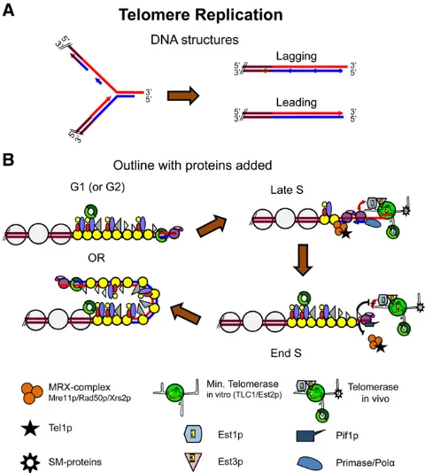
Ty5 is a transposable element found only in heterochro-matin, including subtelomeric DNA. The number of Ty5 elements varies from strain to strain. The S288C strain has eight Ty5 insertions: six near telomeres and two near the Figure 1 DNA structure and major protein components of telomeres.
(A) DNA arrangement at telomeres indicating the subtelomeric X and Y9
elements as well as the terminal repeat sequences. Red strand, G-rich strand with 39overhanging end and blue strand, C-rich strand with 59
HMR silent mating type locus (Zouet al.1995). This chro-mosomal distribution is quite different from that of other classes of Ty elements, which are found close to tRNA genes. Movement of Ty5 to telomeres andHMRis regulated by the targeting domain of the Ty5-encoded integrase that inter-acts directly withSir4p, one of the silencing proteins found in telomeric regions (Xieet al.2001).
Telomeric Chromatin
Telomere binding proteins: direct binders and associated proteins
Table 1 presents a list of proteins that act at telomeres, di-vided into functional categories. Many of these proteins have multiple roles and could be listed in more than one category. The protein complexes associated with telomeres can be subdivided according to the three regions to which they bind: (A) subtelomeric areas containing Y9and X, (B) double-stranded terminal repeat area, and (C) the 39G tail (Figure 1).
(A) Subtelomeric regions are classified into XY9 and X-only ends. While most of the subtelomeric DNA is likely organized in nucleosomes (Wrightet al.1992), the cores of the X elements have a low histone content, and nucleosomes near the Xs have histone modifications characteristic of si-lenced regions such as unacetylated lysine 16 on histone H4 (H4K16) (Zhu and Gustafsson 2009). Consistent with these data, the NAD+-dependent histone deacetylase Sir2p, a H4K16 deacetylase, as well as Sir3p, are also enriched over X repeats (Imai et al. 2000; Zhu and Gustafsson 2009; Takahashiet al.2011), and the area around many X elements is transcriptionally silent (Pryde and Louis 1999). The X elements on XY9telomeres are organized similarly as on X-only telomeres (Takahashi et al. 2011). However, on the distal Y9elements, the overall density of nucleosomes as well as the occurrence of H4K16ac is similar to euchromatic areas. In addition, Sir2pandSir3pare not detected in this region (Zhu and Gustafsson 2009; Takahashi et al. 2011). Collectively, these data suggest that on X-only telomeres, the subtelomeric DNA elements are organized into silenced chromatin that demarcates the terminal area from more in-ternal regions. On XY9telomeres, the distal Y9area is orga-nized into chromatin that resembles that of expressed areas with the X element, again acting as a demarcation zone (Fourelet al.1999; Pryde and Louis 1999; Takahashiet al.
2011). Thus, emerging evidence points toward differences in behavior depending on subtelomeric repeat content and perhaps even individual chromosomal context.
g-H2A, which is generated by Mec1p/Tel1p-dependent phosphorylation, is also enriched in subtelomeric chromatin (Kimet al.2007; Szilardet al.2010). Since this modification normally marks damaged DNA, which activates checkpoints, it is unclear why it persists on telomeres and whether its occurrence has functional consequences. Finally, nucleo-somes in certain areas within subtelomeric DNA contain
the histone H2A variant H2A.Z. Nucleosomes containing H2A.Z often mark gene promoters for efficient activation and perhaps also function as heterochromatin–euchromatin boundary elements (Guillemette et al. 2005; Albert et al.
2007).
Remarkably, there are a few precise matches to the vertebrate telomeric repeat sequence, (TTAGGG)n, within X and Y9DNA, and the essential transcription factorTbf1p
(Brigatiet al.1993) binds these repeatsin vitro(Liu and Tye 1991) and in vivo (Koering et al. 2000; Preti et al.2010; Figure 1A). ThisTbf1pbinding is functionally significant as it participates in telomerase recruitment (Arneric and Lingner 2007). Tbf1pcan also provideRap1p-independent capping on artificial telomeres consisting solely of vertebrate repeats (Alexander and Zakian 2003; Berthiauet al.2006; Bah et al.2011; Ribaudet al.2011; Fukunagaet al.2012). The boundary between subtelomeric DNA and telomeric repeats appears special as it is preferentially accessible to DNases, restriction enzymes, and DNA modifying enzymes (Conradet al.1990; Gottschling 1992; Wrightet al.1992; Wright and Zakian 1995). This behavior suggests a short stretch of DNA that is not strongly associated with proteins. Given this property, limited nuclease digestion can release the distalmost portion of chromosomes containing all telo-meric repeat DNA in a soluble and protein bound form called the telosome (Conrad et al. 1990; Wright et al.
1992). This telosome appears to be histone free and should contain all telomeric repeat binding proteins (Wright and Zakian 1995).
(B) Double-stranded telomeric repeat DNA contains high-affinityRap1pbinding sites every20 bp, which correlates well with the estimate that in vitro assembledRap1 telo-meric DNA contains 1 bound Rap1p molecule in 18 (64)
bp (Gilsonet al.1993; Ray and Runge 1999a,b; Figure 1B). Therefore, given an average telomere length of 300 bp, in-dividual telomeres are probably covered by 15–20 Rap1p
molecules (Wright and Zakian 1995).Rap1pis an abundant nuclear protein of 827 amino acids that wasfirst discovered by its ability to repress or activate gene expression (repres-soractivatorprotein 1) (Shore and Nasmyth 1987). Indeed, given its abundance and the number of telomericRap1 bind-ing sites, most (90%)Rap1p is not telomere associated. DNA consensus sites forRap1p binding are quite heteroge-neous, but those within telomeric DNA are among the high-est affinity sites (Buchmanet al.1988; Longtineet al.1989; Liebet al.2001). Genetic evidence, chromatin immunopre-cipitation (ChIP) and in vivolocalization leave little doubt that Rap1p covers telomeric DNA in living cells (Conrad
et al. 1990; Lustig et al. 1990; Wright and Zakian 1995; Gottaet al.1996; Bournset al.1998). Indeed, the amount of telomere bound Rap1p, along with its binding partners
Rif1/2 somehow establishes the actual telomere length (Marcandet al.1997; Levy and Blackburn 2004).
Although studied extensively, the functional domains for
a BRCT domain, is well tolerated (Moretti et al. 1994; Graham et al. 1999). However, the double myb domain DNA binding module in the middle of Rap1p is essential for all functions of the protein, including those at telomeres (Graham et al. 1999). For example, temperature-sensitive
alleles ofRAP1can cause telomere shortening and telomere-boundRap1pis required to prevent telomere fusions (Conrad
et al. 1990; Lustiget al.1990; Marcandet al. 2008). The C terminus ofRap1pis key for its telomere functions as both the silencing proteins Sir3p/Sir4p and the length regulatory Table 1 Major genes affectingSaccharomyces cerevisiaetelomeres
Gene name aa/MW (KD) Essential (yes/no) Function(s)a
Structural proteins
RAP1 827/92.4 Yes Sequence-specific double-strand DNA binding telomere capping and length regulation, TPE, major transcription factor.
CDC13 Cdc 13 complex 924/104.9 Yes Three-protein complex comprised of Cdc13p, Stn1p, and Ten1p, which binds single-strand TG1-3DNA in sequence-specific manner,
capping, telomerase recruitment.
STN1 494/57.5 No
TEN1 160/18.6 No
RIF1 1916/217.9 No Interacts w. Rap1p; telomerase regulator. RIF2 395/45.6 No Interacts w. Rap1p; telomerase regulator, capping.
YKU70 YKu complex 602/70.6 No Interacts w. TLC1; telomere length regulation; capping; TPE; telomere positioning; nonhomologous end joining.
YKU80 629/71.2 No
SIR2 562/63.2 No Interact w. Sir4p; essential for TPE and HM silencing, histone deacetylase. SIR3 978/111.3 No Interacts with w. Rap1p, Sir4p, histone tails; essential for TPE and HM
silencing.
SIR4 1358/152.0 No Interacts with Yku80p, Sir2p, Rap1p, and histone tails; essential for TPE and HM silencing; telomere positioning.
TBF1 562/62.8 Yes TPE boundary function, telomerase recruitment to short telomeres; transcription factor.
NDJ1 352/40.8 No Meiosis specific, telomere binding, essential for bouquet formation.
Telomere replication
EST1 699/81.7 No Protein subunit of telomerase; recruitment, activation. EST2 884/102.6 No Protein subunit of telomerase; catalytic reverse transcriptase.
EST3 181/20.5 No Protein subunit of telomerase.
TLC1 1157 nt No Telomerase RNA; repeat templating.
PIF1 859/87.6 No DNA helicase, removes telomerase from DNA, also required for maintenance of mitochondrial and nontelomeric nuclear DNA. TEL1 2787/321.5 No Interacts w. Xrs2p, telomere length regulation; telomerase recruitment;
S phase checkpoint kinase.
MRE11 MRX Complex 692/77.6 No Acts as complex in same pathway asTEL1; recruits telomerase; required for type II survivors and other homologous recombination events; Mre 11p is a nuclease, Rad50p has ATPase and DNA binding activity; Xrs2p interacts with Tel1p.
RAD50 1312/152.5
XRS2 854/96.3
MEC1 2368/273.3 Yes Major DNA damage checkpoint kinase; partially redundant function with Tel1p in telomerase recruitment; activated when very short or no telomere;mec1Dsml1Dcells are viable but deficient for both telomere and checkpoint functions of Mec1p
RRM3 723/81.5 No DNA helicase, semiconservative telomere replication; promotes replication at many nontelomeric sites.
Processing and recombination
SGS1 1447/163.8 No DNA helicase, end processing DSBs and telomeres, required for type II survivors; rDNA recombination.
RAD52 471/52.4 No Essential for all homologous recombination, including type I and type II survivors.
RAD51 400/42.9 No Homologous recombination, required for type I survivors. SAE2 345/40.0 No 59strand resection at DSBs and telomeres.
EXO1 702/80.1 No Nuclease, 59end resection at DSBs and telomeres.
DNA2 1522/171.6 Yes Helicase-nuclease; 59end resection at telomeres and DSBs; Okazaki fragment maturation.
POL32 350/40.3 No Subunit of DNA pold; required for break-induced replication and both type I and type II survivors.
Rif1p/Rif2p bind this region (Hardy et al.1992a,b; Moretti
et al.1994; Buck and Shore 1995; Wotton and Shore 1997; Figure 2).
Another key telomere binding protein is the yeast Ku complex (referred to as YKu), composed of Yku70p and
Yku80p (Boulton and Jackson 1996; Porter et al. 1996; Gravel et al. 1998). Given that YKu is essential for DNA repair via non-homologous end joining (NHEJ) and telo-meres are protected from NHEJ, the association of YKu with telomeres is counterintuitive. Nevertheless, this association is critical for telomere function (Gravelet al.1998), not only in yeast but in many organisms (Fisher and Zakian 2005). It is still uncertain where and how Yku associates with chro-mosomal termini, but there is evidence for two pools, one bound directly to telomeric DNA in a mode similar to that used for the nonspecific DNA end binding in NHEJ and an-other being associated with telomeric chromatin via a Yku80p–Sir4pinteraction (Martin et al. 1999; Royet al.
2004). ChIP experiments suggest aSir4p-independent asso-ciation of YKu with some, but not all, core X sequences, and those bound areas also correlate with a high level of tran-scriptional and recombination repression (Marvin et al.
2009a,b). Furthermore, given the ability of YKu to associate
with telomerase RNA, it has also been suggested that YKu functions to recruit telomerase to telomeres (Petersonet al.
2001; Stellwagenet al.2003; Fisheret al.2004; Chanet al.
2008) and/or telomerase trafficking from the cytoplasm to the nucleus (Gallardo et al. 2008, 2011). Consistent with that proposal, YKu association with telomeres is indepen-dent of its association withTLC1RNA and occurs through-out the cell cycle (Fisheret al.2004).
(C) The essential Cdc13p specifically and avidly binds single-stranded TG1-3 DNA of at least 11 nt in vitro (Lin and Zakian 1996; Nugent et al.1996; Hugheset al.2000) and is associated with telomeresin vivo(Bournset al.1998; Tsukamotoet al.2001). The DNA binding domain (DBD) of
Cdc13p is confined to amino acids 497–694 of this 924-amino-acid protein (Figure 2), and this domain reproduces the in vitro DNA binding characteristics of the full-length protein (Hugheset al.2000). Furthermore, structure deter-minations of this DBD bound to a telomeric G strand provide a model for the very high affinity and specificity of this association (Mitton-Fry et al. 2002, 2004). The relatively large N-terminal region (amino acids 1–455) may contain two OB fold domains plus a region defining an interaction with Est1p that is involved in telomerase recruitment (re-cruitment domain, RD) (Nugentet al. 1996; Pennocket al.
2001; Figure 2). A direct Est1–RD interaction is shown by
in vitro experiments (Wu and Zakian 2011). Finally, the N-terminal or first OB fold domain is important for an in-teraction withPol1pand forCdc13pdimerization (Grandin
et al. 2000; Qi and Zakian 2000; Gelinas et al. 2009; Sun
et al.2011).
Two other essential proteins with genetic and biochem-ical interactions with Cdc13p, namely Stn1p and Ten1p, also have a potential for direct interactions with the sin-gle-stranded 39overhangs (Grandinet al.1997, 2001; Gao
et al. 2007). The three-member protein complex composed of Cdc13p/Stn1p/Ten1p has been referred to both as the CST complex or telomeric RPA. Herein, we refer to it as the
Cdc13complex. There are several structural similarities be-tween the three members of the Cdc13 complex and the three proteins making up replication protein A (RPA) (Gao
et al.2007; Gelinaset al.2009), and at least one essential OB fold domain can be swapped betweenRpa2pandStn1p
(Gaoet al.2007).
Stn1pandTen1pmay also act independently ofCdc13p. For example, a Stn1p/Ten1pcomplex when overexpressed can act as a chromosome cap in the absence of Cdc13p
(Petreaca et al. 2006, 2007; Sun et al. 2009). Stn1pcan be divided roughly into two parts, an N-terminal and a C-terminal domain (Petreaca et al. 2006, 2007; Puglisi
et al. 2008; Figure 2). The N-terminal domain, which is necessary for its interaction with Ten1p, is required for its essential functions (Petreacaet al.2007; Puglisiet al.2008). The C-terminal domain interacts with both Cdc13p and
Pol12p, the latter protein a subunit of the DNA Pola com-plex that carries out lagging strand DNA replication (Grossi
et al.2004). Figure 2 Overall domain organizations and interaction areas for major
Telomere dedicated proteins vs. proteins doing double duty
Remarkably, the majority of telomeric proteins have both telomeric and nontelomeric functions (Table 1). For exam-ple, bothRap1pandTbf1pare essential to regulate expres-sion of a large number of genes, many of which are among the most highly transcribed genes in the genome (Pinaet al.
2003; Preti et al. 2010). The Rap1p-associated proteins
Sir2p, Sir3p, and Sir4p promote transcriptional silencing not only at telomeres but also at the silent mating type or HM loci (Ruscheet al. 2003), andRif1phas roles in estab-lishing heterochromatin elsewhere than just at telomeres (Hardy et al. 1992b; Buck and Shore 1995; Buonomo 2010). The YKu complex is essential for NHEJ, in particular during G1 phase of the cell cycle (reviewed in Daleyet al.
2005). The telomerase regulatorPif1paffects maintenance of mitochondrial DNA and replication of nontelomeric loci with the potential to form G-quadruplex structures (Foury and Kolodynski 1983; Schulz and Zakian 1994; Ivessaet al.
2000; Ribeyre et al. 2009; Paeschke et al. 2011). Taken together, at least for budding yeast, it looks as if the proteins important for telomere function by and large are doing dou-ble duty.
How many more genes affect telomere biology?
It is not surprising that a large number of additional genes affect telomere length as genes with general roles in DNA replication, recombination, intra S checkpoint, protein and RNA synthesis pathways would be expected to affect them (Dahlseidet al.2003; Mozdyet al.2008). Indeed, two sys-tematic screens of the deletion collection of nonessential genes confirmed this idea (Askree et al. 2004; Gatbonton
et al. 2006). Of some concern, the gene sets from the two screens show little overlap, and it is not yet clear how many of the genes act directly.
Screens for suppressors of telomere-capping defects also yielded numerous new interactions (Addinall et al. 2008, 2011). For example, members of the KEOPS complex (CGI121,KAE1,BUD32, andGON7) were linked to telomere biology because they were identified by their ability to sup-press the growth defect of cells harboring thecdc13-1allele incubated at slightly elevated temperatures (Downeyet al.
2006). KEOPS genes were also identified via an unrelated screen looking for suppressors of a splicing defect (Kisse-leva-Romanovaet al.2006), and one member of the KEOPS complex is linked to chromosome segregation (Ben-Aroya
et al. 2008). It appears now that the primary function of the KEOPS complex is to add a specific base modification to certain tRNAs (t6A addition; Srinivasanet al.2011). Sim-ilarly,SUA5, a genefirst identified as a translational suppres-sor and then linked to telomere biology (Na et al. 1992; Menget al.2009) is required for the same tRNA modifi ca-tion as the KEOPS complex (Linet al.2010; Srinivasanet al.
2011). How this t6A tRNA modifying activity links with telomere biology is still a puzzle. In summary, with rare
exceptions, we think it likely that all genes affecting yeast telomeres have been identified and would not be surprised if many of the genes identified by genome-wide approaches act indirectly.
The Capping Function
Classical chromosome capping
Arguably the most important function of a telomere is that of providing protection to the end of the chromosome. This capping function is the property that prompted chromosome researchers in the 1930s to name the ends of chromosomes telomeres (Muller 1938; McClintock 1939). Classically, the capping function prevents telomeres from being subject to DNA repair by homologous recombination or NHEJ. More recently, the capping function has expanded to include the concept of protecting telomeres from checkpoints as loss of a single telomere elicits aRad9p-dependent cell cycle arrest (Sandell and Zakian 1993). Loss of these capping functions can be determined by monitoring the integrity of both strands of telomeric DNA, presence of fused chromosome ends, and/or cell cycle arrest. The conservation among eukaryotes of the underlying structure of telomeres, duplex telomeric DNA with G-rich 39overhangs and corresponding sequence-specific duplex and single-strand DNA binding proteins, suggests that the mechanisms of capping are based on conserved principles.
The earliest demonstration that Cdc13p functions in chromosome capping was the discovery that in cells with a temperature-sensitivecdc13-1allele incubated at elevated temperatures, telomeres are degraded in a strand-specific man-ner such that their C strands are lost for many kilobases (Garvik
et al. 1995). In addition, at nonpermissive temperatures, cdc13-1 cells arrest at the G2/M boundary of the cell cycle in aRAD9-dependent fashion (Weinert and Hartwell 1993). These phenotypes also occur incdc13Dcells (Vodenicharov and Wellinger 2006). Therefore, cells lackingCdc13p dis-play the two central hallmarks of telomere uncapping, un-stable chromosome ends, and activation of a DNA damage checkpoint.Cdc13pundergoes cell cycle phase-specific post-translational modifications, including phosphorylation and SUMOylation that may affect capping (Tseng et al. 2006; Li et al.2009; Hanget al.2011). Genetic and biochemical data indicate that these capping activities ofCdc13pinvolve
Stn1pandTen1p, both of which are also essential for capping (Grandinet al. 1997, 2001; Gaoet al. 2007; Petreacaet al.
2007; Xuet al.2009).
An inducible degron allele ofCdc13pcombined with cell cycle synchrony experiments demonstrated that the Cdc13
capping and henceCdc13complex function. This proposal is in line with the fact that during telomere replication, CDK-dependent end processing is at its peak (Ira et al. 2004; Franket al.2006; Vodenicharov and Wellinger 2006). How-ever, given that members of theCdc13complex interact with components of the lagging strand machinery, it is also pos-sible that the capping functions of the Cdc13complex are directly associated with the passage of the replication fork (Nugentet al.1996; Qi and Zakian 2000; Grossiet al.2004; Vodenicharov and Wellinger 2010). In this context it is noteworthy that Cdc13p, although very sequence specific, does not require a physical 39end for its binding, as it can bind single-strand TG1-3 DNA even if the telomeric DNA is on a circular plasmid (Lin and Zakian 1996; Nugent et al.
1996). It thus remains unclear whether the C-strand– specific degradation of telomeres observed when Cdc13
complex-mediated capping is hampered is due to problems at the physical ends or problems associated with terminating replication of telomeric repeats (Figure 3; Anbalaganet al.
2011).
Outside S phase, Rap1p is critical for capping. Rap1p
with C terminus-associated Rif2p, and to a much lesser ex-tent Rif1p, are important for preventing telomere fusions and limiting end resection (Marcand et al. 2008; Bonetti
et al. 2010; Vodenicharov and Wellinger 2010). Further-more,Rif2p(but notRif1p) has a prominent role in prevent-ing the association of Tel1p/MRX complex to telomeres (Hiranoet al.2009; Bonettiet al.2010). MRX is a heterotri-meric complex composed of Mre11p, Rad50p, and Xrs2p
that serves important roles in both DSB recognition,
telo-mere capping, and checkpoint activation (Boulton and Jackson 1998; Nugentet al.1998; Ritchie and Petes 2000; D’Amours
and Jackson 2001; Grenonet al.2001). Most likely there is a nucleolytic activity associated with the complex (Llorente and Symington 2004), and it appears the complex also has the capacity to hold broken chromosome ends in proximity for eventual repair (Kayeet al.2004; Lobachevet al.2004). On the other hand,Rif1p, and to a much lesser extentRif2p, is important to maintain viability in cells whereCDC13 cap-ping is compromised (Addinallet al.2011; Anbalaganet al.
2011). Thus,Rap1pand the associatedRif1pandRif2p pro-teins have important capping functions outside of S phase with
Rif1pandRif2pmaking specific and separable contributions to this capping.
Finally, Yku affects capping in G1 phase (Vodenicharov and Wellinger 2007, 2010; Bonettiet al.2010) as telomeres inykuDcells are resected at this time, even when bound by theCdc13complex. However, the G1 resection inykuDcells is much more modest than, for example, the resection that occurs during late S phase incdc13-1cells at elevated tem-peratures, and this limited resection does not activate a DNA damage checkpoint (Bonettiet al.2010; Vodenicharov and Wellinger 2010).
It is unclear whether telomerase has a capping function that is independent from its telomere elongation activity. Physical assays do not reveal increased end degradation in tlc1D48 or yku80-135i cells (Vodenicharov and Wellinger 2010), mutations that result in reduced Est2p telomere binding (Fisheret al.2004). However,cdc13-1cells that also carry either the tlc1D48or yku80-135i mutation are more temperature sensitive than cdc13-1 cells, suggesting that capping is compromised further by reducedEst2ptelomere binding in these backgrounds (Vegaet al.2007). Moreover, cells lacking telomerase and the recombination protein
Rad52plose telomeric DNA more rapidly than if they lack telomerase alone (Lundblad and Blackburn 1993). One ex-planation for these data are that telomerase protects ends from recombinational lengthening (Leeet al.2007). Alternative ways of capping
While theCdc13p-mediated capping of chromosome ends is essential, situations of telomere capping without Cdc13p
have been described. In all such cases, chromosomes still end in canonical terminal TG1-3 sequences and in some cases, the repeat sequences are still maintained by telomer-ase (Larrivee and Wellinger 2006; Petreaca et al. 2006; Zubko and Lydall 2006; Dewar and Lydall 2010). In one particular case, capping requires the DNA polymerasea -as-sociated Pol12pand overexpression of both an N-terminal part ofStn1pandTen1p(Petreacaet al.2006). In another case, cdc13D cells can be obtained by first deleting key genes involved in exonucleolytic degradation of DNA ends (EXO1, RAD24, and SGS1) and DNA damage check-points (RAD9 and PIF1) (Zubko and Lydall 2006; Dewar and Lydall 2010; Ngo and Lydall 2010). In these cases, telomeres are still maintained by telomerase, if homologous Figure 3 Preventing DNA damage checkpoint signaling at telomeres.
recombination is impossible due to a deletion of RAD52 (Zubko and Lydall 2006). Lastly, if telomere repeat mainte-nance is already accomplished by recombination, as in the survivors that arise in telomerase-deficient cells, then loss of
Cdc13p can be tolerated in a small subset of cells. The fact that only a minor fraction of the culture survives suggests that additional events are required to maintain telomeres in such cells (Larrivee and Wellinger 2006).
DNA structures can also provide an alternative mode of capping. For example, cells that lack both major pathways for telomeric repeat maintenance, i.e., telomerase and ho-mologous recombination, and that are also deficient in
Exo1p, a 59to 39single-stranded exonuclease that processes DSBs, can divide and form colonies (Maringele and Lydall 2004b). Chromosomes in these survivor cells do not end in telomeric repeats but rather in DNA palindromes distal to thefirst essential gene on each chromosome arm.
Crosstalk between DNA damage checkpoint activation and DNA repair
Given that capping protects telomeres from repair and checkpoint activation, it seemed logical to think that proteins involved in DNA repair and checkpoints would not act at telomeres. Paradoxically, many checkpoint and DNA repair proteins associate with telomeres and contribute in important ways to telomeric functions, including capping. For example, the yeast YKu complex, which is critical for NHEJ, is telomere associated (Gravelet al.1998), and in its absence, telomeres are very short and have long G tails throughout the cell cycle (Boulton and Jackson 1996; Porter
et al.1996; Gravelet al.1998; Polotniankaet al.1998). YKu contributes not only to capping but also protects telomeres from recombination, mediates nuclear import and/or reten-tion of telomerase RNA, promotes TPE and telomere tether-ing (Polotniankaet al.1998; Petersonet al.2001; Stellwagen
et al. 2003; Hedigeret al.2006; Ribes-Zamora et al.2007; Gallardo et al. 2008; Marvin et al. 2009a) and is involved in telomere replication (Cosgrove et al. 2002; Gravel and Wellinger 2002).
Mec1p, the most important checkpoint kinase in yeast, has a minor role in telomere length regulation (Ritchieet al.
1999). Consistent with this,Mec1pbinding is only detected at ultrashort telomeres that are probably already nonfunc-tional (Abdallahet al.2009; McGeeet al.2010; Hectoret al.
2012). In fact, Cdc13p inhibits Mec1p binding to a DSB (Hirano and Sugimoto 2007). Moreover, Mec1p prevents telomere formation at DSBs by phosphorylation ofCdc13p, which inhibitsCdc13passociation with the DSB (Zhang and Durocher 2010; Ribaud et al. 2011). In addition, Mec1p
phosphorylation ofPif1pinhibits telomere addition to DSBs (Makovets and Blackburn 2009). Normally, association of
Mec1pto DSBs occurs after end processing and by binding to single-stranded DNA via the replication protein A hetero-trimer (RPA) andDdc2p(Zou and Elledge 2003). An impor-tant issue is whether or not RPA binds the single-stranded G tails generated at the end of S phase (Figure 3). RPA is
detected transiently at telomeres at this time (Schramke
et al. 2003; McGee et al.2010), but this binding could be explained by the RPA that associates with telomeres during semiconservative replication (McGee et al. 2010). Mec1p
binding is not detected at this time, suggesting thatCdc13p
prevents RPA binding so thatMec1p-mediated DNA damage signaling is not elicited by the telomeric single-stranded G tails (Figure 3; Gaoet al.2007; Gelinaset al.2009; McGee
et al.2010).
Although Tel1p associates with DSBs (Nakada et al.
2003; Shima et al. 2005), it has only minor functions in DNA repair. Rather, its major function is telomere length maintenance. Tel1p binds telomeres (Bianchi and Shore 2007b; Hector et al.2007; Sabourinet al.2007) via an in-teraction with the Xrs2p subunit of MRX. Indeed, Tel1p
interacts preferentially with short telomeres and is thought to be involved in telomerase recruitment. However, in con-trast to its binding at a DSB, its association with short telo-meres does not elicit a checkpoint response, a difference that is not fully understood.
Other experiments involving the fate of DSBs made next to telomeric DNA emphasize the interconnections between telomeric DNA and checkpoints. For example, there is some evidence that a tract of telomeric DNA can affect cell cycle progression when it is adjacent to a DSB (Michelson et al.
2005; but note conflicting data in Hirano and Sugimoto 2007). In these experiments, an inducible DSB is created such that one of the ends exposes telomeric repeats and the other does not. The exposure of telomeric DNA does not affect the initial checkpoint response, but it allows for an accelerated recovery from the checkpoint arrest and re-sumption of cell cycle progression (Michelson et al.2005). Intriguingly, this effect could be dependent on keeping the two ends created by the break in close proximity with Rif proteins at the DSB contributing to dampening of the check-point response (Ribeyre and Shore 2012).
Regulated resection
Given that G tails are an essential feature of chromosome ends, they must be regenerated after DNA replication. This processing is particularly a problem for the end replicated by the leading strand polymerase, which is predicted to produce a blunt end (Figure 4A). This problem is solved by postreplication C-strand degradation (Wellinger et al.
Wellinger 2006). The MRX complex acts in the same path-way as Sae2p to generate G tails. Although G tails are shorter in mre11Δcells, they still increase in length in late S/G2 phase in this background (Larriveeet al.2004). How-ever, the nuclease activity of Mre11is not required to gen-erate G tails (Tsukamotoet al.2001). Thus, MRX is not as critical as Sae2p for G-tail generation. Likewise, in sae2Δ cells, C-strand degradation is not eliminated, as there is the second and partially overlapping degradation pathway that requiresSgs1p(Bonettiet al.2009). The fact that mul-tiple nucleases are involved in telomeric end processing is also true at DSBs (Zubko et al. 2004; Gravel et al. 2008; Mimitou and Symington 2008; Zhu et al. 2008). Indeed, on a DSB, a slow MRX-dependent and restrained resection soon gives way to fast and extensive resection carried out by
Exo1porDna2p. At telomeres, theCdc13complex together with the YKu complex seems to inhibit this switch, as deep resection into telomere adjacent unique DNA rarely occurs. Consistent with this idea, there is rampant C-strand resec-tion in cells expressing the temperature-sensitive cdc13-1
allele and growing at high temperatures. The YKu complex also contributes to limiting C-strand resection as cells lacking
YKu have constitutively long G tails, and this phenotype is sup-pressed by deletion of EXO1 (Gravelet al.1998; Polotnianka
et al.1998; Maringele and Lydall 2002). Furthermore,Rap1p
and particularly the associatedRif2pact as inhibitors of MRX-dependent telomere resection (Bonetti et al. 2010). Taken together, these data suggest that telomere processing in late S phase, which occurs right after conventional DNA replica-tion, is triggered similarly at telomeres and DSBs: a Cdk1p -stimulatedSae2p/MRX-mediated activity generates a short G tail. However, at telomeres, further resection is inhibited by a combination of YKu, the Cdc13complex, and the Rif pro-teins such that resection is limited to30–100 nt, occurring only in the distal half of the telomere. Since no deep resection occurs, no unique sequence single-stranded DNA is uncov-ered, and no DNA damage checkpoint activity or cell cycle arrest is elicited. In this scenario, YKu association to telomeres is the primary inhibitor of initiation of resection, while the other factors limit deep resection once resection has begun (Bonetti et al. 2010; Vodenicharov and Wellinger 2010). Telomeres on which resection generates G tails longer than the 10–15 nt must be processed prior to mitosis (Wellinger
et al. 1993a,b). This processing probably involves C-strand resynthesis by conventional DNA replication, but there is also evidence for limited nucleolytic trimming of G tails (Diede
et al.2010).
Telomere Replication
Semiconservative replication of telomeric and subtelomeric DNA
Discussions of telomere replication usually focus on telo-merase, a telomere-specific reverse transcriptase that repli-cates the very end of the chromosome. However, most of the telomeric repeats are replicated by standard semiconserva-tive DNA replication. Conventional replication of telomeric DNA is one of the last events in S phase. Density transfer experiments reveal that Y9repeats and the unique regions adjacent to telomeres replicate very late in S phase (McCar-roll and Fangman 1988; Raghuramanet al.2001). This late replication is due primarily to late activation of origins near telomeres, such as the late firing ARS501 (Ferguson and Fangman 1992). This late firing is independent of origin sequence as an origin that is normally activated in early S phase, such as ARS1 or the origin from the 2-mm plasmid, is activated late in S phase when placed near a telomere (Ferguson and Fangman 1992; Wellingeret al.1993a). Like-wise, ARS501fires in early S phase when moved to a circular plasmid, while linearization of the ARS501 plasmid by telo-mere addition results again in its late activation (Ferguson and Fangman 1992). One possibility is that late originfiring results from the topological freedom enjoyed by unre-strained ends. This model is ruled out by the finding that when a DSB is induced next to an early firing origin, that origin still activates in early S phase (Raghuraman et al.
1994). Thus, telomeres exert a position effect on the timing Figure 4 Molecular models for telomere replication. (A) DNA structures
of origin activation. Late activation of telomere adjacent origins is programmed in G1 phase. Thus, if a telomere prox-imal ARS is excised from the chromosome in late G1 phase, a circular plasmid containing it still replicates in late S phase (Raghuramanet al.1997). Latefiring of telomere adjacent origins is affected by telomere length as origins next to short telomeresfire earlier in S phase than origins near wild-type (WT)–length telomeres (Bianchi and Shore 2007a).
It is tempting to speculate that late activation of telomeric origins is due to the same heterochromatic chromatin structure that causes TPE. However, depleting cells ofSir3p, which eliminates TPE, has little effect on replication timing of telomere adjacent DNA (Stevenson and Gottschling 1999). In contrast, the YKu complex, whose absence causes telomere shortening, long G tails, and reduced TPE, is es-sential for late activation of telomeric origins yet it does not affect activation of more internal origins (Cosgrove et al.
2002). Deletion of Rif1p, which causes telomere lengthen-ing, also results in early replication of telomeric regions (Lianet al.2011).
Perhaps because of late replication, telomere length is particularly sensitive to mutations in conventional replica-tion proteins. For example, telomeres lengthen in cells with temperature-sensitive alleles of several replication proteins, such as DNA polymerase a, DNA replication factor C, and
Rad27p (Carson and Hartwell 1985; Adams and Holm 1996; Parenteau and Wellinger 1999, 2002; Adams Martin
et al.2000; Grossiet al.2004). Since the telomere length-ening in these mutants is telomerase dependent (Adams Martinet al.2000), it likely reflects a competition between semiconservative DNA replication and telomerase extension, both of which occur in late S phase. The key player in this competition is probably the Cdc13 complex, as two of its subunits interact with subunits of the DNA polymerase a
complex, Cdc13p with the catalytic subunit of DNA poly-merasea(Qi and Zakian 2000; Sunet al.2011) andStn1p
withPol12p(Grossiet al.2004).Cdc13palso interacts with
Est1p, a telomerase subunit (Qi and Zakian 2000; Pennock
et al.2001; Wu and Zakian 2011). Thus, when replication proteins are limiting, it may facilitate Cdc13p interaction with telomerase and promote telomere lengthening.
Semiconservative replication of telomeres is a prerequisite for the C-strand degradation that occurs in late S/G2 phase (Wellinger et al.1993a; Dionne and Wellinger 1998). The two telomeres on each chromosome are synthesized differ-ently, and these differences affect their need for C-strand degradation. At one end, the new strand is the product of leading strand synthesis while at the other end, it is the product of lagging strand synthesis (Figure 4A). Theoreti-cally, the telomere replicated by leading strand synthesis can be replicated fully to generate a blunt end, while the other end will be left with a small gap at the 59end of the newly replicated strand after removal of the terminal RNA primer (Figure 4A). Although both ends of at least some DNA mol-ecules are subject to C-strand degradation in a given cell cycle (Wellingeret al.1996), the leading strand and lagging
strand telomeres are treated differently (Parenteau and Wellinger 2002). While both bindCdc13p, only the telomere replicated by the leading strand polymerase binds the MRX complex (Faureet al.2010).
When most people think about difficulties replicating chromosome ends, they think about telomerase and its role in solving the “end replication” problem. However, even semiconservative replication of telomeric DNA poses prob-lems, as replication forks in yeast and other organisms move more slowly through telomeric DNA than through most other regions of the genome (Ivessa et al. 2002; Miller
et al. 2006; Sfeiret al. 2009). This difficulty is thought to arise from the GC-rich nature of telomeric DNA, which gives it a high thermal stability and also allows it to form stable secondary structures, such as G-quadruplex DNA, which can pose problems for DNA replication (Lopes et al. 2011; Paeschkeet al.2011).
The first evidence that telomeric DNA, even at non-telomeric sites, slows replication forks came from two-dimensional gel analyses (Ivessa et al.2002). Additionally, there are multiple other sites in subtelomeric regions, such as inactive replication origins, that slow fork progression. Slow replication of telomeric regions is also seen in genome-wide studies that monitor DNA polymerase II occupancy (Azvolinsky et al. 2009). The yeast replication fork also moves slowly through human telomeric DNA (Bah et al.
2011).
Although fork slowing is detected in telomeric and subtelomeric DNA in wild-type cells, this slowing is 10-fold higher in the absence of Rrm3p, a 59 to 39 DNA helicase (Ivessa et al. 2002; Azvolinskyet al. 2009). The effects of
Rrm3p on fork progression are not limited to telomeres (Ivessa et al.2000; Ivessa et al. 2003) as it promotes fork progression at many nontelomeric loci, such as RNA poly-merase III transcribed genes. All of theRrm3p-sensitive sites are bound by stable protein–DNA complexes whose removal obviates the need forRrm3pduring DNA replication (Ivessa
et al. 2003; Torres et al. 2004). Eliminating any of the si-lencing proteins Sir2p, Sir3p, or Sir4p reduces replication pausing within telomeres in RRM3 cells. However, when both Sir proteins andRrm3pare absent, telomeric pausing is still high (Ivessa et al.2003).Taken together, these data suggest that the sequence, as well as the chromatin struc-ture, of telomeres contribute to their negative effects on fork progression.
Telomere maintenance via telomerase
End replication problems and the discovery of telomerase:
starts with an RNA primer. Removal of the most distal RNA primer leaves a gap of 8–12 nt at the 59 ends of newly replicated strands that cannot befilled in by a conventional DNA polymerase. In the absence of a special end replication mechanism, the product is shorter than the starting tem-plate. This dilemma is the so-called end-replication problem, as classically defined (Watson 1972).
Since eukaryotic chromosomes end with 39 single-stranded G tails that are essential for chromosome stability, there is a second end-replication problem that affects lead-ing strand replication (Llead-ingner et al. 1995). The leading strand DNA polymerase should generate a blunt ended DNA terminus, rather than a G tail (Figure 4A). Postreplica-tion C-strand degradaPostreplica-tion at both ends of chromosomes can solve this problem (Wellingeret al.1996). In this scenario, the 59 ends of the template for leading strand synthesis is degraded to generate long G tails. RNA primed C-strand resynthesis canfill in the C strand, but when the RNA that primes this synthesis is removed, a short G tail will be generated.
In the vast majority of eukaryotes, the continuous loss of DNA due to incomplete replication is solved by telomerase. This activity wasfirst identified by a biochemical approach using extracts from the ciliate Tetrahymena (Greider and Blackburn 1985). Telomerase consists of both protein and RNA subunits (Greider and Blackburn 1987). During DNA extension, telomerase uses a short segment within its inte-gral RNA subunit as the template to extend the 39end of the G-rich strand of the telomere (Greider and Blackburn 1989). Thus, telomerase-generated telomeric repeats are templated not by the chromosome but by telomerase RNA. Once telo-merase extends the 39strand, RNA primed DNA replication by a conventional DNA polymerase can fill in the comple-mentary C strand.
C-strand degradation makes ade factolagging strand-like terminus at the telomere that was lengthened by the leading strand polymerase. This degradation has the benefit of gen-erating a G tail, but it will magnify thefirst end-replication problem as now, in the absence of telomerase, both the lead-ing and the lagglead-ing strand telomeres lose10 nt per S phase (assuming that the average RNA primer is 10 nt). However, the measured loss rate is only half this rate (Lundblad and Szostak 1989; Singer and Gottschling 1994). A possible ex-planation for this discrepancy is that telomerase provides pro-tection from a telomerase-independent lengthening activity, such as recombination. In this model, telomeres in telomer-ase-deficient cells would be lengthened by recombination that would partially compensate for sequence loss by incom-plete replication. This proposal provides an explanation for why telomeric repeats are lost at a faster rate,10 nt/gen-eration, in strains that are both telomerase and recombination deficient compared to a strain deficient for telomerase alone (Lundblad and Szostak 1989; Singer and Gottschling 1994; Leeet al.2007).
Telomerase does not act on blunt-ended DNA molecules. Thus, C-strand degradation of the blunt end produced by
leading strand replication generates not only a G tail for binding of the Cdc13 complex, it also creates a potential substrate for telomerase. With G tails at both ends of a chro-mosome, telomerase could theoretically act on telomeres replicated by either the leading or lagging strand polymerase. However, MRX, which recruitsTel1pand hence telomerase to telomeres, binds preferentially to telomeres replicated by the leading strand polymerase (Faure et al.2010), perhaps be-cause MRX is needed to process blunt ends. MRX also binds preferentially to short telomeres (McGeeet al.2010) and to DSBs next to short (81 bp) but not long (162 bps) tracts of telomeric DNA (Negriniet al.2007; Hiranoet al.2009). Since MRX is needed for efficient recruitment of telomerase, these data predict that telomerase acts preferentially at short telo-meres replicated by the leading strand DNA polymerase.
Biochemical characterization of S. cerevisiae telomerase was slow in coming, perhaps because the enzyme is not abundant. In contrast, genetic analysis of telomerase was pioneered in S. cerevisiae. Thefirst known telomerase sub-unit, EST1 (ever shorter telomeres 1), was identified in a screen for genes with defective telomere function (Lund-blad and Szostak 1989). Although est1D cells are viable, they slowly but progressively lose C1-3A/TG1-3 telomeric DNA. Once telomeres become very short, chromosome loss and cell cycle length go up dramatically. After 50–100 gen-erations, mostest1Dcells die. The combination of progres-sive telomere loss and eventual chromosome instability and cell death is known collectively as theestphenotype (Lund-blad and Szostak 1989).
A similar screen identified an additional three genes whose deletion (EST2andEST3) or mutation (EST4) also yields an
estphenotype (Lendvayet al. 1996). When the wild-type copy of est4was cloned, it was found to be a separation-of-function allele of the previously identified essentialCDC13 gene and renamed cdc13-2(Nugentet al. 1996). Cells with thecdc13-2allele are telomerase deficient but viable because the end protection function of Cdc13p is intact. A separate screen to identify genes whose overexpression interfered with TPE, unexpectedly identified anotherestgene, called TLC1 (telomerasecomponent 1) (Singer and Gottschling 1994). TLC1 encodes a large RNA whose sequence has a 17-nt stretch complementary to the G strand of yeast telomeric DNA. Altering the putative template region inTLC1produced mutant telomeric repeatsin vivo, proving thatTLC1is indeed the templating RNA.Est2pwas identified as the catalytic re-verse transcriptase subunit of yeast telomerase when its se-quence was found to be similar to that of the biochemically purified catalytic subunit of Euplotes aediculatus (a ciliated protozoan) telomerase (Lingneret al.1997).
and does not senesce (Lustig and Petes 1986; Greenwell
et al.1995; Morrowet al.1995). The kinase activity ofTel1p
is required for its role in telomere length maintenance as a kinase dead allele has the same phenotype astel1D(Mallory and Petes 2000). Cells deficient forMec1p, the yeast ATR equivalent and the major checkpoint kinase in yeast, have a very modest decrease in telomere length (Ritchie et al.
1999). Although MEC1 is essential, both its checkpoint and telomere maintenance functions are dispensable for cell via-bility. Its essential function can be bypassed by deletingSML1, an inhibitor of ribonucleotide reductase (Zhao et al.1998). Although neither tel1D normec1D sml1D cells senesce, cells deficient in both kinases have anestphenotype (Ritchieet al.
1999). Cells lacking any one (or all three) of the MRX sub-units act in the same pathway as Tel1pto affect telomere length (Nugentet al.1998). Thus, liketel1Dcells,mrxmutants have short but stable telomeres and anestphenotype in com-bination with loss ofMec1p(Ritchie and Petes 2000). Like-wise, mrx ykuΔcells have an estphenotype (DuBois et al.
2002; Maringele and Lydall 2004a).
Tel1pand the MRX complex are not part of the telomer-ase holoenzyme but have important roles in recruiting telo-merase to telomeres. Consistent with this interpretation, fusion ofCdc13p toEst2p allows telomere maintenance in tel1 mec1cells (Tsukamotoet al.2001). Moreover,tel1 mec1 cells have normal telomerase activity byin vitroassays and can maintain telomeres in arif1D rif2D background (Chan
et al.2001).
Characteristics of components of the telomerase
holoen-zyme:Est1p:TheEST1ORF predicts a 699-amino-acid
pro-tein with no strong structural motifs (Figure 2; Lundblad and Szostak 1989). Est1 binds RNA and single-stranded TG1-3 DNA in vitro (Virta-Pearlman et al. 1996; DeZwaan and Freeman 2009). UnlikeCdc13p,Est1pbinding to TG1-3 DNA requires a 39 OH end. Although Est1p is conserved through mammals, its sequence is divergent, even in fungi (Beerninket al.2003; Reichenbachet al.2003; Snowet al.
2003). Unlike the other telomerase subunits, Est1p abun-dance is cell cycle regulated, low in G1 phase (20 mole-cules/cell) when telomerase is not active and higher in late S/G2 phase (110 molecules/cell) when it is (Taggart
et al. 2002; Wu and Zakian 2011). This cell cycle pat-tern is due primarily to proteasome-dependent cell-cycle– regulated proteolysis (Osterhage et al. 2006), although
Est1 mRNA degradation byRnt1palso contributes to its cell-cycle–regulated abundance (Spellmanet al.1998; Laroseet al.
2006).
Although est1D cells have a classic telomerase-deficient phenotypein vivo, standard primer extension assays for telo-merase activityin vitroare notEst1pdependent (Cohn and Blackburn 1995). Nonetheless, Est1p immunoprecipitates with both TLC1 RNA and telomerase activity, suggesting that it is an integral part of the telomerase holoenzyme (Lin and Zakian 1995; Steiner et al.1996).Est1pbinds di-rectly to a stem-bulge region inTLC1, and disruption of this
interaction confers an est phenotype in vivo (Seto et al.
2002). TheEst1p–TLC1interaction is essential to bring both
Est1pandEst2pto telomeres in late S/G2 phase (Chanet al.
2008).
Genetic evidence using fusion proteins provided thefirst evidence that a Cdc13p–Est1p interaction recruits the telo-merase holoenzyme to telomeres. Est1p is dispensable for telomere maintenance in cells expressing a fusion of the DNA binding domain of Cdc13p (DBDCdc13) and Est2p (DBDCdc13–Est2) (Evans and Lundblad 1999). These results suggest that the critical function of Est1pis to mediate the interaction between telomerase and the telomere. Two-hybrid and coimmunoprecipitation studies support this hy-pothesis by providing physical evidence of an interaction between the two proteins (Qi and Zakian 2000). Moreover, this interaction is direct, as purified Cdc13p andEst1p in-teractin vitroto form a 1:1 complex (Wu and Zakian 2011). The interaction is also specific, asCdc13pdoes not interact with Est3pand is sufficient for recruitingEst1ptoCdc13p -coated TG1-3single-strand DNAin vitro.
The telomerase null phenotypes of certain mutations in CDC13andEST1, such ascdc13-2andest1-60, are proposed to be due to a disruption of the Cdc13p–Est1p interaction (Pennocket al.2001). These particular mutations are charge swap alleles: while each mutation alone confers anest phe-notypein vivo,cdc13-2est1-60cells have short, stable telo-meres and do not senesce. Because the charge interaction between the two proteins is restored in the double mutant, the telomerase proficiency of the double mutant can be explained by restoration of a physical interaction between
Cdc13p and Est1p. Consistent with this interpretation, cdc13-2cells have lowEst1pandEst2pbinding to telomeres (Chanet al.2008) and DSBs (Bianchiet al.2004). However, the strengths of various combinations of interactions (i.e.,
Cdc13p–Est1p, Cdc13–2p–Est1p, and Cdc13p–Est1–60p) are indistinguishable in vitro (Wu and Zakian 2011). The best model to fit all of the data is that these charge swap mutants support wild-type levels ofCdc13p–Est1p interac-tion, but the resulting complex is somehow defectivein vivo
such that it is unable to support wild-type levels of telomerase– telomere interaction or telomerase extension. Indeed, visu-alization of telomerase RNA in living cells suggests that it as-sociates with telomeres incdc13-2cells, but this association is transient (Gallardoet al.2011).
In addition to its role in telomerase recruitment,Est1pis thought to activate telomerase. The best evidence for this model also comes from studies with fusion proteins. Cells expressing aDBDCdc13–Est2fusion protein have hyperelon-gated telomeres, presumably because telomerase is always telomere associated (Evans and Lundblad 1999). However, telomeres are not hyperelongated inest1D cells expressing the fusion. In line with an activating role forEst1p, biochem-ical studies show thatEst1pinteracts directly withEst3p, an interaction that is required for Est3p telomere binding (Tuzon et al. 2011). The role ofEst1p in recruiting Est3p
Est2p:TheEST2ORF predicts an 884-amino-acid protein with motifs found in other reverse transcriptases including three invariant aspartate residues that are essential for ca-talysis (Lingner et al. 1997). Mutation of any one of the conserved aspartates leads to an est phenotype equivalent to that seen in est2D cells and also eliminates telomerase activityin vitro. Thus,Est2pis the catalytic reverse transcrip-tase subunit ofS. cerevisiaetelomerase.
Like other telomerase reverse transcriptases (TERTs), but unlike most other reverse transcriptases, Est2p contains a long basic N-terminal (TEN) domain that is essential for telomerase activityin vivoandin vitro(Friedman and Cech 1999; Figure 2). The TEN domain supports multiple inter-actions within the holoenzyme, including interinter-actions with
TLC1(Friedman and Cech 1999) andEst3p(Friedmanet al.
2003; Talleyet al.2011).Est2pis a low abundance protein (,40 molecules/cell; Tuzon et al.2011), and its levels are
TLC1 dependent (reduced by50% intlc1Dcells; Taggart
et al.2002).
Est3p:TheEST3ORF, which predicts an 181-amino-acid protein, has the unusual property of being generated by a programmed translation frameshift (Figure 2) (Morris and Lundblad 1997). LikeEst1p,Est3pis essential for telomere maintenancein vivo but not for catalysisin vitro(Lendvay
et al. 1996; Lingner et al. 1997). Nonetheless, coimmuno-precipitation shows thatEst3pis part of the telomerase ho-loenzyme (Hughes et al. 2000). The association of Est3p
with telomerase is Est1p dependent (Osterhage et al.
2006), consistent with the direct interaction of purified
Est1pandEst3pseenin vitro(Tuzonet al.2011). By genetic and biochemical criteria, Est3palso interacts with the TEN domain ofEst2p(Friedmanet al.2003; Talleyet al.2011), and Est3p association with telomeres is also Est2p depen-dent, especially in G1 phase (Tuzonet al.2011).
AlthoughEst1pandEst3phave certain similarities, they do not have redundant functions. For example, aDBDCdc13–
Est3 fusion protein can maintain telomeres in est3D but not est1D cells (Hughes et al. 2000). Likewise, an
Est1–DBDCdc13fusion protein does not rescue the telomerase defect of est3D cells, and a DBDCdc13–Est2fusion bypasses the need for Est1p, but not Est3p (Evans and Lundblad 1999).
So far Est3p is found only in budding yeasts. However, a possible key to its function comes from a predicted tural similarity between it and a mammalian telomere struc-tural protein TPP1 (Leeet al.2008; Yuet al.2008). Unlike
Est3p, TPP1 is not a telomerase subunit but rather part of the multiprotein shelterin complex that protects telomeric DNA. However, TPP1 affects telomerase by cooperating with Pot1, a mammalian G-strand binding protein, to increase telomerase processivity (Wanget al.2007; Xinet al.2007).
TLC1: Like the Est proteins, theTLC1 RNA is not abun-dant, present in 30 molecules/cell (Mozdy and Cech 2006). Transcription of TLC1 RNA by RNA polymerase II generates two populations, a slightly longer polyadenylated form (5–10% of total) and a polyA minus form (.90%), the
version in active telomerase (Chaponet al.1997; Bosoyet al.
2003). Akin to snRNAs and snoRNAs, the 59end of theTLC1
RNA has a trimethylguanosine cap (Setoet al.1999; Franke
et al.2008), while generation of the mature nonpolyadeny-lated 39 end occurs via the Nrd1p-dependent noncoding RNA termination pathway (Jamonnak et al. 2011; Noel
et al. 2012). Similar to several fungal telomerase RNAs,
TLC1 is .1000 nt in size, much larger than its ciliate (160 nt) or mammalian (450 nt) counterparts (Singer and Gottschling 1994). However, a TLC1 RNA derivative that reduces the native RNA from 1157 to 384 nt is sufficient to maintain short, but stable yeast telomeresin vivoand to support catalysisin vitro(Zappullaet al.2005). Thus, much ofTLC1RNA is dispensable for enzyme activity.
Although the sequence and size of telomerase RNAs evolve rapidly, conserved secondary structures have been deduced. The structure predicted for the S. cerevisiaeTLC1
RNA centers about a conserved pseudoknot domain that contains the templating region of the RNA and interacts with Est2p(Livengood et al. 2002; Dandjinou et al.2004; Lin et al. 2004; Zappulla and Cech 2004; Qiao and Cech 2008). The remainder of the RNA forms three largely duplex arms that are proposed to act as aflexible scaffold to orga-nize TLC1 RNA interacting proteins (Figure 4). One arm binds Est1p, and this binding is essential for telomerase activity in vivo (Setoet al. 2002). One arm bindsYku80p, an interaction that is not essential for telomere maintenance but brings TLC1 to the nucleus and recruitsEst2p to telo-meres in G1 phase (Stellwagen et al. 2003; Fisher et al.
2004; Vega et al. 2007; Gallardo et al. 2008). The third arm binds the seven-member Sm protein ring, an association that is dispensable for activity but important forTLC1 accu-mulation (Setoet al.1999).
Regulation of telomerase by the cell cycle:Two experiments
using quite different approaches show that telomerase-mediated lengthening is cell-cycle regulated. Thefirst exper-iment followed telomerase action at a DSB induced next to a short stretch of telomeric repeats (Diede and Gottschling 1999). When this break is made in G2/M arrested cells, it is lengthened by telomerase. However, the break is not length-ened in G1-arrested cells, suggesting that telomerase does not act at this time. However,in vitroassays show similar levels of telomerase activity in extracts prepared from cells arrested at these two points in the cell cycle.
The second assay studied the fate of a short telomere in cells with otherwise wild-type length telomeres by using site-specific recombination to generate a single short telo-mere (Marcandet al.2000). The resulting short telomere is preferentially lengthened by telomerase (Marcand et al.
1999), but this lengthening does not occur in G1 or early S phase but rather only in late S/G2 phase (Marcand et al.
2000).
One way to reconcile thefinding that telomerase is active
is inaccessible to telomerase in G1 phase. An obvious way to test this model is to use chromatin immunoprecipitation (ChIP) to detect the presence of telomerase at telomeres as a function of position in the cell cycle. This type of exper-iment yields support both for and against regulated accessi-bility (Taggart et al.2002). Cdc13p is telomere associated throughout the cell cycle, but its binding increases dramat-ically in late S phase, as expected by the occurrence of long G tails at this time (Wellinger et al.1993b). The telomere binding of Est1p (Taggart et al. 2002) and Est3p (Tuzon
et al.2011) is largely limited to late S/G2 phase, consistent with regulated accessibility. However,Est2pis telomere as-sociated throughout most of the cell cycle, including in G1 and early S phase when telomerase does not act (Taggart
et al.2002). Nonetheless, Est2pbinding is not constitutive as there is a second peak of Est2p binding in late S/G2 phase.
The two peak pattern ofEst2ptelomere binding reflects two independent pathways of telomerase recruitment. Both pathways areTLC1dependent as there is no telomere-asso-ciated Est2pin tlc1D cells (Taggartet al. 2002). However,
Est2p telomere association in G1 phase requires a specific interaction between Yku80p and a 48-bp stem-loop struc-ture inTLC1 RNA (Fisheret al.2004) while the late S/G2 phase binding requiresEst1pbinding to a stem-bulge region inTLC1as well as its interaction withCdc13p (Chanet al.
2008). The Est2p that is telomere associated in G1 phase is likely not engaged with the very end of the chromosome as expected for active telomerase as much of it is bound .100 bp from the chromosome end (Sabourin et al.
2007). Consistent with this view, the G1-phase association is not necessary for telomerase action as mutations that eliminate it (tlc1D48;yku80-135i) (Fisheret al.2004) result in only modest telomere shortening (Petersonet al.2001). Even this small reduction in telomere length may not be due to lack of G1-phase telomerase binding as nuclear levels of
TLC1are reduced in the absence of theTLC1–Ku interaction (Gallardoet al.2008; Pfingstenet al.2012). Thus, the short telomeres intlc1D48andyku80-135icells could be a conse-quence of reduced amounts of holoenzyme being imported and/or retained in the nucleus. Recent data indicate that Yku binding to DNA and RNA are mutually exclusive (Pfingstenet al. 2012). Since the binding ofEst2p to telo-meres in G1 phase requires aYku80p–TLC1interaction, it is likely that the Yku that is involved in this interaction asso-ciates with the telomere via protein–protein interactions, not by direct DNA binding.
Cell-cycle–limited telomerase activity at telomeres is also inferred from results in whichTLC1RNA is visualized in individual cells in real time (Gallardo et al.2011). Telo-merase RNA marked with GFP is much more mobile than telomeres in G1 and G2 phases, whereas in late S phase, telomerase RNA movement slows. Thus, TLC1 association with telomeres is more transient in G1 and G2 phases than in late S phase. Genetic experiments argue that the more stably associated TLC1 reflects active telomerase, as these
associations are less frequent in genetic backgrounds where telomerase recruitment is impaired. Thus, results with live cell imaging support previous findings that the association of telomerase with telomeres can occur throughout the cell cycle (Taggart et al. 2002), but only the late S phase telomere-associated Est2pis important for telomere length regulation (Fisheret al.2004). This study also suggests that more than one telomerase complex is present on elongating telomeres as theTLC1complexes, dubbed T-Recs (telomer-ase recruitment clusters), are brighter and larger in late S phase (Gallardoet al.2011).
Est1pis cell cycle regulated with peak abundance in late S/G2 phase (Taggart et al. 2002; Osterhage et al. 2006). Moreover, Est3p telomere binding is Est1p dependent, so its telomere binding also occurs mainly in late S/G2 phase (Tuzon et al. 2011). Thus, telomerase is cell cycle limited at least in part because the telomerase holoenzyme is as-sembled only during a narrow window in the cell cycle (Osterhage et al. 2006). However, even when Est1p is expressed in G1 phase, which results in both Est1p and
Est3p being Est2p–TLC1 associated, telomerase is still not active on telomeres in G1 phase (Osterhage et al. 2006). Thus, Est1p abundance is not the whole answer to cell-cycle–regulated activity. Rif proteins also contribute to lim-iting telomerase action to late S phase as in the absence of either protein, short telomeres can be lengthened in G1 phase (Gallardo et al.2011). Cell-cycle–regulated changes in telomere structure, such as C-strand degradation, which is
Cdk1 dependent, may also contribute to cell cycle limited telomerase action (Frank et al. 2006; Vodenicharov and Wellinger 2006).
Regulation of telomerase by telomere length:Two types of
experiments indicate that short telomeres are preferentially lengthened by yeast telomerase. The first evidence comes from experiments where lengthening of a single short telomere is followed over time (Marcand et al. 1999). It takes 50 generations to return a short telomere to a wild-type length. However, its rate of lengthening changes as it lengthens. When the telomere is at its shortest, it lengthens by 15 nt/generation. This rate progressively decreases until it is only 1 nt/generation when the once short telomere approaches wild-type length.