HIGHLIGHTED ARTICLE
| INVESTIGATION
Identifying Regulators of Morphogenesis Common
to Vertebrate Neural Tube Closure and
Caenorhabditis elegans
Gastrulation
Jessica L. Sullivan-Brown,*,†,1Panna Tandon,* Kim E. Bird,* Daniel J. Dickinson,*,‡Sophia C. Tintori,* Jennifer K. Heppert,* Joy H. Meserve,*,‡Kathryn P. Trogden,* Sara K. Orlowski,* Frank L. Conlon,*,‡,§ and Bob Goldstein*,‡,§ *Department of Biology,‡Lineberger Comprehensive Cancer Center, and§Curriculum in Genetics and Molecular Biology, University of North Carolina at Chapel Hill, North Carolina 27599, and†Department of Biology, West Chester University, West Chester, Pennsylvania 19383
ABSTRACTNeural tube defects including spina bifida are common and severe congenital disorders. In mice, mutations in more than 200 genes can result in neural tube defects. We hypothesized that this large gene set might include genes whose homologs contribute to morphogenesis in diverse animals. To test this hypothesis, we screened a set ofCaenorhabditis eleganshomologs for roles in gastrulation, a topologically similar process to vertebrate neural tube closure. BothC. elegansgastrulation and vertebrate neural tube closure involve the internalization of surface cells, requiring tissue-specific gene regulation, actomyosin-driven apical constriction, and establishment and maintenance of adhesions between specific cells. Our screen identified several neural tube defect gene homologs that are required for gastrulation inC. elegans, including the transcription factorsptf-3. Disruption ofsptf-3inC. elegansreduced the expression of early endodermally expressed genes as well as genes expressed in other early cell lineages, establishingsptf-3as a key contributor to multiple well-studiedC. eleganscell fate specification pathways. We also identified members of the actin regulatory WAVE complex (wve-1,gex-2,
gex-3,abi-1, andnuo-3a). Disruption of WAVE complex members reduced the narrowing of endodermal cells’apical surfaces. Although WAVE complex members are expressed broadly inC. elegans, we found that expression of a vertebrate WAVE complex member,nckap1, is enriched in the developing neural tube ofXenopus. We show thatnckap1contributes to neural tube closure inXenopus. This work identifiesin vivoroles for homologs of mammalian neural tube defect genes in two manipulable genetic model systems.
KEYWORDSMorphogenesis; gastrulation;C. elegans
D
URING development, an embryo transitions from a clus-ter of cells to a well-organized body plan in a process known asmorphogenesis. One important morphogenetic pro-cess is the internalization of cells and tissues from the surface of an embryo. Cells that give rise to internal tissues, such as the intestine and neural tube, are initially borne on the out-side of the embryo and undergo a series of cell shape changes, rearrangements, and migrations that move cells to theirfinal positions within the body.Although diverse cell types undergo internalization, the mechanisms driving these movements are directed in part by a common set of cell behaviors and molecular mechanisms (Solnica-Krezel and Eaton 2003; Rauzi and Lecuit 2009; Sawyeret al.2010). For example, during neural tube forma-tion in vertebrates, the surface neuroepithelium becomes in-ternalized and folds into a closed tube (Figure 1). The initial bending of the surface neuroepithelium into the body depends on apical constriction (reviewed in Schoenwolf and Smith 1990). Apical constriction, which has been observed in diverse animal systems, occurs when the contraction of a cell’s apical actomyosin network results in shrinkage of the apical side (reviewed in Sawyeret al.2010). In vertebrate neuroepithelia, this cell shape change is partially responsible for bending of the neuroepithelium and its resulting invagination into the body. In parallel, cell rearrangements and cell migration act
Copyright © 2016 by the Genetics Society of America doi: 10.1534/genetics.115.183137
Manuscript received September 24, 2015; accepted for publication September 29, 2015; published Early Online October 3, 2015.
Supporting information is available online at www.genetics.org/lookup/suppl/ doi:10.1534/genetics.115.183137/-/DC1
in a coordinated fashion and contribute to formation of the closed neural tube (reviewed in Wallingfordet al.2013).
Genetic studies in the mouse have identified over 200 genes with roles in neural tube closure; however, the func-tions of most of these neural tube defect (NTD) genes in neural tube closure are not well understood (Harris and Juriloff 2007, 2010). Notable exceptions includeShroom3, which encodes an actin binding protein that recruits a myosin activator to specific sites in cells (Hildebrand and Soriano 1999; Haigoet al.2003; Hildebrand 2005; Nishimura and Takeichi 2008), and planar cell polarity genes, which encode proteins with roles in cell rearrangements and apical constric-tion (reviewed in Muñoz-Sorianoet al.2012; Nishimuraet al. 2012; Ossipovaet al.2014).
Given the large number of genes with poorly understood roles in vertebrate neural tube closure, we postulated that some of these genes might have homologs with conserved roles in other model systems. We took a screening approach to identify NTD gene homologs with roles in morphogenesis in other systems. Such an approach has the potential to identify human birth defect genes within vivoroles that can be studied efficiently in nonmammalian model systems, as well as to identify candidates for core mechanisms of morphogenesis that might be shared by multiple animal systems. We probed NTD gene homologs in the roundworm Caenorhabditis ele-gans, a powerful model system for genetics and live imaging studies (Hodgkin 2005; Nanceet al.2005).C. elegansdoes not produce a neural tube, but many cells internalize by api-cal constriction during embryonic development (Nanceet al. 2005; Harrell and Goldstein 2011). In particular, gastrulation— the internalization of endodermal, mesodermal, germline, and sometimes other tissues from an embryo’s surface—involves internalization of scores of cells by apical constriction (Nance et al.2005; Harrell and Goldstein 2011).
We focused on an early event, internalization of endoderm cells during gastrulation. InC. elegans, two endoderm lineage cells (called Ea and Ep, for the endodermal founder cell’s anterior and posterior daughters, and referred to here for convenience asE cells) are borne on the outside of the embryo at the 26-cell stage (Nanceet al.2005). These two cells un-dergo apical constriction, much as apical constriction contrib-utes to vertebrate neurulation (Figure 1). As in vertebrate neurulation, apical constriction in the E cells requires factors driving cell polarization, cytoskeletal factors regulating my-osin localization and activation, and cell adhesion molecules (Nance and Priess 2002; Seversonet al.2002; Lee and Goldstein 2003; Nanceet al.2003; Andersonet al.2008; Roh-Johnson and Goldstein 2009; Grana et al. 2010; Roh-Johnson et al. 2012). Once internalized, the E cells undergo multiple divisions, giving rise to the entire intestine.
Here we report the screening of a large set ofC. elegansNTD gene homologs for roles in gastrulation using a two-step RNA interference (RNAi) enhancer screen developed previously (Sawyer et al. 2011). Our screen succeeded in identifying four NTD homologs with roles in C. elegans gastrulation. One of these genes,sptf-3, encodes a transcription factor with
homology to the Sp family of transcription factors (Ulmet al. 2011; Hirose and Horvitz 2013). We found thatsptf-3is re-quired for normal expression of genes involved in endoder-mal lineage specification. Our screen also identified two members of the WAVE complex of actin regulators, abi-1/ Abi1andgex-3/Nckap1. The requirement for these genes ap-pears to reflect a requirement for the WAVE complex more generally because we found that all five C. elegans genes encoding members of the WAVE complex (wve-1/Wave1,
gex-2/Sra1, gex-3/Nckap1, abi-1/Abi1, and nuo-3a/Brick1) have roles in C. elegansgastrulation. Although WAVE com-plex members appear to be expressed in all or most tissues inC. elegansembryos (Patelet al.2008), we show that in the vertebrate Xenopus,nckap1 messenger RNA (mRNA) is en-riched in the developing neural tube. We report that as in mouse (Rakeman and Anderson 2006; Yokota et al.2007), neural tube closure in Xenopus depends on Nckap1/Nap1. Our results identifyin vivoroles for homologs of mammalian NTD genes in two model systems, forming a platform for fu-ture work on mechanisticin vivoroles for these genes.
Materials and Methods
C. elegans strains and worm maintenance
C. eleganswere cultured and handled as described in Brenner (1974). Experiments were performed using the following
strains: wild-type BristolN2, LP77end-3(ok1448);ced-5(n1812) (Sawyeret al.2011),JJ1317zuIs3[end-1::GFP],JR2274 wIs137[rol-6;end-3::END-3[P202L]::GFP],MS126irIs16 [tbx-35::NLS::GFP;unc-119(+)], JJ1531zuIs70[end-1::GFP::CAAX;
unc-119(+)] (Wehman et al.2011), end-1(ok558), end-3(ok1448), LP54 PH::mCherry; NMY-2::GFP, MT2547 ced-4(n1162), MT2551 ced-4(n1162), dpy-17(e164), MT5287 ced-4(n1894),MT5816ced-4(n2273),MT5851ced-4(n2274),
WM43gex-3(zu196), OX232wve-1(zu469)unc-101(m1)/hin1 (gift from the Soto laboratory), OX309 wve-1(zu469)
unc-101(m1)/hin1(gift from the Soto laboratory),VC1180 gex-2(ok1603)/dpy-9(e12), LP163 unc-119(ed3) III;
nuo-3(cp14[nuo-3aD+unc-119(+)]) IV/nT1[qIs51], LP418
sptf-3(cp154[mNG^SEC^3xFlag::sptf-3])I, LP419 spft-3(cp155[mNG^3xFlag::sptf-3]) I, and LP362 gex-3(cp114[mNG^3xFlag::gex-3])IV (Dickinsonet al.2015).
RNAi screening and quantification of embryonic lethality
Primary screen: RNAi-by-feeding:RNAi-by-feeding was per-formed by a protocol outlined previously (Sawyeret al.2011) with minor modifications. Three tofive L4larvae from a
wild-type background or LP77end-3(ok1448);ced-5(n1812) gastrulation-sensitized background were placed on RNAi plates seeded with double-stranded RNA (dsRNA)–producing bacterial strains (Timmons and Fire 1998; Kamathet al.2001). Bacterial feeding strains were obtained from a dsRNA feed-ing library from the Medical Research Council (MRC) Gen-eservice (Kamath and Ahringer 2003). Bacterial clones were confirmed using the sequencing primer L4440F (59 -GAGTGAGCTGATACCGC-39). Worms were transferred to fresh plates approximately every 12 hr. After48 hr, worms were removed from the plates. The F1embryos and larvae
were counted at least 24 hr after the embryos were laid, and embryonic lethality (percent of embryos that failed to hatch) was determined. dsRNA-targeting GFP was used to determine embryonic lethality in the wild-type and LP77end-3(ok1448);
ced-5(n1812)gastrulation-sensitized backgrounds. Enhance-ment of lethality for each gene tested was determined by sub-tracting the percent embryonic lethality (percent of embryos that failed to hatch) of the LP77end-3(ok1448);ced-5(n1812) gastrulation-sensitized background from the wild-type back-ground. We used 1 SD above the mean of lethality in the LP77
end-3(ok1448); ced-5(n1812) gastrulation-sensitized back-ground, which was 8.43%, as a threshold for defining en-hancers: any gene whose dsRNA produced lethality in the gastrulation-sensitized background of more than 8.43% above the lethality produced by the same dsRNA in the wild-type background was considered an enhancer and was carried for-ward to the secondary screen. All RNAi-by-feeding experi-ments were performed at 20°.
Secondary screen: RNAi-by-injection: PCR templates for in vitrotranscription of dsRNA were generated from a plasmid isolated either from the bacterial feeding library or from com-plementary DNA (cDNA) isolated from embryos (SuperScript
III First-Strand Synthesis System, Life Technologies). For plas-mid PCR, we used a primer recognizing the T7 promoter se-quence; for cDNA PCR, we used a two-step PCR strategy outlined previously (Sawyeret al.2011). All PCR products were gel purified and used as a template in anin vitro tran-scription reaction (T7 RiboMAX Express RNAi System, Promega) according to the manufacturer’s instructions. dsRNA integrity was assessed by gel electrophoresis, and dsRNA concentration was determined by spectrophotometry. dsRNA was injected at a concentration of 1 mg/ml into adult her-maphrodites using a Narishige injection apparatus and a Parker Instruments Picospritzer II mounted on a Nikon Eclipse TE300 microscope. dsRNA was stored in one volume of 100% ethanol at280°.
Genome engineering
Thenuo-3gene produces an mRNA that encodes two distinct polypeptides. The 59nuo-3aORF (Wormbase accession num-ber Y57G11C.12a) encodes a BRICK homolog, while the 39 nuo-3b ORF (Wormbase accession number Y57G11C.12b) encodes an NADH-ubiquinone oxidoreductase a-subunit. We generated knockouts ofnuo-3aalone by Cas9-triggered homologous recombination (Dickinsonet al.2013). We con-structed a homologous repair template in which the 727 bp of intergenic sequence betweennuo-3 and the next upstream gene,arl-8, was fused directly to thenuo-3bORF such that the nuo-3a ORF at the 59 end of thenuo-3 transcript was deleted, butnuo-3bwas still under the control of the native
nuo-3 promoter. The unc-119(+) selectable marker was inserted upstream of the 727-bp arl-8/nuo-3 intergenic re-gion. To avoid disruptingarl-8expression, a second copy of the arl-8/nuo-3 intergenic region was placed upstream of
unc-119(+). Finally, the repair template plasmid contained 2.0 and 1.7 kb of unmodified sequence for homologous re-combination at the 59and 39ends of the construct, respec-tively. The complete sequence of this plasmid is available on request. The Cas9 expression plasmid pDD162 (Dickinson et al.2013) was modified by insertion of the guide sequence 59-tcctttagttgttgttgaa-39. The Cas9 plasmid, repair template, and co-injection markers were prepared and injected into young adults of strainHT1593unc-119(ed3) IIIas described previously (Dickinsonet al.2013). We isolated four indepen-dent alleles with deletions ofnuo-3a. All four alleles caused a maternal-effect lethal phenotype as homozygotes, consis-tent with the known phenotypes of other WAVE complex subunits (Patelet al.2008). To facilitate strain maintenance, these alleles were placed over the GFP-marked balancer
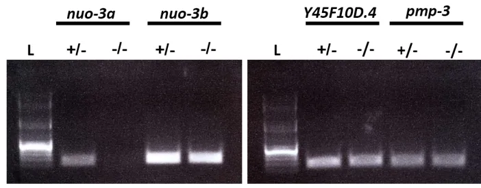
nT1[qIs51] (IV; V). Using RT-PCR, we confirmed that nuo-3bmRNA was still expressed in embryos homozygous for the nuo-3adeletion (Supporting Information,Figure S2).
pDD268. The 59homology arm comprised 642 bp upstream of theSPTF-3start codon, and the 39homology arm comprised thefirst 560 bp of theSPTF-3coding region (including thefirst exon and part of thefirst intron). A silent c/t mutation was introduced at nucleotide residue 33 of theSPTF-3coding re-gion to prevent cleavage of the repair template by Cas9. The complete sequence of the repair template plasmid is available on request. The Cas9 plasmid and repair template were in-jected into young adults of strainN2, and knock-in animals were isolated by selecting on Hygromycin. We found that the mNG^SEC^3xFlag::sptf-3insertion caused a maternal-effect lethal phenotype, so the insertion allele was maintained in heterozygotes. To isolatemNG^SEC^3xFlag::sptf-3 homozy-gotes (which produce embryos lackingsptf-3expression), we singled L4rollers and checked for the presence of viable
prog-eny a day later. Animals that had laid only dead eggs were dissected and their embryos used for RNA sequencing (RNA-seq). To isolate an mNG^3xFlag::sptf-3 protein fu-sion, the L1/L2 offspring of animals heterozygous for
mNG^SEC^3xFlag::sptf-3 were heat shocked at 32° for 5 hr. Five days after heat shock, marker-excised animals were identified by the combination of a wild-type pheno-type and mNG fluorescence. We found that two of nine
filmedmNG^3xFlag::sptf-3embryos were gastrulation de-fective, consistent with partial rescue and suggesting that the fusion protein does not fully rescue function.
Microscopy, image quantification, mRNA quantification
Differential interference contrast (DIC) imaging:C. elegans embryos were mounted on poly-L-lysine-coated coverslips at
the one- to four-cell stage mounted on a 2.5% agarose pad with egg buffer (as in Sawyeret al.2011). Four-dimensional (4D) DIC video microscopy was performed on a Nikon Eclipse 800 microscope using a Diagnostic Instruments SPOT2 cam-era. Images were acquired with a 603 oil objective every 60–90 sec at 1-mm optical sections. Images were analyzed with MetaMorph software (MetaMorph, Inc.).
Fluorescence microscopy:The spinning-disk confocal images of JJ1531 zuIs70; JJ1317 zuIs3 [spe-26(+)/end-1::GFP],
JR2274 wIs137 [end-3::END-3(P202L)-GFP; rol-6],MS126 unc-119(ed4) III; irIs16 [tbx-35::NLS::GFP]; and Xenopus membrane (CAAX)::GFP RNA were acquired on a Nikon Eclipse Ti-E microscope equipped with a CSU-X1 Yokogawa head and a Hamamatsu ImagEM EMCCD camera. The Nikon Inverted Eclipse TE2000-U microscrope equipped with a Yokogawa Spinning Disk Confocal Scanner Unit CSU10 and a Spot Insight 2-megapixel camera was used to acquire im-ages of the LP54 PH::mCherry; NMY-2::GFP strain.
Quantification of expression from transgenes: Embryos from the following backgrounds were analyzed as controls or injected withsptf-3dsRNA:JJ1317zuIs3[spe-26 (+)/end-1::GFP],JR2274wIs137[end-3::END-3(P202L)-GFP;rol-6], andMS126unc-119(ed4) III;irIs16[tbx-35::NLS::GFP]. Em-bryos at the two- to four-cell stage were mounted on agarose
pads 24–30 hr after injection andfilmed by DIC microscopy until near the time of P4cell division. Images were acquired
once every 3 min at 4-mm optical sections through 20 mm total with the 603oil objective. A 488-nm laser was used at 15% power for imaging end-1::GFP andend-3::END-3-GFP embryos and at 25% power fortbx-35::GFP embryos (expo-sure times 200 msec). Fluorescence intensities were calcu-lated by subtracting the average pixel values of off-embryo background from the average pixel values of E cell nuclei, with pixel values measured using MetaMorph software.
RNA-seq and analysis:Four replicates offive to six embryos were collected at the 8AB stage for each genotype:N2and
sptf-3(cp155). For each replicate, we weakened eggshells with chitinase (Edgar and Goldstein 2012) and lightly dis-rupted them mechanically by aspiration. Weflash froze these embryos in liquid nitrogen and stored them at 280°. We prepared cDNA (Clontech SMARTer Legacy Kit) and RNA-seq libraries (Nextera XT Kit) according to the manufacturers’ guidelines. We multiplexed these libraries and sequenced them on a single HiSequation 2500 lane for 50 cycles from a single end.
Identical reads were collapsed using fastx_collapser (FASTX-Toolkit v. 0.0.13.2; http://hannonlab.cshl.edu/fastx_toolkit/) and aligned to the ce10 genome using TopHat2 v. 2.0.11 (Kimet al. 2013). Countfiles were generated using HTseq-count-0.5.3p3 (Anders et al. 2015) and a WBcel235.78.gtf referencefile (http://ftp.ensembl.org/pub/release-78/gtf/ caenorhabditis_elegans/). To analyze lineage-restricted genes that in our results were robustly expressed without excessive variation between replicates, we included in our analysis all genes whose mRNAs had (1) lineage-restricted expression in Supplementary Table 1 in Hashimshony et al. (2015), (2) a minimal mean of 10 RPKM in our results, and (3) total RPKM values that were greater than total 95% confidence intervals between replicates from our results.
NMY-2::GFP accumulation and apical membrane length analysis:For NMY-2::GFP accumulation analysis, LP54 PH:: mCherry; NMY-2::GFP embryos andgex-3(RNAi); LP54 em-bryos were analyzed as described previously (Roh-Johnson and Goldstein 2009). Images were acquired once every 60 sec at 1.5-mm optical sections (10mm total).
For analysis of apical membrane lengths, JJ1531 zuIs70
[pJN152:end-1::gap::caax;unc-119(+)] (provided by J. Nance) embryos andgex-3(RNAi);zuIs70embryos were mounted on agarose pads at the two- to four-cell stage 30–36 hr after in-jection andfilmed by DIC microscopy until near the time of P4cell division. Images were acquired once every 90 sec at
the case ingex-3(RNAi)embryos), the length of the apical mem-brane in contact with eggshell was determined.
Movies of membranes blebs were filmed with LP54 PH::mCherry; NMY-2::GFP (uninjected orwve-1(RNAi)) em-bryos mounted in a ventral position. Images were acquired every 3 sec at 1-mm optical sections (through 5–8mm total).
Contact length ratios:P1cells were isolated from wild-type
andWM43gex-3(zu196)embryos at the two-cell stage using the same method of isolation described in Wertset al.(2011). The cells were mounted in Shelton’s growth medium (Shelton and Bowerman 1996) either as P1cells or, after one division, as
P2-EMS cell pairs. Cells were imaged using 4D DIC video
mi-croscopy on a Nikon Eclipse 800 microscope with a Diagnostic Instruments SPOT2 camera. The contact length ratios are rep-resented as contact length/radius, as in Granaet al.(2010). Measurements were taken using Image J.
Xenopus laevis (Xenopus) manipulations
In situ hybridization:Xenopusembryos were staged accord-ing to Nieuwkoop and Faber (1967). Whole-mount in situ hybridization was carried out as described by Harland (1991). The probe fornckap1 in situhybridization was generated from nckap1; IMAGE clone ID: 4970729.
Xenopus plasma membrane imaging experiments:For im-aging membranes,Xenopusembryos were injected at the one-cell stage with 100 ng of membrane (CAAX)::GFP RNA or with 100 ng of membrane (CAAX)::GFP RNA together with 45 ng of Nckap1 MO1. The pCS2_mem(CAAX)::GFP plasmid, kindly provided by the Wallingford laboratory, was digested with Not1 and used as the template for RNA preparation. RNA was prepared by in vitro transcription of CAAX::GFP using the Ambion mMessage Kit with the Sp6 polymerase. Neurula-stage embryos (stage 16) were mounted in glass-bottom dishes in 2% agarose submerged in 0.13MBS buffer and oriented using eyebrow hairs glued to toothpicks. Images were acquired once every 2 min at 10-mm optical sections with the 103objective and a 1.53 magnifying lens using the Nikon Eclipse Ti-E microscope. The 488-nm laser was used at 50% power (exposure time 150 msec).
Morpholino injections: Two nonoverlapping translation-blocking morpholinos (MOs) were designed against the start site of nckap1 and upstream 59 UTR region (Gene Tools) (Figure S6 shows MO target sequences). Xenopusembryos were injected with the following range of Nckap1 MO con-centrations: control mismatch MO (43–45 ng), MO1 (43–45 ng for NTD phenotype; 25–30 ng for suboptimal dosage), and MO2 (25–30 ng for NTD phenotype; 20 ng for suboptimal dosage). MO injections were performed as described previ-ously (Tandonet al.2012).
Western blotting:The 166-bp 59UTR ofXenopus nckap1was cloned together with 123 bp of 59cds (41 aa) from stage 25 whole-embryo cDNA using the iProof High Fidelity PCR Kit (Bio-Rad) with 45°annealing temperature and 10-sec extension.
This PCR product (289 bp) was purified (QIAquick PCR
Puri-fication Kit, Qiagen) and digested with HindIII and BamHI restriction enzymes (NEB) for insertion into the pEGFP-N1 vector (Clontech). This allowed the fusion of eGFP in frame at the 39end of the PCR product. This nckap159UTR-GFP fusion (1023 bp) was then digested from the vector usingXhoI and NotI restriction enzymes and ligated into the modified pSP64TxB vector (withb-globin UTR elements). The new vec-tor was linearized (XmaI), purified, and transcribed to make capped mRNA for injections (mMessage mMachine SP6 Tran-scription Kit, Invitrogen). FertilizedXenopusembryos were in-jected with a range of Nckap1 MO1, MO2, and control MO concentrations and then subsequently injected with 1 ng of nckap1 59 UTR-GFP RNA. Embryos were cultured in 0.13 MBS solution at 16° until stage 11, when they were then resuspended in lysis buffer for protein analysis, as performed previously (Tandonet al.2012, 2013; Sojkaet al.2014). West-ern blots were probed with anti-mouse-JL8 GFP (Clontech, 632381, 1:10,000 dilution) to ascertain blocking of GFP-RNA translation by the MO and anti-mouse-SHP2 (BD Transduction, 610622, 1:2500 dilution) as a loading control, followed by anti-mouse HRP (Jackson, 1:10,000) and ECL substrate to de-velop the blot.
Data availability
Strains are available upon request.Table S1contains infor-mation about the genes we identified asC. eleganshomologs to mammalian neural tube defect genes and includes acces-sion numbers.
Results
Identifying NTD gene homologs in C. elegans
Mammalian NTD genes were compiled using a previously generated list of NTD genes (Harris and Juriloff 2007, 2010), which we augmented by additional literature searches for mouse and human NTD genes. We also included a set of folic acid metabolism genes because mutations in some folic acid metabolism genes have been shown to affect neural tube closure in mouse embryos (Harris 2009).C. eleganshomologs were identified by BLAST searches using protein sequences. C. elegansBLAST hits with anE-value score below a threshold (131029) were considered candidate NTD gene homologs.
In addition, fourC. elegansgenes (ced-4/Apaf-1,brc-1/Brc1,
our screen genes likely to have diverged recently by gene du-plication. In total, we searched 234 candidate mammalian NTD genes by BLAST against theC. elegansgenome, including 3 human genes with known SNPs as risk factors for NTDs and 5 folic acid metabolism genes. We identified 191 C. elegans genes with homology to mammalian NTD genes (Table S1).
Identifying genes with roles in C. elegans embryonic development by an RNAi enhancer screen
Our laboratory previously developed a two-step screen as an efficient method to identify genes with roles in C. elegans gastrulation from among candidate genes (Sawyer et al. 2011), and we applied that screen here to the NTD homologs. In brief, wefirst used an RNAi-by-feeding approach to disrupt gene functions (Kamath et al.2001; Kamath and Ahringer 2003), recording percent embryonic lethality in the wild-type and gastrulation-sensitized end-3(ok1448); ced-5(n1812) backgrounds. We used these results to identify genes in which the RNAi phenotype resulted in increased embryonic lethality in the gastrulation-sensitized background (Figure 2). For convenience, we refer to these genes as enhancers (see Materials and Methods for details). We next targeted the enhancers in wild-type worms using RNAi-by-injection because this method can induce a stronger loss of function than RNAi-by-feeding,filming embryonic development to di-rectly identify genes that are required for gastrulation. We included in our primary screen 104 of the 191 NTD gene homologs, including genes that were available in an RNAi-by-feeding library and whose feeding clones we could
con-firm by sequencing (Table S2).
For each of the 104 genes, RNAi-by-feeding was performed in triplicate in the wild-type and gastrulation-sensitized back-grounds. As controls for RNAi effectiveness in each trial, we used bacterial strains targetingpar-6(used as a positive con-trol for effective RNAi, which gave an average of 99.88 6 0.39 and 100.0060.00% embryonic lethality in the wild-type and gastrulation-sensitized backgrounds, respectively) and gfp (negative control, 0.40 6 0.40 and 5.92 6 2.5% embryonic lethality in the wild-type and gastrulation-sensitized backgrounds, respectively). We also included
mom-5, a homolog of the Wnt receptor Frizzled, as a positive control for the effectiveness of the gastrulation-sensitized background (5.48 6 3.1 and 56.33 6 13.49% embryonic lethality in the wild-type and gastrulation-sensitized back-grounds, respectively).mom-5was previously implicated in gastrulation (Leeet al.2006), and dsRNAs targetingmom-5
were previously shown to enhance lethality in the gastrulation-sensitized strain (Sawyeret al.2011).
Screening NTD gene homologs in the wild-type and gastrulation-sensitized backgrounds, we identified 22 NTD gene homologs as enhancers, which we considered as candidate C. elegansgastrulation genes (Figure 2 andTable S2). Among the 22 enhancers, we identified three Wnt pathway genes that were previously known to function inC. elegansgastrulation:
mom-2/Wnt,dsh-2/Dsh, andmig-5/Dsh(Leeet al.2006). We view this result as validating our approach as a method for
identifyingbonafidegastrulation genes among NTD gene ho-mologs. We also identified a predicted heparan sulfotransferase gene,hst-1, that is predicted to function with Wnt and other signaling pathways (Yan and Lin 2009) and a well-studied G protein a-subunit gene with known embryonic roles, goa-1
(Bastiani and Mendel 2006). The functions of these proteins in gastrulation were not pursued further here.
Secondary screen for genes with roles in C. elegans gastrulation
To determine which of the enhancer genes are required for gastrulation inC. elegans, we injected dsRNAs targeting each of the remaining 17 enhancers into wild-type adult worms and monitored the development of embryos produced at least 24 hr after injection. In our experience, injection of dsRNAs often results in penetrant phenotypes, phenocopying null mutants more often than does RNAi-by-feeding (prelim-inary unpublished observations). 4D DIC microscopy of living embryos was used to observe development at the 26- to 28-cell stage, looking specifically for gastrulation defects. A gastrulation defect was defined by the failure of the E cells to internalize before their division. Embryos were scored as gastrulation defective if one or both the E cells divided on the surface of the embryo (Figure 3 and Figure 4, asterisks). Among gastrulation-defective embryos, we further distin-guished two phenotypic classes: late internalization (if Ea and/or Ep failed to internalize, but all four of their daughter cells internalized before dividing) and failed internalization (if some of the four daughter cells failed to internalize before dividing) (Figure 3 and Figure 4, asterisks). We found that RNAi targeting four genes (sptf-3,abi-1, gex-3, andced-4) resulted in embryos consistently showing gastrulation de-fects (Figure 4 andFile S1,File S2,File S3,File S4, andFile S5). We also identified three genes, not considered further here, in which only 1 embryo of 13 or more analyzed showed a gastrulation-defective phenotype after RNAi:sec-24.2(which also showed cytokinesis defects),epi-1, andcka-2(Figure 3). Among the four genes consistently showing gastrulation de-fects,ced-4(RNAi)had the weakest effect on E cell internaliza-tion (Figure 3). We analyzed four different mutant alleles of
ced-4, including the null alleleced-4(n1162)(Yuan and Horvitz 1992), and found similarly weak but consistent gastru-lation defects (Figure S1). Taken together, these results identify four NTD gene homologs—sptf-3,abi-1,gex-3, andced-4—as having direct or indirect roles inC. elegansgastrulation. Given the low penetrance of gastrulation defects after targeting
ced-4, we did not consider it further here. Later we probe the basis for gastrulation defects insptf-3(RNAi),abi-1(RNAi), and
gex-3(RNAi)embryos.
sptf-3 affects the length of E cell cycles and endoderm cell fate and encodes a protein that is enriched in nuclei during gastrulation
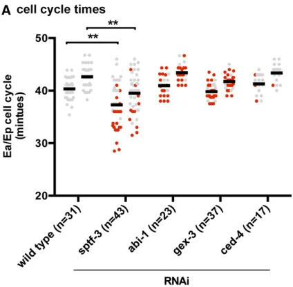
(Edgar and McGhee 1988). In wild-type embryos, the cell cycle length of the Ea cell (40.33 6 0.39 min) is slightly but consistently shorter than that of the Ep cell (42.64 6 0.39 min), as observed previously (Boeck et al. 2011). To determine whether sptf-3, abi-1, or gex-3 regulates E cell cycle lengths, we injected dsRNAs targeting these genes. Tar-getingabi-1orgex-3by RNAi did not affect cell cycle lengths (P.0.05) (Figure 5A) (ced-4gave similar results). However, RNAi againstsptf-3reduced E cell cycle lengths significantly (P,0.01; Ea, 37.2860.65 min; Ep, 39.5160.57 min). Most cells that failed to internalize insptf-3(RNAi)embryos had a shortened cell cycle. However, there were some cells that failed to internalize despite having normal cell cycle timing, indicating that the reduced cell cycle alone does not fully explain the gastrulation defects ofsptf-3(RNAi)embryos. Genes that regulate endoderm cell fate are known to affect the timing of E cell division and E cell internalization during gastrulation. Endodermal cell fate is marked by the expression of two partially redundant GATA transcription factors,end-1
andend-3(Maduroet al.2005, 2007; Owraghiet al.2010;
Boecket al.2011). When the endoderm cell fate regulators
end-1andend-3are disrupted, the E cells have a shorter cell cycle and divide prematurely on the outside of the embryo, before internalization (Zhuet al.1997; Maduroet al.2005; Boeck et al.2011). Thesptf-3 gene encodes an Sp1 family transcription factor with multiple recognized roles inC. elegans, including vulval development, oocyte production, embryonic development, and specification of apoptotic cell fate (Ulm et al. 2011; Hirose and Horvitz 2013). Given the previously recognized role ofsptf-3in cell fate specification and our data showing thatsptf-3affects cell cycle length in the E cells, we hypothesized thatsptf-3plays a role in specifying E cell fate. Because sptf-3 has diverse functions in different cells in C. elegans, we predicted thatsptf-3 also might contribute to other early cell lineages. Consistent with this prediction, we found that afluorescentSPTF-3fusion protein resulting from tagging the endogenous locus localized to all nuclei in gastrulation-stage embryos (Figure 5B).
To assess the effects on E cell fate specification, we examined GFP reporters ofend-1andend-3expression insptf-3(RNAi)
Figure 2 Primary RNAi screen. dsRNAs targeting 104 NTD gene homo-logs were fed into wild-type and gastrulation-sensitized backgrounds. Shown are bar graphs indicating percent embryonic lethality in wild-type (gray) and gastrulation-sensitized backgrounds (green), performed in trip-licate,695% confidence intervals. (A) Putative enhancers. RNAi targeting genes listed in this category resulted in increased embryonic lethality be-yond a threshold in the gastrulation-sensitized background (seeMaterials and Methods). Controls: dsRNA
tar-geting par-6,gfp, andmom-5. (B
embryos. sptf-3(RNAi) resulted in a significant reduction of end-1::GFP reporter intensities throughout the Ea and Ep cell cycle (Figure 6, A and B).end-3expression is apparent earlier in the Ea/Ep cell cycle than isend-1expression (Maduroet al. 2007; Boeck et al. 2011). We found that sptf-3(RNAi)also resulted in decreased levels ofend-3::GFP (Figure 6C). In ad-dition to theend-1andend-3endodermal markers, we ana-lyzed thefluorescence intensities of an MS cell reporter when
sptf-3was disrupted to determine whether cell fates are af-fected more generally. The MS lineage gives rise to multiple tissue types, including muscle and neurons but not endoderm cells (Sulstonet al.1983). Unlike the endoderm markers, sptf-3(RNAi)did not significantly decrease the expression of tbx-35::GFP, a marker of MS cell fate (Broitman-Maduroet al. 2006), in mesoderm progenitor cells (P.0.05) (Figure 6D). To determine whether sptf-3 affects gene expression more broadly beyond the E lineage, we performed transcriptome profiling from wild-type andsptf-3knockout 15-cell-stage em-bryos and analyzed mRNA levels of genes whose mRNAs were shown in previous quantitative experiments to be lineage re-stricted (Hashimshonyet al.2015).sptf-3knockout resulted in the expected loss ofsptf-3mRNA, little change to three actin mRNAs expressed at distinct levels, and effects on end-1,
end-3, andtbx-35mRNA levels that were consistent with the earlier reporter construct results for each of these genes. Anal-ysis of additional lineage-specific markers demonstrated that
sptf-3knockout decreased levels of specific mRNAs from the MS and AB lineages (Figure 6E). Thus,sptf-3is likely to affect endodermal internalization at least in part by regulating the expression of genes that determine early endodermal fate and regulate gastrulation, andsptf-3also has broader roles in reg-ulating expression of genes in multiple cell lineages.
Althoughsptf-3(RNAi) had detectable effects on the ex-pression of certain early endoderm cell fate markers earlier,
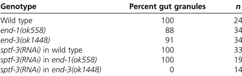
sptf-3 had little effect on expression levels of the endoder-mally expressed genes nhr-57orelt-7(Figure 6E), nor did they affect a marker of terminal differentiation of the endo-derm. We found thatsptf-3(RNAi)embryos were capable of forming birefringent rhabditin granules, sometimes called gut granules(Table 1)(Babu 1974; Lauferet al.1980). How-ever, disruption ofsptf-3by RNAi enhanced the disruption of intestinal development inend-3(ok1448)worms (and not in
end-1(ok558)worms) (Table 1).
Taken together, these results indicate thatsptf-3has critical roles in the expression of early endodermal cell fate genes and the subsequent internalization of the endoderm cells, as well as gene expression in other cell lineages. The results establishsptf-3as a broad regulator of gene expression that, among other roles, makes important contributions to the well-studiedC. elegansendodermal specification pathway.
The WAVE complex is required for gastrulation in C. elegans
Our secondary screen revealed that genes encoding two members of the WAVE complex of actin regulators,abi-1/Abi1 andgex-3/Nckap1, are required for gastrulation inC. elegans. The WAVE complex is highly conserved, regulating actin-dependent
Figure 3 Secondary RNAi screen. dsRNAs targeting the enhancers iden-tified in the primary screen were injected into wild-type animals. Embryos
from each background werefilmed by 4D DIC microscopy. Films were
analyzed for internalization of the endoderm cells (see Figure 4 for exam-ples of gastrulation defects indicated here).
processes in plants, Dictyostelium, yeast, Drosophila, C. ele-gans, and vertebrate animal systems (reviewed in Takenawa and Suetsugu 2007). Wave proteins were discovered ini-tially by homology to an actin regulatory domain in the Wiskott-Aldrich syndrome proteins (WASPs): WAVE is an abbreviation of WASP family verprolin-homologous (Miki et al.1998). Activation of the WAVE complex is regulated by multiple stimuli, including Rac GTPases (Mikiet al.1998, Eden et al.2002), resulting in a tightly controlled, polarized actin assembly mediated via Arp2/3 (Machesky and Insall 1998; Macheskyet al.1999). The WAVE complex is composed offive
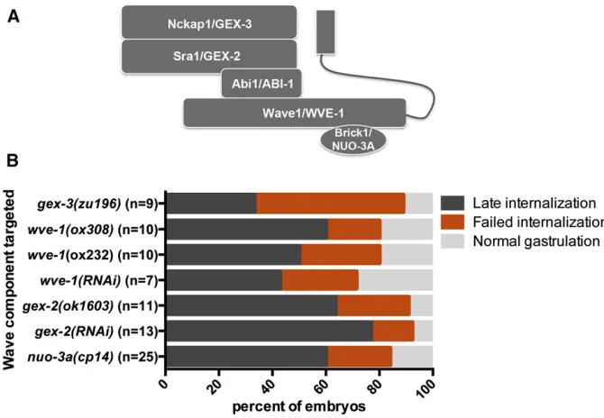
proteins (Eden et al. 2002), whose C. elegans homologs areGEX-3/Nckap1,GEX-2/Sra1,ABI-1/Abi1,WVE-1/Wave1, and NUO-3A/Brick1 (Figure 7A). InC. elegans,GEX-3,GEX-2,
ABI-1, andWVE-1exist in a protein complex, and as in other organisms, disruption of one component results in degradation of the other subunits (Patelet al.2008). To determine whether the role we found forABI-1/Abi1 andGEX-3/Nckap1 in gastru-lation reflects a role for the entire WAVE complex in gastrulation, we disrupted gex-2 and wve-1using RNAi and also analyzed phenotypes in existing null mutants (Soto et al. 2002; Patel et al.2008). Results from RNAi and mutant analyses indicate that bothgex-2andwve-1are required for gastrulation (Fig-ure 7B). In addition, a genetic null ofgex-3 validated the RNAi gastrulation-defective phenotype (Figure 7B).
The smallest member of the WAVE complex, nuo-3a/ Brick1, has been difficult to study inC. elegansby traditional RNAi approaches because thenuo-3gene produces an RNA that encodes two distinct polypeptides: the BRICK homolog (thenuo-3aORF) and the NADH-ubiquinone oxidoreductase a-subunit (nuo-3b ORF). As a result, dsRNA targeting the
nuo-3 transcript should affect the RNA levels of both poly-peptides. To overcome this challenge, we used CRISPR/Cas9-triggered homologous recombination (Dickinsonet al.2013) to generate a knockout ofnuo-3a/Brick1while leaving the nuo-3b NADH-ubiquinone oxidoreductasecoding sequence in-tact (Figure S2). We found that like the other members of the WAVE complex, thenuo-3a/Brick1homolog was required for gastrulation inC. elegans(Figure 7B).
We analyzed the E cell cycle timing ofgex-3,wve-1,gex-2, andnuo-3amutants. We found that E cell cycle lengths were similar in these backgrounds as in wild-type worms, showing only a modest decrease in the Ea cell cycle length ingex-2
mutants and a modest increase in the Ea cell cycle length in nuo-3a mutants (P,0.05) (Figure S3). We conclude that WAVE complex members are unlikely to affect gastrulation via effects on cell cycle length.
Apical narrowing and cell compaction are affected by gex-3
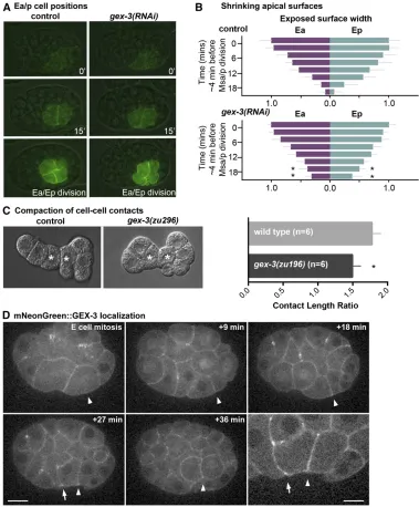
We hypothesized that mutations in gex-3 may affect apical constriction in the E cells. When members of the WAVE com-plex were disrupted, we noticed that plasma membranes formed blebs (File S6), similar to the phenotype observed pre-viously when members of the Arp2/3 complex, a downstream target of the WAVE complex, were disrupted (Roh-Johnson and Goldstein 2009). Furthermore, previous studies have in-dicated that Arp2/3 has critical roles inC. elegansgastrulation (Seversonet al.2002; Roh-Johnson and Goldstein 2009). To determine whether members of the WAVE complex affect E cells’apical constriction, we analyzed the narrowing of Ea/Ep cells’ apical domains over time using an end-1::GFP::CAAX background (Wehmanet al.2011) in which the plasma mem-branes of the endoderm precursor cells are labeled. To disrupt the WAVE complex, we usedgex-3(RNAi)because disruption of gex-3is known to reduce levels of other members of the WAVE complex (Patelet al.2008). We measured the maximal
anterior-to-posterior length of the exposed apical surfaces over time during the Ea/Ep cell cycle. We found that the Ea and Ep apical lengths ingex-3(RNAi)embryos began to narrow simi-larly to control embryos, but the final stages of endoderm internalization were affected, with more apical surface left exposed ingex-3(RNAi)embryos late in the cell cycle than in wild-type embryos (P , 0.05) (Figure 8, A and B). These results suggest that while initiation of apical constriction appeared normal, later stages were disrupted, similar to what was observed in Arp2/3 knockdown embryos (Roh-Johnson and Goldstein 2009). Although shrinkage of apical cell sur-faces was affected, we found that myosin became apically
enriched ingex-3(RNAi)embryos to a similar level as in control embryos (Figure S4), much as found in Arp2/3(RNAi) em-bryos previously (Roh-Johnson and Goldstein 2009). We con-clude thatgex-3is likely to affect apical constriction without strong apparent effects on myosin distribution.
Because the WAVE complex has been shown to affect the formation of cell junctions inC. elegans(Bernadskayaet al. 2011), and because we observed blebbing after targeting the WAVE complex (File S6), we predicted that the WAVE com-plex affects cell shapes. To examine cell shapes, we re-moved eggshells from two-cell-stage embryos, separated the cells, and followed development of these isolates.C. elegans
Figure 6 Endoderm cell fate marker
expression was reduced insptf-3
(RNAi)embryos. (A)end-1::GFP DIC overlay from video stills. White dot-ted line is drawn around the E cells. (B) Fluorescence intensity from the end-1::GFP reporter strain was re-duced insptf-3(RNAi) embryos. (C)
Fluorescence intensity ofend-3::
end-3-GFP was reduced insptf-3 (RNAi)embryos. (D) Fluorescence in-tensity of the mesodermal MS line-age markertbx-35::GFP in MSa/p’s daughters was not significantly re-duced insptf-3(RNAi)embryos (P. 0.05). In B, C, and D, 100%
repre-sents the maximumfluorescence
embryos with eggshells removed are known to undergo com-paction, expanding cell contacts and resulting in an increas-inglyflattened appearance at the embryo surface where cells meet (Granaet al.2010). Previous studies have shown that when proteins involved in cell adhesion (SAX-7/L1CAM and
HMR-1/cadherin) were depleted, compaction of the Ea/Ep cells was reduced, as measured by a decreased length of cell contact between the Ea and Ep cells (Granaet al.2010). We performed similar experiments in control and gex-3mutant embryos and observed a modest but statistically significant decrease in contact length ratios in gex-3 mutant embryos (Figure 8C), suggesting that the WAVE complex has a modest effect on cell shapes in vivo. A functional mNG-tagged form of GEX-3 (Dickinson et al. 2015) showed cortical localiza-tion with some enrichment at apical borders between Ep and MS granddaughters (Figure 8D), a known site of Arp2/3-dependent transient F-actin enrichment (Roh-Johnson and Goldstein 2009). Thefindings from ourin vivostudies—that the WAVE complex affects apical narrowing, cell compaction, and gastrulation inC. eleganswithout strong effects on cell cycle timing or myosin localization—are consistent with the known effects of the WAVE complex, which affects F-actin architecture generally and cell junctions of other cells in C. elegans specifically (Takenawa and Suetsugu 2007; Bernadskayaet al.2011).
Nckap1 contributes to neural tube closure in Xenopus
We hypothesized that genes implicated in mammalian neu-ral tube closure and C. elegans morphogenesis might have broadly conserved roles in morphogenesis. To test whether genes we found by screening in C. eleganshave conserved cellular functions, we turned toXenopusembryos, which al-low for imaging neural tube closure at the level of individual cells.Xenopusis a valuable vertebrate model in NTD research (see,e.g., Wallingford and Harland 2002; Haigoet al.2003; Grayet al.2009; Liuet al.2011; Ossipovaet al.2014) owing in part to its external development, which facilitatesin vivo imaging of neural tube closure (Kiesermanet al.2010). To determine whetherXenopusmight serve as a model for prob-ing the WAVE complex’s roles in neural tube formation, we tested whether a homolog of gex-3, nckap1, affects neural tube closure inXenopus. Wefirst examined whethernckap1 was expressed in embryos during neural tube closure at stage 18. Using RNAin situhybridization, we found that nckap1
mRNA was expressed at neural tube closure stages and that the mRNA was enriched in the neural tube and surrounding neural tissues (Figure S5).
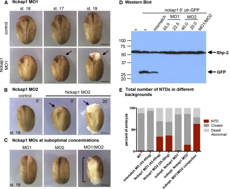
To test whether Nckap1 has a role in neural tube closure, we designed two nonoverlapping MOs to the 59UTR ofnckap1 (MO1 and MO2; for MO design, see Figure S6) to disrupt Nckap1 expression. Both MOs disrupted neural tube closure (Figure 9, A and B). Co-injection of Nckap1 MO1 and MO2 together at suboptimal concentrations (concentrations at which each MO alone had little or no effect) resulted in an increased incidence of NTDs (x2test,P,0.01), suggesting
that the MOs are specific to Nckap1 (Figure 9, C and E). Disruption of Nckap1 was confirmed by co-injecting GFP-taggednckap1-59UTR RNA with various concentrations of Nckap1 MOs (Figure 9D). Using a Western blot, we were unable to observe a GFP signal from MO1- and MO2-injected embryos. Further validating the specificity of the MOs, we showed that injection of a 5-bp mismatch MO resulted in reduced but not absent GFP signal (Figure 9D), and embryos injected with 45 ng of the 5-bp mismatch MO did not develop NTDs (data not shown).
Injection of the MO1 and MO2 at 43–45 and 25–30 ng, respectively, resulted in embryos that appeared superficially normal prior to neural tube closure stages. However, a delay in neural tube closure was evident between stages 16 and 19 (Figure 9, A and B). At various points during neural tube closure, lesions or small tears formed in the ectoderm, often beginning as a small lesion (Figure 9A, arrow, stage 17) that expanded in the neural tube (Figure 9A, arrow, stage 19). These lesions were observed at various sites along the neural tube in different embryos, often in the cranial and posterior regions. At later stages, the neural tube remained open, and additional lesions often were seen in the ventral surface (Figure S7). To examine the morphology of individual cells in Nckap1 MO-injected embryos, we injected embryos with GFP-CAAX mRNA to mark plasma membranes and filmed neural tube closure. At times when the neural folds converge in control embryos, cells were seen to round up and then become detached from the neuroepithelium in the Nckap1 MO-injected embryos at stage 16 (File S7,File S8, andFigure S8). Cells were seen to dissociate from the lesions, suggesting loss of cell adhesion. Although the loss of adhesion observed inXenopusand the more moderate effect on compaction in C. elegansappear superficially similar, we note that either de-fect could be a primary efde-fect of loss of a WAVE complex mem-ber or secondary consequences of unknown additional defects.
Discussion
Using a two-step enhancer screen, we discovered NTD gene homologs as newly recognized contributors toC. elegans gas-trulation. These genes include the transcription factorsptf-3
and members of the WAVE complex of actin regulators. These
findings suggest that some NTD gene homologs may have broadly shared roles in diverse animal systems in internaliz-ing surface tissues. We speculate that such roles might reflect
Table 1 Birefringent gut granules failed to form in sptf-3(RNAi);
end-3(ok1448)embryos
Genotype Percent gut granules n
Wild type 100 24
end-1(ok558) 88 34
end-3(ok1448) 91 34
sptf-3(RNAi)in wild type 100 33
sptf-3(RNAi)inend-1(ok558) 100 19
sptf-3(RNAi)inend-3(ok1448) 0 14
ancient and conserved roles retained inC. elegansand verte-brates from common ancestors. Alternatively, such roles could reflect convergent uses of similar genes in similar mor-phogenetic processes. Our approach supports the value of screening in C. elegansto identify genes with shared roles in morphogenesis in diverse animal systems.
sptf-3, an Sp factor, is a novel gastrulation gene in C. elegans
Sp proteins are a family of transcription factors with three conserved C2H2 zincfinger binding domains preceded by a buttonhead (BTD) box, a distinguishing feature of Sp proteins (Suske et al. 2005). There are nine Sp-family members in humans and mice, grouped into three clades from phyloge-netic sequence analysis (Schaeper et al. 2010). Four Sp-related factors have been identified inC. elegans(trp-1,sptf-1,
sptf-2, andsptf-3); however, only two genes,sptf-2andsptf-3, contain the three canonical C2H2 zinc finger repeats (Ulm et al.2011). Phylogenetic analyses of DNA binding domain sequences suggest that Sp factors inC. elegansare associated with the Sp1–Sp4 clade (Ulmet al.2011), while Sp8 is found within the Sp6–Sp9 clade (Schaeperet al.2010). InC. elegans, the only Sp factor identified thus far with roles in embryonic development issptf-3(Sunet al.2007; Ulmet al.2011; Hirose and Horvitz 2013; and our data).
Disruption ofsptf-3inC. eleganshas been linked to a range of developmental defects in embryonic morphogenesis, vulva specification, and oocyte production (Ulm et al. 2011). In addition,sptf-3regulates distinct apoptotic programs of spe-cific cells in the developing worm, controlling both caspase-dependent and -incaspase-dependent pathways (Hirose and Horvitz 2013). In the mouse, Sp8 appears to negatively regulate ap-optosis because disruption of Sp8 results in increased apopto-sis in neural progenitor and neural crest cells (Zembrzycki et al.2007; Kasberget al.2013).
Our data support an essential role forsptf-3in embryonic morphogenesis because we have identified a role forsptf-3in cell internalization during the first stages of gastrulation. When we examined the endoderm fate markers end-1 and
end-3 in the sptf-3(RNAi) background, we observed a de-crease in their expression, suggesting that, among several distinct roles,sptf-3plays a role in regulating cell fate deci-sions in the E cell lineage. Results from published ChIP-seq experiments withSPTF-3did not identifyend-1orend-3as direct targets ofSPTF-3. However, the ChIP-seq results did identify skn-1 as a target of SPTF-3 (Hirose and Horvitz 2013). skn-1 is a known upstream activator of the end-3
andend-1genes (reviewed in Maduro and Rothman 2002). Thus, these data suggest thatSPTF-3is part of the network of transcription factors regulating endoderm fate specification inC. elegans.
Disruption of the vertebrate Sp8 results in exencephaly (NTDs in the cranial region) and spina bifida (NTDs in the posterior region) in the mouse and limb truncation defects in the mouse, chick, zebrafish, andX. tropicalis(Bellet al.2003; Treichel et al. 2003, Kawakami et al. 2004, Chung et al. 2014). Sp8 is part of a transcriptional program that patterns neural progenitors into distinct domains (Grieselet al.2006; O’Learyet al. 2007; Sahara et al. 2007; Zembrzycki et al. 2007; Kasberg et al.2013); however, a transcriptional net-work involving Sp8 during neural tube closure has not yet been identified. It will be interesting to determine whether there are shared targets ofsptf-3and Sp8. In support of this, disruption of Sp8 in zebrafish embryos has been shown to result in decreased expression ofgata2, a GATA transcription factor, in the neuroepithelium (Penberthyet al. 2004). The role ofgata2during neural tube closure is not clear because mutations in mice result in early embryonic lethality; how-ever, evidence suggests thatgata2may affect early neuronal differentiation (Nardelli et al. 1999; Craven et al. 2004).
Identification of conserved targets during cell internalization inC. elegansgastrulation and neural tube closure may help to identify conserved transcriptional networks that contribute to morphogenesis.
The WAVE complex has early roles in endoderm internalization
The gex mutants (gex-1/wve-1,gex-2, andgex-3) andabi-1
were characterized initially by a failure of endoderm tissue to remain internalized. The termgexrefers to gut on the exterior arising from failed hypodermal (epidermal) migration, resulting in a large ventral cleft and exposed endoderm cells at later stages of morphogenesis (Sotoet al.2002; Withee et al.2004; Patelet al.2008). Here we provide evidence that WAVE complex mutants have earlier defects in endoderm internalization, which occurs at the 26- to 28-cell stage. We
show that knockdown of all known WAVE components resulted in similar endoderm internalization defects, exhibit-ing late-internalization or failed-internalization pheno-types. The failed-internalization phenotype is a less frequently observed phenotype among gastrulation-defective mutants (Sawyer et al. 2011) and could be influenced by subtle cell positioning defects, as described previously for otherC. elegans cells in WAVE complex mutants (Sotoet al.2002; Patelet al. 2008).
Arp2/3, an actin nucleator that can function downstream of the WAVE complex, has essential roles in C. elegans gas-trulation (Seversonet al.2002; Roh-Johnson and Goldstein 2009). The small GTPase Rac1 is a known upstream regula-tor of the WAVE complex, and mutations inced-10/Rac1 re-sult in ventral enclosure defects similar to the WAVE complex mutants previously characterized (Soto et al. 2002; Patel
Figure 8 Ea/Ep cell apical surface shrinking and cell compaction
are affected bygex-3(RNAi)and
mNeonGreen::GEX-3 localization. (A) E cell positions over time;
im-ages are from end-1::CAAX-GFP
et al. 2008).ced-10/Rac1 has been implicated inC. elegans gastrulation previously (Roh-Johnsonet al.2012), although whether it functions with the WAVE complex and Arp2/3 during gastrulation is not known.
Studies inC. eleganshave suggested that the Arp2/3 com-plex and members of the WAVE comcom-plex have important roles in cell adhesion via the maturation and maintenance of intestinal epithelial junctions (Bernadskayaet al.2011). Thus, the WAVE complex may have roles in establishing api-cal junctions in Ea and Ep cells. Apiapi-cal junctions may contrib-ute to a “clutch” mechanism in which contractions of the apical actomyosin cytoskeleton gradually become coupled to membranes, driving shrinkage of the apical domains of cells (Roh-Johnsonet al.2012). Failure of the E cells to in-ternalize when WAVE complex members are disrupted could feasibly represent a defect in the proposed clutch mechanism. Disruption of the Arp2/3 complex results in a reduction of
apical membrane shrinkage, similar to phenotypes observed ingex-3(RNAi)embryos (Roh-Johnson and Goldstein 2009). It is possible that these defects arise because apical junctions are not established correctly or are not attached properly to the actin cortex. In support of WAVE complex members af-fecting cell adhesion in the E cells, we show that cell com-paction is reduced ingex-3mutant cell isolates.
The WAVE complex has important but poorly understood roles during neural tube closure
Nckap1,Abi1, andRac1have been implicated in neural tube closure in mouse as disruption of these genes results in NTDs (Rakeman and Anderson 2006; Yokotaet al.2007; Migeotte et al.2011; Dubieleckaet al.2011). Mutations inWave1and Wave2do not result in NTDs, and mutations inWave3have not been reported (Soderling et al.2003; Yamazaki et al. 2003; Yanet al.2003). Although the mechanisms regulated
Figure 9 Targetingnckap1affected neural tube closure inXenopus. (A) Nckap1 MO1 (Figure S5shows MO target sites) resulted in a NTD and cell dissociation in the neural tube (arrows). (B) A second, nonoverlapping Nckap1 MO (MO2) also resulted in a NTD (arrows). (C and E) Suboptimal concentrations of MO1 and MO2 did not result a NTD; however, co-injecting MO1/MO2 at suboptimal concentrations resulted in NTDs (brackets). (D) Western blot from protein lysates of embryos injected with Nckap1 59UTR-GFP RNA, probed with anti-GFP. Nckap1 59UTR-GFP levels were decreased in the mismatch control (fold change 0.614), while levels were undetectable in the MO1 and MO2 samples. Shp-2 was used as a loading control. All
by these WAVE complex members during neural tube closure are not clear, defects in apical actinfilaments in the neuro-epithelium (Yokotaet al.2007) and lack of lamellipodia pro-trusions from primary neuroepithelium have been observed (Rakeman and Anderson 2006; Yokotaet al.2007).Nckap1 andRac1also have earlier roles during mouse gastrulation: disruption ofNckap1andRac1affect the migration of meso-dermal cells away from the primitive streak. This defect is linked to the cardia bifida phenotypes observed in Nckap1 and Rac1 mouse mutants and may also be a contributing factor to the open neural tube in these embryos (Rakeman and Anderson 2006; Migeotteet al.2011).
Here we present evidence that Nckap1 is required for neural tube closure inXenopus. Whennckap1function was depleted, neural tube closure was significantly delayed com-pared to control embryos, and often the neural tube failed to close. The tissue integrity of Nckap1-depleted embryos appeared compromised because lesions or tears in the neural plate tissue formed, and cells dissociated from the neuroepi-thelium. These phenotypes are similar to those described in cysteine-rich motor neuron protein (Crim1)–depleted Xeno-pus embryos. Like Nckap1, disruption of Crim1 resulted in cells that dissociated from the neural tube. Crim1 was shown to affect the junctional localization of E- and C-cadherin in the neural plate and the ability ofb-catenin to stably asso-ciate with junctional complexes (Ponferrada et al. 2012). Although future studies will be needed to determine whether Nckap1 affects the localization or expression of functional adhesion proteins in the neuroepithelium, it seems likely that Nckap1 affects cell adhesion either directly or indirectly. We hope that the use ofC. eleganscan contribute to identifying candidate cellular mechanisms that might be regulated by nckap1and other NTD genes during neural tube closure.
Acknowledgments
We thank members of the Goldstein and Conlon laboratories and Gregg Phares for critical reading of and comments on the manuscript. Some strains were provided by the Caeno-rhabditis Genetics Center, which is funded by National Institutes of Health Office of Research Infrastructure Pro-grams (P40-OD010440). This work was supported by Na-tional Institutes of Health grants R01-GM083071 to B.G. and T32-CA009156 and a Howard Hughes postdoctoral fellow-ship from the Helen Hay Whitney Foundation to D.J.D., National Science Foundation Graduate Research Fellowships to S.C.T. and J.K.H., National Institutes of Health grant R21-HD073044 to F.L.C., American Heart Association Postdoc-toral Fellowship 13POST16950044 to P.T., and National Institutes of Health grant F32-GM090447 to J.S.B.
Literature Cited
Anders, S., P. T. Pyl, and W. Huber, 2015 HTSeq—a Python framework to work with high-throughput sequencing data. Bio-informatics 31: 166–169.
Anderson, D. C., J. S. Gill, R. M. Cinalli, and J. Nance, 2008 Polarization of the C. elegans embryo by RhoGAP-mediated exclusion of PAR-6 from cell contacts. Science 320: 1771–1774.
Babu, P., 1974 Biochemical genetics of Caenorhabditis elegans. Mol. Gen. Genet. 135: 39–44.
Bastiani, C., and J. Mendel, 2006 Heterotrimeric G proteins in
C. elegans. WormBook 2006: 1–25.
Bell, S. M., C. M. Schreiner, R. R. Waclaw, K. Campbell, S. S. Potter
et al., 2003 Sp8 is crucial for limb outgrowth and neuropore closure. Proc. Natl. Acad. Sci. USA 100: 12195–12200. Bernadskaya, Y. Y., F. B. Patel, H. T. Hsu, and M. C. Soto,
2011 Arp2/3 promotes junction formation and maintenance in theCaenorhabditis elegansintestine by regulating membrane association of apical proteins. Mol. Biol. Cell 22: 2886–2899. Boeck, M. E., T. Boyle, Z. Bao, J. Murray, B. Mericle et al.,
2011 Specific roles for the GATA transcription factors end-1 and end-3 duringC. elegansE-lineage development. Dev. Biol. 358: 345–355.
Boulton, S. J., J. S. Martin, J. Polanowska, D. E. Hill, A. Gartner
et al., 2004 BRCA1/BARD1 orthologs required for DNA repair inCaenorhabditis elegans. Curr. Biol. 14: 33–39.
Brenner, S., 1974 The genetics ofCaenorhabditis elegans. Genetics 77: 71–94.
Broitman-Maduro, G., K. T. Lin, W. W. Hung, and M. F. Maduro, 2006 Specification of the C. elegans MS blastomere by the T-box factor TBX-35. Development 133: 3097–3106.
Chung, H. A., S. Medina-Ruiz, and R. M. Harland, 2014 Sp8 reg-ulates inner ear development. Proc. Natl. Acad. Sci. USA 111: 6329–6334.
Craven, S. E., K. C. Lim, W. Ye, J. D. Engel, F. de Sauvageet al., 2004 Gata2 specifies serotonergic neurons downstream of sonic hedgehog. Development 131: 1165–1173.
Dickinson, D. J., J. D. Ward, D. J. Reiner, and B. Goldstein, 2013 Engineering the Caenorhabditis elegans genome using Cas9-triggered homologous recombination. Nat. Methods 10: 1028–1034.
Dickinson, D. J., A. M. Pani, J. K. Heppert, C. D. Higgins, and B. Goldstein, 2015 Streamlined genome engineering with a self-excising drug selection cassette. Genetics 200: 1035–1049. Dubielecka, P. M., K. I. Ladwein, X. Xiong, I. Migeotte, A. Chorzalska
et al., 2011 Essential role for Abi1 in embryonic survival and WAVE2 complex integrity. Proc. Natl. Acad. Sci. USA 108: 7022– 7027.
Eden, S., R. Rohatgi, A. V. Podtelejnikov, M. Mann, and M. W. Kirschner, 2002 Mechanism of regulation of WAVE1-induced actin nucleation by Rac1 and Nck. Nature 418: 790–793. Edgar, L. G., and B. Goldstein, 2012 Culture and manipulation of
embryonic cells. Methods Cell Biol. 107: 151–175.
Edgar, L. G., and J. D. McGhee, 1988 DNA synthesis and the control of embryonic gene expression in C. elegans. Cell 53: 589–599.
Grana, T. M., E. A. Cox, A. M. Lynch, and J. Hardin, 2010 SAX-7/ L1CAM and HMR-1/cadherin function redundantly in blasto-mere compaction and non-muscle myosin accumulation during
Caenorhabditis elegansgastrulation. Dev. Biol. 344: 731–744. Gray, R. S., P. B. Abitua, B. J. Wlodarczyk, H. L. Szabo-Rogers, O.
Blanchardet al., 2009 The planar cell polarity effector Fuz is essential for targeted membrane trafficking, ciliogenesis and mouse embryonic development. Nat. Cell Biol. 11: 1225–1232. Griesel, G., D. Treichel, P. Collombat, J. Krull, A. Zembrzyckiet al., 2006 Sp8 controls the anteroposterior patterning at the midbrain-hindbrain border. Development 133: 1779–1787.
Harland, R. M., 1991 In situ hybridization: an improved whole-mount method for Xenopus embryos. Methods Cell Biol. 36: 685–695.
Harrell, J. R., and B. Goldstein, 2011 Internalization of multiple cells during C. elegansgastrulation depends on common cyto-skeletal mechanisms but different cell polarity and cell fate reg-ulators. Dev. Biol. 350: 1–12.
Harris, M. J., 2009 Insights into prevention of human neural tube defects by folic acid arising from consideration of mouse mu-tants. Birth Defects Res. A Clin. Mol. Teratol. 85: 331–339. Harris, M. J., and D. M. Juriloff, 2007 Mouse mutants with neural
tube closure defects and their role in understanding human neural tube defects. Birth Defects Res. A Clin. Mol. Teratol. 79: 187–210.
Harris, M. J., and D. M. Juriloff, 2010 An update to the list of mouse mutants with neural tube closure defects and advances toward a complete genetic perspective of neural tube closure. Birth Defects Res. A Clin. Mol. Teratol. 88: 653–669.
Hashimshony, T., M. Feder, M. Levin, B. K. Hall, and I. Yanai, 2015 Spatiotemporal transcriptomics reveals the evolutionary history of the endoderm germ layer. Nature 519: 219–222. Hildebrand, J. D., 2005 Shroom regulates epithelial cell shape via
the apical positioning of an actomyosin network. J. Cell Sci. 118: 5191–5203.
Hildebrand, J. D., and P. Soriano, 1999 Shroom, a PDZ domain-containing actin-binding protein, is required for neural tube morphogenesis in mice. Cell 99: 485–497.
Hirose, T., and H. R. Horvitz, 2013 An Sp1 transcription factor coordinates caspase-dependent and -independent apoptotic path-ways. Nature 500: 354–358.
Hodgkin, J., 2005 Introduction to genetics and genomics, in WormBook, ed. C. elegans Research Community,http://www. wormbook.org.doi/10.1895/wormbook.1.17.1.
Kamath, R. S., and J. Ahringer, 2003 Genome-wide RNAi screen-ing inCaenorhabditis elegans. Methods 30: 313–321.
Kamath, R. S., M. Martinez-Campos, P. Zipperlen, A. G. Fraser, and J. Ahringer, 2001 Effectiveness of specific RNA-mediated in-terference through ingested double-stranded RNA in Caeno-rhabditis elegans. Genome Biol. 2: RESEARCH0002.
Kasberg, A. D., E. W. Brunskill, and S. Steven Potter, 2013 SP8 regulates signaling centers during craniofacial development. Dev. Biol. 381: 312–323.
Kawakami, Y., C. R. Esteban, T. Matsui, J. Rodriguez-Leon, S. Kato
et al., 2004 Sp8 and Sp9, two closely related buttonhead-like transcription factors, regulate Fgf8 expression and limb out-growth in vertebrate embryos. Development 131: 4763–4774. Kieserman, E. K., C. Lee, R. S. Gray, T. J. Park, and J. B. Wallingford,
2010 High-magnification in vivo imaging of Xenopus embryos for cell and developmental biology. Cold Spring Harb. Protoc. 2010: pdb prot5427.
Kim, D., G. Pertea, C. Trapnell, H. Pimentel, R. Kelley et al., 2013 TopHat2: accurate alignment of transcriptomes in the presence of insertions, deletions and gene fusions. Genome Biol. 14: R36.
Laufer, J. S., P. Bazzicalupo, and W. B. Wood, 1980 Segregation of developmental potential in early embryos ofCaenorhabditis elegans. Cell 19: 569–577.
Lee, J. D., N. F. Silva-Gagliardi, U. Tepass, C. J. McGlade, and K. V. Anderson, 2007 The FERM protein Epb4.1l5 is required for or-ganization of the neural plate and for the epithelial-mesenchymal transition at the primitive streak of the mouse embryo. Develop-ment 134: 2007–2016.
Lee, J. Y., and B. Goldstein, 2003 Mechanisms of cell positioning duringC. elegansgastrulation. Development 130: 307–320. Lee, J. Y., D. J. Marston, T. Walston, J. Hardin, A. Halberstadtet al.,
2006 Wnt/Frizzled signaling controlsC. elegansgastrulation by activating actomyosin contractility. Curr. Biol. 16: 1986–1997.
Liu, W., Y. Komiya, C. Mezzacappa, D. K. Khadka, L. Runnelset al., 2011 MIM regulates vertebrate neural tube closure. Develop-ment 138: 2035–2047.
Machesky, L. M., and R. H. Insall, 1998 Scar1 and the related Wiskott-Aldrich syndrome protein, WASP, regulate the actin cytoskeleton through the Arp2/3 complex. Curr. Biol. 8: 1347– 1356.
Machesky, L. M., R. D. Mullins, H. N. Higgs, D. A. Kaiser, L. Blanchoin
et al., 1999 Scar, a WASp-related protein, activates nucleation of actinfilaments by the Arp2/3 complex. Proc. Natl. Acad. Sci. USA 96: 3739–3744.
Maduro, M. F., and J. H. Rothman, 2002 Making worm guts: the gene regulatory network of the Caenorhabditis elegans endo-derm. Dev. Biol. 246: 68–85.
Maduro, M. F., R. J. Hill, P. J. Heid, E. D. Newman-Smith, J. Zhu
et al., 2005 Genetic redundancy in endoderm specification within the genusCaenorhabditis. Dev. Biol. 284: 509–522. Maduro, M. F., G. Broitman-Maduro, I. Mengarelli, and J. H. Rothman,
2007 Maternal deployment of the embryonic SKN-1/MED-1,2 cell specification pathway inC. elegans. Dev. Biol. 301: 590–601. Migeotte, I., J. Grego-Bessa, and K. V. Anderson, 2011 Rac1
me-diates morphogenetic responses to intercellular signals in the gastrulating mouse embryo. Development 138: 3011–3020. Miki, H., S. Suetsugu, and T. Takenawa, 1998 WAVE, a novel
WASP-family protein involved in actin reorganization induced by Rac. EMBO J. 17: 6932–6941.
Munoz-Soriano, V., Y. Belacortu, and N. Paricio, 2012 Planar cell polarity signaling in collective cell movements during morpho-genesis and disease. Curr. Genomics 13: 609–622.
Nance, J., and J. R. Priess, 2002 Cell polarity and gastrulation in
C. elegans. Development 129: 387–397.
Nance, J., J. Y. Lee, and B. Goldstein, 2005 Gastrulation in C. elegans. WormBook 2005: 1–13.
Nance, J., E. M. Munro, and J. R. Priess, 2003 C. elegansPAR-3 and PAR-6 are required for apicobasal asymmetries associated with cell adhesion and gastrulation. Development 130: 5339– 5350.
Nardelli, J., D. Thiesson, Y. Fujiwara, F. Y. Tsai, and S. H. Orkin, 1999 Expression and genetic interaction of transcription fac-tors GATA-2 and GATA-3 during development of the mouse central nervous system. Dev. Biol. 210: 305–321.
Nieuwkoop, P. D., and J. Faber, 1967 Normal Table of Xenopus laevis. North Holland, Amsterdam.
Nishimura, T., and M. Takeichi, 2008 Shroom3-mediated recruit-ment of Rho kinases to the apical cell junctions regulates epi-thelial and neuroepiepi-thelial planar remodeling. Development 135: 1493–1502.
Nishimura, T., H. Honda, and M. Takeichi, 2012 Planar cell po-larity links axes of spatial dynamics in neural-tube closure. Cell 149: 1084–1097.
O’Leary, D. D., S. J. Chou, and S. Sahara, 2007 Area patterning of the mammalian cortex. Neuron 56: 252–269.
Ono, S., and G. M. Benian, 1998 TwoCaenorhabditis elegansactin depolymerizing factor/cofilin proteins, encoded by the unc-60 gene, differentially regulate actin filament dynamics. J. Biol. Chem. 273: 3778–3783.
Ossipova, O., K. Kim, B. B. Lake, K. Itoh, A. Ioannouet al., 2014 Role of Rab11 in planar cell polarity and apical constriction during vertebrate neural tube closure. Nat. Commun. 5: 3734.
Owraghi, M., G. Broitman-Maduro, T. Luu, H. Roberson, and M. F. Maduro, 2010 Roles of the Wnt effector POP-1/TCF in the
C. elegansendomesoderm specification gene network. Dev. Biol. 340: 209–221.
Patel, F. B., Y. Y. Bernadskaya, E. Chen, A. Jobanputra, Z. Pooladi
et al., 2008 The WAVE/SCAR complex promotes polarized cell movements and actin enrichment in epithelia duringC. elegans