Malignant transformation in
polyneuropathy associated with
monoclonal gammopathy
M. Eurelings, MD; H.M. Lokhorst, MD, PhD; S. Kalmijn, MD, PhD; J.H.J. Wokke, MD, PhD;
and N.C. Notermans, MD, PhD
Abstract—Objective: To assess the frequency of hematologic malignancies at diagnosis and to determine the incidence and predictors of malignant transformation during follow-up in patients with polyneuropathy associated with monoclonal gammopathy. Methods: Potential predictors of malignant transformation from medical history, hematologic, neurologic, and laboratory examination performed each 6 months were evaluated by univariable and multivariable Cox proportional hazard analysis.Results:Of 193 patients with polyneuropathy associated with monoclonal gammopathy, 17 patients had a hematologic malignancy at diagnosis. The incidence rate of malignant transformation in 176 patients without a malig-nancy at diagnosis was 2.7/100 patient years. Weight loss, progression of the polyneuropathy, unexplained fever or night sweats, and M-protein level were independent predictors.Conclusions:Since hematologic malignancies occur frequently in polyneuropathy associated with monoclonal gammopathy, the authors suggest that all patients should be screened at diagnosis and subsequently during follow-up if malignant transformation is suspected.
NEUROLOGY 2005;64:2079 –2084
Monoclonal gammopathy of undetermined
signifi-cance (MGUS) occurs in 0.1 to 3% of the normal
population and prevalence rises with age.
1MGUS is
characterized by the presence of a monoclonal
pro-tein (M-propro-tein) in serum or urine without an
under-lying hematologic malignancy. Criteria for MGUS
include a serum M-protein level less than 30 g/L, a
bone marrow plasma cell infiltration of less than
10%, and absence of lytic bone lesions, anemia,
hy-percalcemia, or renal insufficiency, and stability of
the M-protein level.
2,3Persons with MGUS are
asymptomatic and have no signs of hematologic
ma-lignancies, including fatigue, weight loss, bone pain,
or susceptibility to infections. However, 1% of
pa-tients with MGUS develop a hematologic malignancy
per year, and the risk of malignant transformation
persists even after 30 years of follow-up.
4In IgG
MGUS malignant transformation to a plasma cell
malignancy has been reported to be 1% per year,
5and in IgM MGUS malignant transformation to a
malignant lymphoid disorder like lymphoma or
im-munocytoma occurs in 1.5% of patients per year.
6The relation between polyneuropathy and MGUS
is supported by epidemiologic, pathologic, and
pas-sive transfer studies.
7-11In a retrospective cohort
analysis we found that 22% of patients with
polyneu-ropathy associated with MGUS developed a
hemato-logic malignancy during long-term follow-up of median
6 years.
12This suggests a higher frequency of
hemato-logic malignancies in MGUS associated with
polyneu-ropathy than in MGUS without polyneupolyneu-ropathy.
Therefore, we prospectively studied 193
non-selected patients with polyneuropathy associated
with monoclonal gammopathy with a mean follow-up
duration of 3 years, and assessed the frequency of
underlying hematologic malignancies at diagnosis
and the incidence of malignant transformation
dur-ing follow-up. In addition, we analyzed which factors
predicted malignant transformation in 104 patients
without a hematologic malignancy at diagnosis in
whom bone marrow examination was performed at
the beginning and the end of follow-up.
Methods. Patients. From January 1995 to August 2004, 193 patients with polyneuropathy associated with MGUS were identi-fied at the Department of Neuromuscular Diseases of the Univer-sity Medical Center Utrecht. These patients were referred to our clinic from all over the country for diagnosis and screening for causes. Before inclusion none of the patients was treated with chemotherapeutics. All patients with polyneuropathy and M-protein without other causes of the neuropathy were included after signed informed consent.13In all patients the initial workup
included 1) medical history, 2) neurologic examination, 3) routine laboratory analysis, 4) electrophysiologic studies, including nerve conduction and concentric needle examination using standardized techniques, identifying a predominantly axonal or demyelinating neuropathy according to the criteria of the American Academy of Neurology (AAN),14,155) a survey for M-protein by immunofixation
of serum and 10 x concentrated urine, 6) antibody reactivity,16,177)
physical examination by a hematologist, 8) skeletal X-ray, 9) X-ray of the lungs, 10) sonography or CT scan of the abdomen (on indication), and 11) bone marrow investigation. Bone marrow
as-From the Department of Neurology, Rudolf Magnus Institute of Neuroscience (Drs. Eurelings, Wokke, and Notermans), Department of Hematology (Dr. Lokhorst), Julius Center for Health Sciences and Primary Care (Dr. Kalmijn), University Medical Center, Utrecht, The Netherlands.
M.E. is supported by the Netherlands Organization for Scientific Research (NWO), project no. 940 –37– 030. Received January 3, 2005. Accepted in final form March 14, 2005.
pirates and biopsies were obtained from the crista iliaca posterior and viewed by a pathologist. Additional investigations included immunophenotyping with specific plasma cell, B cell, and T cell monoclonal antibodies. Congo red staining was performed for the detection of amyloidal deposits. In patients suspected of amyloid-osis, i.e., painful axonal neuropathy or autonomic function disor-der, without amyloidal deposits in bone marrow, Congo red staining of a rectal biopsy or a sural nerve biopsy was performed. During follow-up patients were examined each 6 months, includ-ing 1) medical history, 2) physical examination, 3) neurologic ex-amination, 4) routine laboratory analysis, and 5) determination of the level of M-protein. Duration of follow-up was defined by the time between the first hematologic screening and the diagnosis of an endpoint or the end of the study. We obtained the following variables: fatigue, defined by difficulty in performing normal daily activities; unexplained bone pain, defined as consistent localized pain other than joint or muscle pain; infections; B-symptoms, de-fined by unexplained fever, night sweats, unexplained weight loss (⬎5 kg/6 months); progression of the polyneuropathy, defined by deterioration of the neuropathy leading to disability (graded with decrease of the Rankin disability score of one point) or decrease of sensory function or strength of one point in 6 months.18,19Strength
was measured with MRC grading system in six muscles of both arms (deltoid, biceps and triceps brachii, finger extensors, finger flexors, and first interosseus) and both legs (iliopsoas, quadriceps femoris, hamstrings, anterior tibial, gastrocnemius, and peroneal) leading to a maximum score of 120. Sensory functions, both touch and pinprick sense, were graded as normal⫽4, abnormal distal to wrist/ankle ⫽ 3, distal half forearm/leg⫽2, distal to elbow/ knee⫽1, distal to axilla/groin⫽0. Vibration sense studied with tuning fork perception (128 Hz) was graded as present on middle finger/hallux ⫽ 4, ulnar styloid/medial malleolus ⫽ 3, elbow/ knee ⫽ 2, clavicula/crista iliaca ⫽ 1, no perception ⫽ 0. Joint position sense was graded as present of middle finger/hallux⫽2, diminished⫽1, absent⫽0. Summation of all sensory modalities could lead to a maximum score of 56. We obtained the following laboratory variables: M-protein isotype; M-protein level (g/L), sig-nificant rise of M-protein level (⬎25% if M-protein levelⱖ5 g/L with an absolute rise of⬎5 g/L, minimal 5 g/L if M-protein level ⬍ 5 g/L),20measured at least at two different time points; and
anemia (hemoglobin level⬍7.4 mmol/Lⱕ12 g/L in women and ⬍8.6 mmol/Lⱕ14 g/L in men).
Outcome. The main outcome of the study was malignant transformation to a hematologic malignancy, i.e., multiple myeloma, plasmacytoma, amyloidosis, immunocytoma, Non-Hodgkin’s lym-phoma, Castleman’s disease, and POEMS (polyneuropathy, organo-megaly, endocrinopathy, M-protein, and skin symptoms). Criteria for multiple myeloma and plasmacytoma were defined according to Kyle and Greipp21and multiple myeloma patients were
classi-fied according to Salmon and Durie.22Indolent myeloma and
smol-dering myeloma fulfilling the criteria of Stage I myeloma were classified as Stage 1 myeloma. Immunocytoma was defined based on WHO criteria, i.e., infiltration of the bone marrow of⬎25% by mature B-cells. Non-Hodgkin lymphoma was defined according to the REAL classification.23MGUS was defined according to Kyle.2
Statistical analysis. The incidence of malignant transforma-tion during follow-up was assessed in all patients without a hema-tologic malignancy at diagnosis. The analyses of predictors for malignant transformation during follow-up were performed on pa-tients without a hematologic malignancy at the initial hematologic screening at baseline (at diagnosis), with complete data on impor-tant predictive factors and bone marrow examination at the begin-ning and the end of follow-up. The association between each predictor and malignant transformation was first assessed by uni-variable Cox proportional hazards analysis, with the hazard ratio (HR) and 95% CI as measures of association.
Variables that were univariately associated with hematologic malignancies at follow-up (p⬍0.1) were then included in a multi-variable Cox proportional hazards analyses model to evaluate their independent contribution in the prediction of malignancies. First we included variables that can be obtained by history taking and physical examination, thereafter we included laboratory vari-ables. Model reduction was performed by excluding variables that were not significantly related to hematologic malignancies (HR withp⬍0.05) from the overall model by the step-down method. The relationship between malignancy (at baseline or at follow-up) and mortality was studied using a statistical model relating a determinant whose status changes over time to survival type (cen-sored) outcome data, i.e., Cox regression with time-varying covari-ates.24 The results were expressed as HR (and 95% CI). The
relationship between survival and type of monoclonal gammopa-thy was analyzed using Kaplan-Meier survival analysis.
Table 1Clinical characteristics of 193 patients with polyneuropathy associated with monoclonal gammopathy in percentage unless otherwise specified
At diagnosis During follow-up
MGUS, n⫽176 Malignancy, n⫽17 MGUS, n⫽159 Malignancy, n⫽17
Age at onset, mean (SD), y 60 (10) 57 (10) 60 (10) 56 (10)
Follow-up, mean (SD), mo 38 (32) 55 (39)
Sex, male 68 75 69 61
Fatigue 21 50* 18 44*
Bone pain 6 13 5 17
Infections 1 6 1 6
Night sweats/unexplained fever 2 13 1 11*
Weight loss 10 44† 8 24*
Kidney dysfunction 3 6 3 11
Progression of polyneuropathy 44 89‡
Autonomic function disorder 7 17 7 0
*⫽p⬍0.05. †⫽p⬍0.01. ‡⫽p⬍0.001.
MGUS⫽monoclonal gammopathy of undetermined significance; malignancy⫽hematologic malignancy; kidney dysfunction⫽serum creatinine level⬎20 mg/L.
Results. Baseline and neurologic characteristics. For this study 193 patients underwent neurologic and hemato-logic screening. Most patients were men (69%) with a mean age of 60 years (table 1). Thirty-four patients had monoclonal IgM anti-MAG antibodies (29% of the patients with IgM monoclonal gammopathy or IgM/IgG biclonal gammopathy, table 2). Twenty-three patients had a slowly progressive demyelinating polyneuropathy associated with monoclonal IgM anti-MAG antibodies with ataxia or senso-rimotor symptoms and signs. Eleven patients with anti-MAG antibodies had a more progressive disease course. Of the 83 patients with IgM monoclonal gammopathy (or bi-clonal gammopathy with IgM) without anti-MAG antibody reactivity 64% had a demyelinating polyneuropathy with predominance of sensory symptoms and signs. Of the 66 patients with IgG monoclonal gammopathy 73% had ax-onal polyneuropathy with predominance of sensory symp-toms and signs. Nine patients had IgA monoclonal gammopathy with axonal sensory polyneuropathy.
Frequency of underlying hematologic malignancies. Of the 193 patients, 17 (9%) had a hematologic malignancy at first screening (3 multiple myeloma [2 stage I and 1 stage III multiple myeloma], 3 plasmacytoma, 2 amyloidosis, 4 immunocytoma, 1 non-Hodgkin’s lymphoma and multiple myeloma, 1 Castleman’s disease, 3 POEMS). Characteris-tics of patients are summarized in tables 1 and 2.
Incidence of malignant transformation during follow-up. The incidence of malignant transformation during follow-up was assessed in 176 patients with
polyneurop-athy associated with MGUS in whom no hematologic ma-lignancy was found after extensive hematologic screening at baseline. Bone marrow examination was performed in 139 patients (79%). After a mean follow-up of 3 years, 17 patients (incidence rate 2.7 per 100 person-years) devel-oped a hematologic malignancy (4 multiple myeloma [3 stage I and 1 stage III multiple myeloma], 1 plasmacy-toma, 10 immunocyplasmacy-toma, 1 Non-Hodgkin’s lymphoma, and 1 POEMS). Five patients with polyneuropathy associated with IgM anti-MAG antibodies and a progressive disease course had malignant transformation (4 immunocytoma, 1 Non-Hodgkin’s lymphoma).
Predictive factors for malignant transformation. The analyses of predictive factors for malignant transformation during follow-up were restricted to 104 patients without a hematologic malignancy at diagnosis and complete data on important predictors including bone marrow examination at the beginning and end of follow-up (88 with MGUS and 16 with a hematologic malignancy at the end of follow-up). Of the 104 patients included in the analyses of potential predictors more patients had a progressive polyneuropathy and a hematologic malignancy than in the 72 patients who were not included in these analyses. Of these 72 patients 45 patients were lost to follow-up. Other characteristics did not differ. The HRs for the associations between the potential predictors and hematologic malignancy deter-mined with univariable analysis are presented in table 3. In multivariable analysis unexplained weight loss, pro-gression of the polyneuropathy, unexplained fever or night
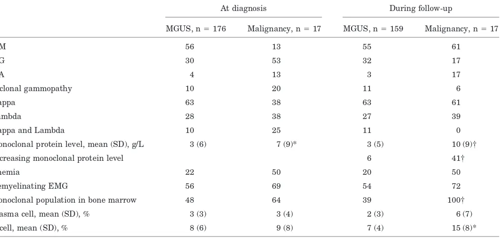
Table 2Laboratory characteristics of 193 patients with polyneuropathy associated with monoclonal gammopathy in percentage unless otherwise specified
At diagnosis During follow-up
MGUS, n⫽176 Malignancy, n⫽17 MGUS, n⫽159 Malignancy, n⫽17
IgM 56 13 55 61
IgG 30 53 32 17
IgA 4 13 3 17
Biclonal gammopathy 10 20 11 6
Kappa 63 38 63 61
Lambda 28 38 27 39
Kappa and Lambda 10 25 11 0
Monoclonal protein level, mean (SD), g/L 3 (6) 7 (9)* 3 (5) 10 (9)†
Increasing monoclonal protein level 6 41†
Anemia 22 50 20 50
Demyelinating EMG 56 69 54 72
Monoclonal population in bone marrow 48 64 39 100†
Plasma cell, mean (SD), % 3 (3) 3 (4) 2 (3) 6 (7)
B cell, mean (SD), % 8 (6) 9 (8) 7 (4) 15 (8)*
*p⬍0.05. †p⬍0.001.
sweats, and M-protein level were independent prognostic variables significantly associated with malignant transfor-mation (table 4). Among patients who had or who devel-oped a hematologic malignancy, three patients died (pneumonia) compared to five patients of those without a malignancy (breast cancer, kidney cancer, pulmonary em-bolism after aortic bypass surgery, pneumonia, and cardiac disease, HR for mortality 8.3, 95% CI: 2.1 to 33.7). We did not find a difference in survival of patients with IgG/IgA or IgM type of the monoclonal gammopathy.
Discussion.
In this prospective study of 193
pa-tients with polyneuropathy associated with
monoclo-nal gammopathy 34 patients were diagnosed with a
hematologic malignancy at initial screening or
dur-ing follow-up (18%). At diagnosis 9% of the patients
with polyneuropathy associated with monoclonal
gammopathy had an underlying hematologic
malig-nancy. During 3 years of follow-up in 17 of the 176
remaining patients with polyneuropathy associated
with MGUS malignant transformation occurred. The
incidence of malignant transformation in
polyneu-ropathy associated with MGUS (2.7/100
patient-years, 95% CI 1.52/100 to 4.22/100 patient years)
appears to be higher than in MGUS without
poly-neuropathy (1/100 patient-years, 95% CI 0.85/100 to
1.24/100 patient years,
p
⬍
0.05).
5Unexplained
weight loss, progression of the polyneuropathy,
unex-plained fever or night sweats, and M-protein level
were independent predictive factors of malignant
transformation.
This study confirms the findings of our previous
retrospective cohort study in which 22% of the
pa-tients with polyneuropathy associated with MGUS
developed a hematologic malignancy during
long-term follow-up.
12The high frequency of hematologic
malignancies could have been due to selection bias,
because patients with polyneuropathy associated
with monoclonal gammopathy and a progressive
dis-ease course may be referred earlier to a tertiary
clinic than patients with slow progression of the
dis-Table 3Univariable analysis of the potential predictors for malignant transformation
MGUS, n⫽88 Malignancy, n⫽16 HR (95% CI)
Age at onset, mean (SD), y 59 (10) 56 (9) 1.0 (1.0–1.0)
Fatigue 20 40 2.9 (1.1–8.0)*
Bone pain 6 13 3.6 (0.9–14)*
Infections 1 0 0.7 (0.1–5.9)
Night sweats/unexplained fever 0 7 10.9 (2.0–60)†
Weight loss 11 20 74.1 (8–684)†
Progression of polyneuropathy 51 94 4.8 (0.8–36)*
IgG/IgA monoclonal gammopathy 38 21 2.0 (0.4–11)
Monoclonal protein level, mean (SD), g/L 2 (5) 11 (9) 1.1 (1.0–1.2)‡
Increasing monoclonal protein level 7 43 2.9 (1.0–8.3)‡
Anemia 14 50 121 (0.1–106267)
Anti-MAG antibodies 28 46 1.2 (0.4–3.9)
Values are percentages, unless otherwise specified, with hazard ratios (95% CI).
*p⬍0.1. †p⬍0.01. ‡p⬍0.05.
MGUS⫽monoclonal gammopathy of undetermined significance; malignancy⫽hematologic malignancy; anemia⫽hemoglobin level ⬍12 g/L (⬍7.4 mmol/L) in women,⬍14 g/L (⬍8.6 mmol/L) in men; anti-MAG antibodies⫽anti-myelin-associated-glycoprotein antibodies.
Table 4Multivariable analysis of clinical and laboratory predictors of malignant transformation
Model 1 Model 2 Model 3
Clinical determinants, HR (95% CI)
Laboratory determinants, HR (95% CI)
Clinical and laboratory determinants, HR (95% CI)
Weight loss 226 (12–4381) 226 (12–4381)
Progression of the polyneuropathy 118 (3.0–4638) 118 (3.0–4638)
Unexplained fever or night sweats 27 (1.8–403) 27 (1.8–403)
Monoclonal protein level (per 1 g/L) 1.1 (1.0–1.2) 1.0 (0.9–1.1)
Values are hazard ratios (95% CI).
ease. In addition, the high frequency of underlying
hematologic malignancies could have been due to
re-ferral bias, since patients with a very slowly
progres-sive polyneuropathy followed in a non-university
hospital may not be screened for the presence of an
M-protein and once they have a progressive
polyneu-ropathy these patients are referred to an university
hospital where the malignant monoclonal
gammopa-thy is found. However, a high number of patients
without a hematologic malignancy at initial
screen-ing developed a hematologic malignancy durscreen-ing
pro-spective follow-up. To determine the incidence of and
predictors for malignant transformation during
follow-up, patients with polyneuropathy associated
with MGUS were screened at regular intervals. The
group of patients in the follow-up study for analyses
of predictive factors included more patients with a
progressive polyneuropathy and more patients with
a hematologic malignancy than the group of patients
not included. Patients without a progressive disease
course were frequently lost to follow-up. This leads
to under-representation of patients with chronic
polyneuropathy in the analysis, and may affect the
association of progression of the polyneuropathy and
malignant transformation.
Several prognostic factors for malignant
transfor-mation for patients with MGUS without
polyneurop-athy have been reported in previous retrospective
studies, i.e., M-protein level,
25,26increase of
M-protein level during follow-up, light-chain M-
protein-uria, age
⬎
70 years,
27kappa light chain,
26and IgA
isotype.
28In the past 2 years, two large studies
re-ported M-protein level as the most important
inde-pendent predictor for malignant transformation in
both IgG and IgM MGUS.
5,6Patients with
polyneu-ropathy associated with MGUS in this prospective
multivariate study were comparable to patients with
MGUS in the reported studies with respect to age,
M-protein level, and bone marrow infiltration and we
confirmed the findings in MGUS without
polyneu-ropathy.
5,6In addition to reported findings, we
iden-tified weight loss, unexplained fever or night sweats,
and progression of the polyneuropathy as predictive
factors for malignant transformation. This is
im-portant since specialists in neurology, hematology,
and internal medicine should be aware of the
un-derlying risk of a hematologic malignancy in
pa-tients
with
polyneuropathy
associated
with
monoclonal gammopathy. Unexpectedly, the
inci-dence of malignant transformation in patients
with a demyelinating polyneuropathy associated
with IgM anti-MAG antibodies is similar to the
high incidence found in the total group of patients
with polyneuropathy associated with MGUS. Also in
the patients with anti-MAG antibodies, who
nor-mally have a slowly progressive disease course,
pro-gression of the polyneuropathy was associated with
malignant transformation.
22,29Frequent bone marrow examination enables early
detection of patients with a hematologic malignancy
and may prevent serious complications by adequate
timing of treatment. In this and other studies the
neuropathy symptoms improved with the
acknowl-edged therapy of the underlying malignancy.
18,30-32References
1. Axelsson U, Bachmann R, Hallen J. Frequency of pathological proteins (M components) in 6,995 sera from an adult population. Acta Med Scand 1966;179:235–247.
2. Kyle RA. Monoclonal gammopathy of undetermined significance. Natu-ral history in 241 cases. Am J Med 1978;64:814 – 826.
3. Durie BG, Kyle RA, Belch A, et al. Myeloma management guidelines: a consensus report from the scientific advisors of the International My-eloma Foundation. Hematol J 2003;4:379 –398.
4. Kyle RA, Therneau TM, Rajkumar SV, Larson DR, Plevak MF, Melton LJ, III. Long-term follow-up of 241 patients with monoclonal gammopa-thy of undetermined significance: the original Mayo clinic series 25 years later. Mayo Clin Proc 2004;79:859 – 866.
5. Kyle RA, Therneau TM, Rajkumar SV, et al. A long-term study of prognosis in monoclonal gammopathy of undetermined significance. N Engl J Med 2002;346:564 –569.
6. Kyle RA, Therneau TM, Rajkumar SV, et al. Long-term follow-up of IgM monoclonal gammopathy of undetermined significance. Blood 2003; 102:3759 –3764.
7. Kelly JJ, Kyle RA, O’Brien PC, Dyck PJ. Prevalence of monoclonal protein in peripheral neuropathy. Neurology 1981;31:1480 –1483. 8. Mendell JR, Sahenk Z, Whitaker JN, et al. Polyneuropathy and IgM
monoclonal gammopathy: studies on the pathogenic role of anti-myelin-associated glycoprotein antibody. Ann Neurol 1985;17:243–254. 9. Latov N, Hays AP, Sherman WH. Peripheral neuropathy and anti-MAG
antibodies. CRC Crit Rev Neurobiol 1988;3:301–332.
10. Tatum AH. Experimental paraprotein neuropathy, demyelination by passive transfer of human IgM anti-myelin-associated glycoprotein. Ann Neurol 1993;33:502–506.
11. Ellie E, Vital A, Steck A, Boiron JM, Vital C, Julien J. Neuropathy associated with “benign” anti-myelin-associated glycoprotein IgM gam-mopathy: clinical immunological, neurophysiological, pathological find-ings and response to treatment in 33 cases. J Neurol 1996;243:34 – 43. 12. Eurelings M, Notermans NC, Van de Donk NW, Lokhorst HM. Risk
factors for hematological malignancy in polyneuropathy associated with monoclonal gammopathy. Muscle Nerve 2001;24:1295–1302.
13. Notermans NC, Wokke JH, Franssen H, et al. Chronic idiopathic poly-neuropathy presenting in middle or old age: a clinical and electrophys-iological study of 75 patients. J Neurol Neurosurg Psychiatry 1993;56: 1066 –1071.
14. Research criteria for diagnosis of chronic inflammatory demyelinating polyneuropathy (CIDP). Report from an Ad Hoc Subcommittee of the American Academy of Neurology AIDS Task Force. Neurology 1991;41: 617– 618.
15. Notermans NC, Franssen H, Eurelings M, van der Graaf Y, Wokke JHJ. Diagnostic criteria for demyelinating polyneuropathy associated with monoclonal gammopathy. Muscle Nerve 2000;23:73–79.
16. Pestronk A, Li F, Griffin J, et al. Polyneuropathy syndromes associated with serum antibodies to sulfatide and myelin-associated glycoprotein. Neurology 1991;41:357–362.
17. Van den Berg LH, Hays AP, Nobile Orazio E, et al. Anti-MAG and anti-SGPG antibodies in neuropathy. Muscle Nerve 1996;19:637– 643. 18. Notermans NC, Lokhorst HM, Franssen H, et al. Intermittent
cyclo-phosphamide and prednisone treatment of polyneuropathy associated with monoclonal gammopathy of undetermined significance. Neurology 1996;47:1227–1233.
19. Notermans NC, Wokke JH, Lokhorst HM, Franssen H, van der Graaf Y, Jennekens FG. Polyneuropathy associated with monoclonal gam-mopathy of undetermined significance. A prospective study of the prog-nostic value of clinical and laboratory abnormalities. Brain 1994;117: 1385–1393.
20. Lokhorst HM, Segeren CM, Verdonck LF, et al. Partially T-cell-depleted allogenic stem-cell transplantation for first-line treatment of multiple myeloma: a prospective evaluation of patients treated in the phase III study HOVON 24 MM. J Clin Oncol 2003;21:1728 –1733. 21. Kyle RA, Greipp R. Plasma cell dyscrasias: current status. Crit Rev
Oncol Hematol 1988;8:93–152.
22. Durie BGM, Salmon SE. A clinical staging system for multiple my-eloma. Correlation of measured myeloma cell mass with presenting clinical features, response to treatment, and survival. Cancer 1975;36: 842– 854.
23. Harris NL, Jaffe ES, Diebold J, et al. World Health Organization clas-sification of neoplastic disorders of the hematopoietic and lymphoid tissues: report of the Clinical Advisory Committee Meeting—Airlive House, Virginia, November 1997. J Clin Oncol 1999;17:3835–3849. 24. Cox DR. Regression models and life tables. J Royal Stat Soc 1972;34:
187–220.
26. Van der Poel MHW, Coebergh JWW, Hillen HFP. Malignant transfor-mation of monoclonal gammopathy of undetermined significance among out-patients of a community hospital in Southeastern Netherlands. Br J Haematol 1995;91:121–125.
27. Baldini L, Guffanti A, Cesana BM, et al. Role of different hematologic variables in defining the risk of malignant transformation in monoclo-nal gammopathy. Blood 1996;87:912–918.
28. Blade J, Lopez-Guillermo A, Rozman C, et al. Malignant transforma-tion and life expectancy in monoclonal gammopathy of undetermined significance. Br J Haematol 1992;81:391–394.
29. Nobile Orazio E, Manfredini E, Carpo M, et al. Frequency and clinical correlates of anti-neural IgM antibodies in neuropathy associated with IgM monoclonal gammopathy. Ann Neurol 1994;36:416 – 424. 30. Dalakas MC, Flaum MA, Rick M, Engel WK, Gralnick HR. Treatment
of polyneuropathy in Waldenström’s macroglobulinemia: role of para-proteinemia and immunologic studies. Neurology 1983;33:1406 –1410. 31. Latov N. Pathogenesis and therapy of neuropathies associated with
monoclonal gammopathies. Ann Neurol 1995;37 Suppl 1:S32– 42. 32. Ropper AH, Gorson KC. Neuropathies associated with paraproteinemia.
N Engl J Med 1998;338:1601–1607.
Neuro
Images
Thermogram of idiopathic segmental
anhidrosis
Y. Nakazato, MD; N. Tamura, MD; A. Ohkuma, MD; K. Yoshimaru, MD; and K. Shimazu, MD, Saitama, Japan
A 45-year-old man presented with an 8-year history of left hemifacial hyperhidrosis induced by exercise or hot environments. He began to experience episodes of heat intolerance and noticed anhidrosis over the right face, right neck, left trunk, and right
lower leg (figure). Physical, neurologic, and laboratory examina-tions revealed no abnormalities. He had no diabetes mellitus. Tonic pupils and hyporeflexia were absent. Idiopathic segmental anhidrosis (ISA) was diagnosed.1We consider that ISA is a forme
fruste of Ross syndrome plus,2in which multiple lesions
associ-ated with sympathetic ganglion damage are suggested to occur.
Copyright © 2005 by AAN Enterprises, Inc.
1. Nakazato Y, Shimazu K, Tamura N, Hamaguchi K. Idiopathic segmental anhidrosis. Clin Neurol 1991;38:940 –943.
2. Shin RK, Galetta SL, Ting TY, Armstrong K, Bird SJ. Ross syndrome plus: beyond Horner, Holmes-Adie, and harlequin. Neurology 2000;55: 1841–1846.
Figure. Thermography (Thermoviewer JTG 5310, JEOL Co., Japan) shows a “patchwork pattern” of body surface temperature distribu-tion after exercise. Body surface temperature was higher in the anhidrotic area (red) than in the sweating area (blue).
Address correspondence and reprint requests to Dr. Yoshihiko Nakazato, De-partment of Neurology, Saitama Medical School, 38 Morohongo, Moroyama, Iruma-gun, Saitama, 350-0495, Japan; e-mail: nakachan@saitama-med.ac.jp