Scholarship@Western
Scholarship@Western
Electronic Thesis and Dissertation Repository
3-5-2018 2:00 PM
Object processing in the medial temporal lobe: Influence of object
Object processing in the medial temporal lobe: Influence of object
domain
domain
Anna BlumenthalThe University of Western Ontario
Supervisor Köhler, Stefan
The University of Western Ontario Graduate Program in Psychology
A thesis submitted in partial fulfillment of the requirements for the degree in Doctor of Philosophy
© Anna Blumenthal 2018
Follow this and additional works at: https://ir.lib.uwo.ca/etd
Part of the Cognitive Neuroscience Commons
Recommended Citation Recommended Citation
Blumenthal, Anna, "Object processing in the medial temporal lobe: Influence of object domain" (2018). Electronic Thesis and Dissertation Repository. 5311.
https://ir.lib.uwo.ca/etd/5311
This Dissertation/Thesis is brought to you for free and open access by Scholarship@Western. It has been accepted for inclusion in Electronic Thesis and Dissertation Repository by an authorized administrator of
i
Abstract
We live in a rich visual world, surrounded by many different kinds of objects. While we
may not often reflect on it, our ability to recognize what an object is, detect whether an
object is familiar or novel, and bring to mind our general knowledge about an object, are
all essential components of adaptive behavior. In this dissertation, I investigate the neural
basis of object representations, focusing on medial temporal lobe (MTL) structures,
namely, perirhinal cortex, parahippocampal cortex, and hippocampus. I use what type of
thing an object is, or more specifically, the broader category (e.g., “face” or “house”) or domain (e.g., “animate or “inanimate”) to which an object belongs to probe MTL
structures. In the Chapter 2, I used fMRI to explore whether object representations in
MTL structures were organized by animacy, and/or real-world size. I found domain-level
organization in all three MTL structures, with a distinct pattern of domain organization in
each structure. In Chapter 3, I examined whether recognition-memory signals for objects
were organized by category and domain in the same MTL structures. I found no evidence
of category or domain specificity in recognition memory-signals, but did reveal a
distinction between novel and familiar object representations across all categories.
Finally, in Chapter 4, I used a neuropsychological approach to discover a unique
contribution of the hippocampus to object concepts. I found that an individual with
developmental amnesia had normal intrinsic feature knowledge, but less extrinsic, or
associative feature knowledge of concepts This decreased extrinsic feature knowledge led
to abnormalities specific to non-living object concepts. These results show that the
hippocampus may play an important role in the development of object concepts,
potentially through the same relational binding mechanism that links objects and context
in episodic memory. Taken together, these findings suggest that using object category or
domain to probe the function of MTL structures is a useful approach for gaining a deeper
understanding of the similarities and differences between MTL structures, and how they
ii
Keywords
Medial Temporal Lobe, Perirhinal Cortex, Parahippocampal Cortex, Hippocampus,
fMRI, Representational Similarity Analysis, Developmental Amnesia, Semantic
Memory, Category-Specificity, Domain-Specificity, Animacy, Real-world Size, Object
iii
Co-Authorship Statement
The projects reported in the current thesis were carried out under the supervision of Dr.
Stefan Köhler. The study presented in Chapter 2 is under revision for resubmission to
Human Brain Mapping. Dr. Bobby Stojanoski is a co-first author on the paper, he
contributed to design, data collection, and analyses. The paper also benefited from Dr.
Chris Martin’s theoretical and technical expertise, as well as Dr. Rhodri Cusack’s
technical expertise, reflected in their co-authorship. Chapter 3 is currently being prepared
for publication with Jane Kouptsova, Dr. Martin, and Dr. Köhler. Dr. Martin and Jane
contributed to data analysis. Lastly, the research presented in Chapter 4 is published in
Neuropsychologia (Blumenthal et al., 2017). Dr. Devin Duke is a co-first author, he
designed and implemented data collection, while analysis was done by him, myself, and
Dr. Ben Bowles. Writing and revisions were done by myself, advised by Dr. Köhler and
Dr. McRae. Dr. McRae’s input at all stages of the project was crucial, reflected in his
position as senior author. Additionally, Dr. Rosenbaum and Dr. Gilboa collaborated on
this project, providing theoretical guidance. Lastly, all projects comprising this thesis
were advanced through insights from fellow lab members, including Jane Kouptsova,
Jordan DeKraker, HY Yang, and Kayla Ferko. The general introduction and discussion in
this dissertation are largely my own ideas and writing, but were shaped by input from my
iv
Acknowledgments
First, I would like to thank my supervisor, Dr. Stefan Köhler. Stefan’s mentorship over
the last four years has been invaluable to my growth as a scientist. His passion for
understanding the neural basis of human memory, and remarkably in depth knowledge of
the literature has inspired me to pursue a career in this field. Stefan’s meticulous
approach to science and healthy skepticism has trained me to do my best work, and to
always communicate my ideas precisely. Under his mentorship, I have become a stronger
critical thinker, writer, speaker, and an engaged member of the cognitive neuroscience of
memory community. I am grateful to have had this training opportunity, and look
forward to continued collaboration!
I would also like to thank my advisory committee members: Dr. Jody Culham, Dr.
Rhodri Cusack, and Dr. Ken McRae. Each provided valuable scientific and pragmatic
advice. In addition to their official role on my committee, I had the opportunity to
collaborate closely with both Dr.Cusack and Dr. McRae. These experiences truly made
me understand the common claim that “no-one should ever do science alone.” I learned
an incredible amount from both of them, while always having a great time.
On the topic of not doing science alone, I am deeply grateful to all of the scientists at the
BMI. The BMI has been a wonderful place to grow, thanks to the supporting,
enthusiastic, and collaborative nature of its members. In particular, I would like to thank
two BMI members who served as collaborators and mentors, Dr. Chris Martin and Dr.
Bobby Stojanoski. Chris has been a role model for me, as his combination of theoretical
knowledge and technical prowess are unmatched. His down to earth attitude has made
him an excellent mentor and friend, always there to offer wise calmly-delivered advice
(with a sprinkle of humor thrown in). Complimentary to Chris’s even-toned advice
throughout the years, has been Bobby’s exuberant optimism, and endless energy for
science. His support has kept me going through many challenges, and has shaped me into
a better scientist and person. I am so grateful for his belief in me, and look forward to
v
I am also grateful to Dr. Jessica Grahn, for her career mentorship and encouragement of
women in science more generally. I thank my lab members, in particular Jordan
Dekraker, who is always willing to drop whatever he is doing to talk about the
hippocampus, and Kayla Ferko, for being a fantastic mentee and the most entertaining
data collection partner.
Finally, I would like to thank my family and friends for their support and enthusiasm
throughout the years. Especially the unwavering support from those who didn’t really
understand why I was still in school, and in Canada nevertheless. My family has always
understood and fostered my inner dork. They have encouraged me to pursue knowledge
for the sake of knowledge, not let me complain too much, and are always there to remind
me that studying the brain is pretty much the coolest thing ever. I couldn’t have done this
vi
Table of Contents
Abstract ... i
Co-Authorship Statement... iii
Acknowledgments... iv
Table of Contents ... vi
List of Tables (where applicable) ... x
List of Figures (where applicable) ... xi
List of Appendices (where applicable) ... xiii
Chapter 1 ... xiii
1 Introduction ... 1
1.1 Category and domain specific deficits in neuropsychological research ... 5
1.2 Category and domain organization: anatomical space ... 6
1.3 Category and domain organization: Representational space ... 8
1.4 Representational-Hierarchical theory ... 10
1.5 Binding of Items in context model... 13
1.6 Object processing in the medial temporal lobes ... 17
1.7 Feature-based object concept models ... 20
1.8 Summary of literature review ... 22
1.9 Goals of the current dissertation ... 23
1.10 References ... 24
Chapter 2 ... 33
2 Animacy and real-world size shape object representations in the medial temporal lobes ... 33
2.1 Introduction ... 33
vii
2.2.1 Participants ... 39
2.2.2 Stimuli ... 39
2.2.3 Experimental procedure ... 40
2.2.4 Image acquisition ... 42
2.2.5 Neuroimaging analyses ... 43
2.3 Results ... 46
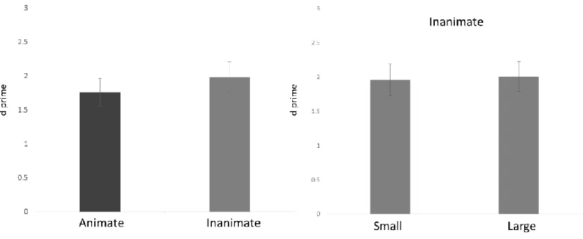
2.3.1 Behavioural ... 46
2.3.2 fMRI ... 49
2.4 Discussion ... 54
2.4.1 PrC ... 56
2.4.2 PhC ... 58
2.4.3 HpC ... 60
2.4.4 Conclusion ... 61
2.5 References ... 62
Chapter 3 ... 70
3 Organization of recognition memory signals for objects in the medial temporal lobes ... 70
3.1 Introduction ... 70
3.1.1 Category-specific memory signals ... 72
3.1.2 Coding of memory signals ... 74
3.1.3 Goals of the current study ... 76
3.2 Materials and methods ... 78
3.2.1 Neuroimaging analyses ... 78
3.3 Results ... 85
3.3.1 Behavioral ... 85
viii
3.4 Discussion ... 94
3.4.1 Conclusion ... 99
3.5 References ... 102
Chapter 4 ... 107
4 Abnormal semantic knowledge in a case of developmental amnesia ... 107
4.1 Introduction ... 107
4.2 Participants ... 115
4.2.1 HC ... 115
4.2.2 NB ... 121
4.2.3 Control participants ... 121
4.3 Experiment 1: Feature production ... 122
4.3.1 Materials ... 122
4.3.2 Procedure ... 122
4.3.3 Results ... 123
4.4 Experiment 2: Typicality ratings ... 127
4.4.1 Materials ... 127
4.4.2 Procedure ... 129
4.4.3 Results ... 129
4.5 Discussion ... 133
4.5.1 Conclusion ... 141
4.6 References ... 142
Chapter 5 ... 150
5 General Discussion... 150
5.1 Object processing in the MTL: influence of object domain ... 150
ix
5.3 Organization of recognition memory signals for objects in the MTL ... 154
5.4 Abnormal semantic knowledge in case of development amnesia ... 157
5.5 Benefits of exploring representational space and future directions ... 160
5.6 Conclusions and future directions ... 161
Appendices ... 165
x
List of Tables
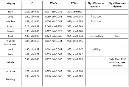
Table 2.1 Behavioural performance on the continuous recognition memory task by object
category 61
Table 3.1. Results from category specific linear contrast tests on the recognition-memory
signal in each MTL structure tested 100
Table 4.1 Neuropsychological profile of patient HC 129
Table 4.2 Number of exemplars used in each object category in the typicality rating
xi
List of Figures
Figure 1.1. Category and domain level anatomical organization in the ventral visual
stream 20
Figure 1.2. Category and domain level organization of representational space in the
ventral visual stream 22
Figure 1.3. Representational-Hierarchical theory 26
Figure 1.4. Binding of items-in-context (BIC) model 27
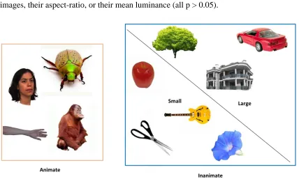
Figure 2.1. Stimuli from 12 different object categories, separated by animacy and
real-world size for inanimate objects 53
Figure 2.2. Continuous recognition memory task 55
Figure 2.3. Behavioral performance: continuous recognition memory task 60
Figure 2.4. Representational space for object-evoked responses in the medial temporal
lobe 64
Figure 2.5. Organization of object-evoked responses in the medial temporal lobe 65
Figure 2.6. Visualization of representational space for object-evoked responses in the
medial temporal lobe 66
Figure 3.1. Recognition-memory signal representational similarity analysis for MTL
structures 100
Figure 3.2. Example of linear models used to test within category, within domain, and
across category distinctions between perceived novel and familiar object stimuli. 103
Figure 3.3. Across category distinction between perceived novel and familiar object
stimuli 104
xii
Figure 3.5. Representational dissimilarity matrices averaged across category for novel
and familiar object stimuli, after pattern-based demeaning. 107
Figure 4.1. Coronal slice of T1-weighted MR image showing bilateral hippocampal
volume reduction in HC and a representative control participant in Olsen et al. (2013).
128
Figure 4.2. Mean intrinsic and extrinsic feature production for HC, NB, and their control
participants 139
Figure 4.3. Typicality judgements for living and nonliving object concepts for HC, NB,
xiii
List of Appendices
Appendix A: Documentation of ethics approval for Chapter 2 and 3
Chapter 1
1
Introduction
We live in a rich visual world, surrounded by many kinds of objects. While we may not
often reflect on it, our ability to recognize what an object is, detect whether an object is
familiar or novel, and bring to mind our general knowledge about an object, are essential
components of adaptive behavior. Ultimately, these abilities guide how we interact with
objects in our world. Our perception and interaction with objectsis guided by what type
of thing an object is, or more specifically, the broader category (e.g., “face” or “house”) or domain (e.g., “animate” like a face, or “inanimate” like a house) to which that object
belongs. For example, imagine you are at the local park to meet your friends for a picnic.
You are surrounded by many types of objects, including animate objects (dogs, people),
small inanimate objects (your friend’s guitar, silverware for the picnic), and large
inanimate objects (trees, park benches). When encountering an animate object, you may
want to know if it is moving towards you or away from you, or whether it appears hostile
and friendly, whereas with a small inanimate object you might want to pick it up, and use
it to make music, or eat lunch. This is quite different from a large inanimate object, such
as a tree, which you might use as a landmark to meet your friends for the picnic.
Interestingly, not only do object category and domain appear to guide our perception and
behavior, but they are also reflected in our neural organization. One of the most striking
examples is that brain damage can cause category or domain specific deficits (for a
review see Capitani et al., 2003). Additionally, measuring activity in the healthy brain
while participants view objects has revealed category and domain level organization
across the ventral visual stream (VVS), a neural pathway involved in object processing
running from occipital cortex through ventral to anterior lateral and medial temporal
cortex (Mishkin & Ungerleider, 1982; Mishkin et al., 1983; Goodale & Milner, 1992;
Goodale et al., 2008; Kravitz et al., 2013). Within the VVS, there are contiguous regions
that show more activity for objects from a certain category than objects from other
categories. Perhaps most well-known is the fusiform face area (FFA), a region on the
right fusiform gyrus that shows higher neural activity for faces than for other object
(parahippocampal place area, Epstein et al., 1999), body parts (extrastriate body area,
Downing et al., 2001), and words (visual word form area, McCandliss et al., 2003).
Further, these category preferring regions are not randomly strewn throughout the ventral
visual stream, but fall within a broader functional organization by domain. For example,
the FFA falls within a larger zone of lateral cortex that shows more activity for animate
objects than inanimate objects, whereas the PPA falls within a medial zone that prefers
inanimate objects (for a review see Grill-Spector & Weiner, 2014). More recently,
Konkle et al. (2013, 2012) have shown that this medial inanimate preference zone is for
objects whose real-world size is large, and there is a separate zone located more
dorsolaterally that shows a preference for small inanimate objects. This distinction by
real-world size is not present in the animate domain, leaving this organizational principle
to be referred to as the “tripartite distinction”. In addition to finding continuous regions of
cortex that show more activation on average for one category or domain over another,
similar category and domain level organization is observed when taking a less
anatomically specific approach than looking for continuous preference zones, and
examining the representational space of object-evoked responses across the VVS.
Specifically, patterns of activity across the VVS are more densely clustered, or more
similar to each other for certain categories of objects (e.g., faces), and category clustering
is embedded within a broader domain organization, where animate objects are
represented more similarly to each other than to inanimate objects, and vice versa
(Kriegeskorte et al., 2008; Proklova et al., 2016). Real-world size also appears to matter
in representational space, with large inanimate objects evoking more similar patterns of
activity to each other than to small inanimate objects and vice versa (Julien et al., 2016).
Research on category and domain specificity in the VVS has focused on the more
posterior and lateral portions of the temporal lobe, and typically has not included the full
extent of the medial temporal lobe (MTL). MTL structures include perirhinal cortex
(PrC), parahippocampal cortex (PhC), entorhinal cortex (EC), and the hippocampus
(HpC). It should be noted that some studies have included a portion of the PhC, given the
anterior aspect of the PPA is often functionally localized to both the lingual gyrus and the
posterior portion of PhC. However, PhC in its entirety as defined anatomically has not
is likely due to the fact that a prominent view has held that MTL structures are part of a
system dedicated to declarative memory, and distinct from the VVS (Squire et al., 1991).
However, evidence primarily from studies focused on the PrC has challenged this view.
Specifically, evidence suggests that PrC is crucial for recognition memory of objects as
well as fine-grained object perception (Murray and Bussey, 1999; Bussey et al., 2007).
According to one theory, the representational hierarchical theory (R-H theory), PrC is
crucial for both object memory and object perception because it represents the
convergence of low-level visual features into holistic object representations (Murray and
Richmond, 2001; Barense et al., 2012; Erez et al., 2015; for a review see Cowell et al.,
2010).
The view that PrC is the apex of object processing in the VVS can also be seen in
memory focused theories of the MTL. For example, according to the Binding of Items in
Context model, or BIC model of episodic memory, PrC sends object information to the
hippocampus where it is bound to context information to form an episodic memory
(Eichenbaum et al., 2007; Diana et al., 2007 Davachi, 2006). In the BIC model, PhC
represents context information, including spatial scene context. This notion aligns with
work from the vision literature showing that the more posterior portion of PhC is often
functionally localized by scenes (Epstein & Kanwisher, 1998). However, the picture
becomes more complicated when considering that posterior PhC also shows a preference
for buildings and other large objects (Aguirre et al., 1998; Epstein & Kanwisher, 1998;
Bar & Aminoff, 2003; Mullally & Maguire, 2011; Konkle & Oliva, 2012; Troiani et al.,
2012; Magri et al., 2016). Further, Martin et al. (2013, 2016) has shown category-specific
recognition memory signals for objects in PhC. Thus, it remains an open question as to
whether PrC is involved in object processing in a domain-general manner, or whether
both PrC and PhC contribute to object processing, but differ by the category or domains
to which they are sensitive. The HpC is generally not considered as being involved in
object processing, or to show category or domain level organization, but this has not been
thoroughly tested. Specifically, the HpC is thought to receive both object and spatial
context information, and bind that information together, resulting in representations that
therefore category and domain general (for a review on the BIC model see Eichenbaum et
al., 2007).
In the current dissertation, I used object domain as a tool to explore how MTL structures
contribute to object perception, memory, and semantic knowledge of objects. To
investigate these questions, I used two approaches, fMRI in healthy adults, and
behavioral testing in individuals with selective damage to MTL structures. Specifically,
in Chapter 2, I explore how object representations are organized by category and domain
across MTL structures using fMRI. In Chapter 3, I interrogate the same fMRI data to ask
whether object recognition-memory signals themselves are organized by category or
domain in MTL structures, and whether memory is coded by average changes in signal,
or by pattern based changes in representation. In Chapter 4, I explore a unique way that
one MTL structure, the HpC, may contribute to object knowledge specific to the
nonliving domain.
To motivate the following chapters, in the remainder of the Introduction I first provide a
brief overview of the evidence for category and domain specific neural organization in
three parts: 1) category and domain specific deficits in neuropsychological research; 2)
fMRI evidence for category and domain specific VVS organization in anatomical space;
and 3) fMRI evidence for category and domain specific VVS organization in
representational space. I then briefly cover R-H theory because it provides motivation for
viewing MTL regions as a continuous part of the VVS, and therefore for exploring
organization in a similar manner. Additionally, I introduce how R-H theory models of
PrC motivated us to investigate whether memory signaling in this region was category or
domain specific. I then provide a brief overview of the BIC model, which further
motivated exploring content differences across MTL structures, and whose HpC model
formed the basis for our investigation of how the HpC might contribute to semantic
knowledge of objects. I then discuss current fMRI work on content differences, in
particular category specificity, across different MTL structures. Finally, I provide a brief
1.1
Category and domain specific deficits in
neuropsychological research
One well-known type of category-specific impairment is prosopagnosia, or “face
blindness”, which is a neurological disorder in which individuals are impaired
specifically at recognizing faces. Prosopagnosia can be acute (acquired) or congenital
(Behrmann & Avidal, 2005). This disorder generally occurs when there is damage to the
fusiform face area (Gruter et al., 2008), or to connections from the FFA to other brain
areas (Thomas et al., 2009). Selective impairments have also been reported for
knowledge of tools (see Johnson-Fray, 2004 for a review). While to our knowledge there
are no cases of specific impairments for buildings, cases of landmark agnosia have been
reported, in which individuals can no longer recognize or orient themselves in their
environment by familiar landmarks, generally after damage to the PPA (Takahashi et al.,
2002; Claessen et al., 2017). Overall, these results suggest that there is some category
specificity in the neural organization of object knowledge, at least for some categories.
At the broader domain level, a number of studies have described individuals with domain
specific deficits in object knowledge due to focal brain damage (for a review see Capitani
et al., 2003). The earliest report of disproportionate impairments in certain categories of
knowledge over others were those by Warrington, Shallice, and McCarthy (1984). They
reported 4 patients who had made a partial recovery from herpes encephalitis, all of
whom showed extensive bilateral temporal damage on CT scans. Interestingly, each
patient showed significantly more impairment for knowledge of living things and foods
than for nonliving objects. These impairments occurred in both the verbal and visual
domain. This pattern of more severe impairment for living objects relative to inanimate
holds in the majority of cases. Capitani et al., (2003) reviewed 79 published case studies
of category specific deficits, and found 61 cases with individuals who had a
disproportionate impairment for biological categories relative to artefacts, and 18
showing a disproportionate impairment for artefacts. Further, they argue that patterns of
category selective impairment can be fractionated into animate objects, inanimate
biological objects (fruits and vegetables), and artefacts. While there are some cases of
broader domain, with an obvious distinction between living things and artefacts. What
about evidence for a distinction between large and small inanimate objects? This is
somewhat less clear in the current literature, as usually objects used in testing are smaller
(tools) so these dimensions have not been thoroughly pit against each other in the
neuropsychology literature.
1.2
Category and domain organization: Anatomical
space
One of the most well-known examples of category-specific neural organization was the
discovery of the fusiform face area (FFA). Kanwisher et al. (1997) showed participants
images of faces as well as images of other object types, and found that a univariate
contrast to evaluate whether any regions show higher average activity for faces revealed a
contiguous region in the right fusiform gyrus appears fairly robustly across individuals.
This finding has since been widely replicated, and has sparked a lively debate as to
whether the FFA is a module dedicated to face processing (Grill-Spector et al, 2004;
Grill-Spector et al., 2006; Gauthier et al., 1999, 2000). Importantly, it is possible to
decode patterns of activity specific to other object categories within the FFA (Haxby,
2001), suggesting that it may not be exclusively involved in processing faces. However, a
region that prefers faces, even if not dedicated, does suggest some category-level
functional organization of visual cortex. Additionally, a number of other regions have
been found in occipital and temporal cortex that show differential increase in activity for
specific categories, including, for example, for scenes, places, and buildings
(parahippocampal place area, PPA) (Epstein & Kanwisher, 1998; Epstein et al., 1999),
for bodies (extrastriate body area) (Downing et al., 2001), and for tools (Chao et al.,
1999).
Interestingly, regions with preferential responses to specific categories are not organized
randomly in their spatial relationships. Instead, they fall within broader functional zones,
fusiform sulcus, and animate objects more strongly activate the area lateral to the sulcus
(for a review see Grill-Spector and Weiner, 2014). In other words, the FFA and EBA are
embedded within a larger animacy zone, and the PPA within a larger inanimate zone.
More recently, fMRI based work by Konkle et al. (2012, 2013) has shown that this
inanimate medial zone is specialized for inanimate objects whose real-world size is large,
whereas there is a separate zone located more dorsal and lateral to the animate zone that
prefers objects whose real-world size is small (including a tool-preferring region). There
is no evidence for an equivalent divide in zones by real-world size for animate objects,
leaving this organization schema to be referred to as “tripartite” (Konkle et al., 2013).
Importantly, these domain preference zones can be identified in a variety of tasks,
whether it be passive viewing of images, low level perceptual tasks, or object
categorization, but the majority of work in this area has used fairly low-level perceptual
tasks to ensure subjects are paying attention. Some work has shown that task might
impact the involvement of more posterior or anterior VVS regions, for example, Taylor et
al. (2012) show more posterior areas are involved in naming object domain, whereas
naming a specific object engages more anterior areas. It is an open question therefore, as
to whether anterior areas such as the MTL which may be more involved in processing
individual objects, still show category and domain organization.
1.3
Category and domain organization:
Representational space
Evidence from univariate analyses, as described above, has shown contiguous regions in
the VVS that show more activity for certain categories or domains of objects. In this
approach, activity is averaged across voxels for each category or domain, and then
average activity levels are contrasted between categories or domains, and thresholded by
looking for continuous clusters of voxels that show a similar response. Both category and
domain level organization of visual cortex can also be seen using a quite different
analysis approach, representational similarity analysis (RSA; for a review see
Kriegeskorte et al., 2008b). In RSA, brain activity is measured by assessing how
fine-grained patterns of activity evoked by different objects relate to one another. RSA,
therefore, does not require that voxels in the same areas across subjects show the same
response patterns, as stimuli are now compared in “representational space”, where a “representation” is defined by the pattern of activity evoked across voxels in a given
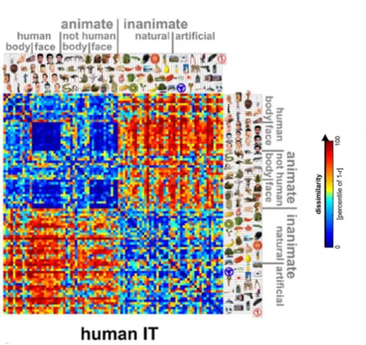
region in response to a distinct stimuli. For example, Kriegeskorte et al. (2008a) showed
participants a large number of objects spanning a number of categories and domains,
while participants performed a low-level visual task (press a button when the fixation
cross appears red). Patterns of activity evoked across the entire VVS for each object were
compared to the patterns of activity evoked for each of the other objects using a
dissimilarity measurement (1-Pearson’s correlation), and were then plotted in a
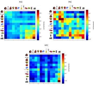
representational dissimilarity matrix (RDM) (see fig. 3). From this data driven approach
one can see both category and domain level organization of object evoked responses. For
example, there is a dark blue square indicating the responses for face stimuli compared to
other face stimuli are more highly similar to each other than they are to other object
categories. Perhaps even more striking is the domain level organization across all stimuli,
in which animate objects are more similar to each other than they are to inanimate
objects, and vice versa. Interestingly, a similar representational space can be revealed in
nonhuman primates when comparing patterns of activity in neural firing across visual
Using a similar RSA approach, a recent study showed that real-world size is an
organizing principle across representational space in visual cortex (Julien et al., 2016).
Participants were scanned while they viewed inanimate objects from 20 different
categories, whose real world size was large or small (while retinal size was controlled
for). They were instructed to memorize each object to ensure that they paid careful
attention. Patterns of activity evoked by large objects were more similar to other large
objects than they were to small objects and vice versa, when evaluated across a large
region spanning temporo-parietal-occipital cortex, similar to the large cortical volume
examined by Kriegeskorte et al. (2008). This pattern of organization was also apparent
when evaluating representations in smaller functionally defined regions known to show
average increased activity for scenes or large objects (PPA, retrosplenial cortex, occipital
place area), as well as object responsive regions that do not show increased average
activity for large objects (lateral occipital cortex, posterior fusiform gyrus), but this
organization was not present in early visual cortex.
1.4
Representational-Hierarchical theory
Research on category and domain-level functional organization of object evoked
responses in the brain has centered on the occipital lobe, and on the posterior and lateral
temporal lobe, generally leaving out the anterior medial temporal lobe, which includes
PhC, PrC, EC, and the HpC. The posterior portion of PhC is sometimes included in these
studies because the anterior portion of the PPA is localized here. However, the entire
structure defined anatomically, as well as the PrC, EC and HpC, are typically not
examined. Some of the reasons for this might be historical, as MTL structures were
generally considered to be part of a dedicated declarative memory system (Squire et al.,
1991), and therefore outside of the purview of object recognition. Indeed, category and
domain organization has been mostly studied in passive viewing paradigms or during
low-level perceptual tasks. However, at least one theory, the representational-hierarchical
theory (R-H theory)has challenged the view that MTL structures should be considered as
dedicated to memory, and argued for the inclusion of these structures as an extension of
the VVS (Murray & Bussey, 1999; Murray & Richmond, 2001; Saksida & Bussey, 2010;
Cowell et al., 2010). The R-H theory has guided this research by motivating us to extend
category and domain specific mapping to MTL structures, and to ask questions about
links between memory signaling and representational content. The central tenet of R-H
theory is that, in general, there are no brain regions dedicated to one specific
psychologically defined process, such as memory or perception. Instead, functional
differences between brain regions are better characterized by their representational
content, or the form of information they carry. This content may be used in different
processes, e.g., perception or memory. In what follows, I will describe evidence for the
According to R-H theory, the key difference across VVS regions, from early visual
cortex through HpC, is that representations become more highly conjunctive as you move
along the posterior to anterior axis of occipital and ventral temporal cortex. Therefore,
brain damage to a particular region should disrupt performance on any task for which
representations at that level of complexity and specificity are required. For example, in
pairwise visual discrimination tasks of objects, PrC lesions in non-human primates and
rodents do not impair performance when the objects can be discriminated by a single
low-level feature, but impair performance when feature conjunctions must be used (i.e.,
when feature ambiguity is high, or individual features are highly overlapping) (Buckley
and Gaffan, 1998; Bussey et al., 2002, 2003; Bartko et al., 2007). The same pattern is
seen in the HpC, for even more highly conjunctive content than single objects, for
example, in the discrimination of complex scenes with spatial object conjunctions (Lee et
al., 2006; 2012). Overall, a role of the MTL in visual perception has been reported in
rodents, non-human primates, and humans, using a variety of methodologies, providing
strong evidence for a causal role of MTL structures in perception, as opposed to just
memory, although there is still an avid debate (for reviews see, Graham et al., 2010;
Saksida and Bussey, 2010; Murray et al., 2007; but see Squire & Zola-Morgan, 1991;
Squire et al., 2004; Suzuki et al., 2004, Suzuki, 2009a, 2009b).
Research guided by the R-H view has also accounted for patterns of memory impairment
in cases of MTL damage. One powerful approach has been to test predictions derived
from computational models based solely on a network organized by conjunctive
representations. Cowell et al. (2006) created a connectionist model in which inputs were
object features, and a lower layer represented caudal, or more posterior VVS areas with
feature-based representations, and an upper layer corresponding the PrC, which
represented the conjunctions of those features (whole objects). When the PrC layer of the
model was lesioned, susceptibility to interference was increased, as there was no way to
disambiguate objects with overlapping features relying solely on the caudal layer. The
same susceptibility to interference was shown in rats with PrC lesions (Bartko et al.,
2010, but see Clark et al., 2011), and humans with amnesia (Barense et al., 2012, but see
Kim et al., 2011; Suzuki, 2009). Similarly, increased impairments in object recognition
during the delay period. This feature interference from stimuli is thought to be akin to
real-life situations, during which there is almost always a continuous stream of visual
information incoming. This computational evidence provides an explanation for
delay-dependent impairments on discrimination tasks in human and non-human animals
(Meunier et al., 1993; Mumby and Pinel, 1994). This model also generated a new
prediction - which impairment in amnesia should actually be due to detecting novel
stimuli as familiar, because without high-level conjunctive representations, the high
amount of feature overlap in novel stimuli should lead to familiarity signals. McTighe et
al. (2010) tested this prediction by familiarizing rodents with an object during a study
period, and then by putting rodents in a dark cage (no visual interference), or a regular
cage (visual interference) for a delay period. Rodents were tested to determine how they
explored novel or familiar stimuli (rodents spend more time exploring novel stimuli
naturally). Rodents in the visual interference condition treated novel stimuli as familiar,
but this pattern of results was not present in the group with no visual interference. These
results suggest that delay-dependent memory impairments can be accounted for by
considering representations in PrC alone. Without high-level object representations, there
is a high amount of feature interference, which causes novel objects to appear familiar.
Importantly, this changes the mainstream conception of amnesia, as an inability to
recognize things as familiar, to a discrimination problem where everything feels familiar.
These findings provide strong evidence for R-H theory, and have motivated consideration
of MTL structures as a part of the VVS in the current thesis. If one considers MTL
structures to be a part of the VVS, it is therefore important to explore the organization of
object representations in these regions along the same category and domain lines that
have previously been explored in more posterior and lateral VVS regions. Further,
computational R-H models provide a mechanism by which representations in these
regions can lead to memory signals, thereby directly motivating our analyses in Chapter
3, where we asked if memory signals were organized by category and domain.
Specifically, connectionist models represent objects with overlapping features more
similarly, and recognition memory signals come directly from activation of these
particular form of content, closely in line with our finding in the final chapter that HpC
contributes to semantic memory (and not only episodic memory) through creating
representations of items linked to contextual information.
Figure 1.5. Representational-Hierarchical theory (Barense et al., 2012)
1.5
Binding of items in context model
While R-H theory has focused on an inclusion of MTL structures as an extension of the
VVS, with an emphasis on content, characterizations of MTL structures with a focus on
their contribution to long term memory share some overlap with R-H theory. In
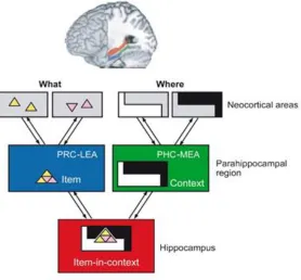
particular, the binding of items in context model, or BIC model, has focused on how
MTL structures differ in their contributions to long term memory in terms of the types of
information each structure contributes. Like the R-H model, the BIC model also places
PrC at the apex of the VVS, containing high level object representations. Similar to much
of the empirical work on object processing in PrC, the model treats PrC as being domain
general, leaving an investigation of domain and category specificity unexamined. In
terms of the PhC, the BIC model argues that this structure represents spatial context, a
lab and in the current dissertation addresses. Finally, the BIC model has focused on the
role of the HpC as binding item and context information for the service of episodic
memory, a role which we expand in the current thesis to domain specific object
knowledge.
The BIC model was proposed Eichenbaum et al. (2007), although a similar model
(without the addition of functional differentiation between lateral and medial entorhinal
cortex) was proposed by Davachi (2006). The model describes two pathways converging
in the HpC, the VVS pathway that culminates in the PrC and lateral entorhinal cortex,
which contains high-level item information, and a dorsal stream pathway that culminates
in the PhC and medial entorhinal cortex containing context, including “where”
information. Because both the lateral and medial entorhinal cortex provide input to the
HpC, these two streams converge in the HpC. It is thought that a key function of the HpC
is to bind this item and context information into a holistic representation – the basis for
rich coherent episodic memory (Eichenbaum et al., 2007; Davachi, 2006; Diana et al.,
2007, first proposed by Mishkin, 1983). Importantly, the model includes the assumption
that information flow is not purely hierarchical; there are projections from the
hippocampus through the EC back to PrC and PhC, which have their own back
projections to more posterior visual areas. It should be noted that emphases on the lateral
and medial EC initially came from animal work, given EC is difficult to image in
humans, although recent work has found initial evidence for a functional distinction in
humans (Maass et al., 2015). According to this model, the functional role of
back-projections is to reinstate previously encountered information, providing the neural basis
for episodic recollection. For example, a single cue, such as an object, can activate a
previously stored HpC pattern, which can then cause reinstatement of an episode by
reactivating the original object and context information in the ventral and dorsal visual
stream. In what follows, I overview some evidence for the BIC model of MTL function,
and then discuss some of the shortcomings of the model. Finally, I discuss this model in
relation to the R-H model and the questions about MTL structures posed in this
dissertation.
Evidence supporting the BIC model comes from fMRI, human MTL lesion studies, as
well as non-human animal models. Some of the evidence arose from attempts to
understand whether MTL regions contribute differentially to two subjective experiences
of remembering, termed familiarity and recollection. Familiarity refers to the experience
of feeling a sense of familiarity for an object or entity (a face you see on the bus), while
recollection refers to recalling contextual information specific to your initial encounter
with that object or entity (recalling that the person on the bus is a butcher, and a time you
went to buy a pork-chop in the shop last week). Notably, the content of these two
experiences differ, in the case of familiarity you have only an item or object, and in the
case of recollection you re-experience an episode with that object/entity, but also
contextual information (and often multiple other objects or entities). These two types of
remembering involve different MTL regions, patients with selective HpC damage have
more difficulty with recollection, but preserved item familiarity (Yonelinas et al., 2002;
Aggleton et al., 2005), and a patient with PrC/EC damage and a preserved HpC showed
abnormalities in familiarity but not recollection (Bowles et al., 2007), although it should
be noted that the damage also included anterior lateral temporal cortex. Interestingly,
while most evidence suggests a dichotomy between HpC and PrC, one patient with
damage more posterior, in PhC, also showed more impairments in recollection than
familiarity (Cipolotti et al., 2006). Similar dissociations are seen in non-human animals.
For example, monkeys and rodents with PrC lesions can no longer perform
delayed-non-match-to-sample tasks in which they are required to identify which object is novel after a
delay period, whereas those with damage restricted to the HpC still can (Nemanic et al.,
2004; Mumby, 2001). PhC and HpC do impair object recognition, but only when the
novelty consists of putting the object in a new location or context (Eacott and Norman,
2004; Mumby et al., 2002). Lastly, electrophysiology shows neurons in PrC are sensitive
to changes in the amount of experience with an object, whereas HpC and PhC neurons do
not show the same selectivity (but show stronger spatial coding) (for a review see Brown
and Xiang, 1998).
In terms of fMRI studies, there is also evidence to suggest that PrC is involved primarily
in object/item processing, PhC context, and the HpC in the binding of items in content.
examples. At encoding, HpC activity is higher for items that are later recollected, and is
also higher during recollection (for review see Eichenbaum et al., 2007). PrC, on the
other hand, has been shown to track familiarity at encoding, by showing more activity for
items later associated with a stronger familiarity response (Ranganath et al., 2004), and
has also shown decreased responding at retrieval that tracks the strength of subjective
item familiarity (Yassa and Stark, 2008). This PrC BOLD pattern of response is similar to
the pattern of responses in neurons recorded in monkey and rodent PrC. HpC activity has
been found when participants remember whether two items were presented together
(Kirwan et al., 2004; Jackson et al., 2004; Sperling et al., 2003), remembering the context
an item was presented in (Davachi et al., 2003; Ranganath et al., 2004), or remembering
the location an item was presented in (Staresina et al., 2006). A key difference seems to
be whether the encoding of information is within the object, or associative by nature. For
example, Diana et al. (2009) had subjects either encode color as part of an object “The elephant is red because it is sunburned” or external to the object “The elephant stopped at the light because it was red”. At retrieval, HpC and PhC activity correlated with recalling
the associative encoding, whereas PrC activity correlated with successful retrieval only
when the color was encoded as a feature of the object.
Unlike the R-H model, the BIC model is framed specifically around explaining how MTL
structure contribute to different types of memory. At first glance this focus on the
division between PrC and HpC in terms of their contributions to recollection or
familiarity may seem antithetical to an R-H view, given that the R-H view stresses that
brain structures are not modules dedicated to processes. However, a link can be made
when considering how the representations in PrC and HpC might differently contribute to
the subjective states of recollection and familiarity. Specifically, representations of
objects/entities in PrC contribute to familiarity, which is by definition based on a signal
from an object or entity alone. On the other hand, associative or items-in-context
representations in HpC necessarily contribute to recollection because it is defined by the
experience of this type of information (see Graham et al., 2010; Cowell et al., 2010).
Indeed, the idea that moving beyond process memory based distinctions to evaluating
MTL structures based on content or representations can be clearly understood from the
“Future work should focus on revealing the nature of representational capacities of
the MTL cortical input structures” (Davachi, 2006, pg. 698).
Figure 1.6. Binding of items-in-context (BIC) model (Eichenbaum et al., 2007).
1.6
Object processing in the medial temporal lobes
In light of characterizing MTL structures based on their differential content, a number of
questions still remain, one of which pertains to PhC content. In the BIC model, PhC has
been characterized as being involved in context (i.e., remembering a scene that is
associated with a face), and spatial information more generally. One obvious challenge to
this characterization of PhC is that in the vision literature the PPA (which partially covers
posterior PhC) shows sensitivity to buildings and other large objects (Magri et al., under
review). Martin et al. (2013, 2017) have shown recognition memory signals for objects
from some categories in PhC, namely buildings, furniture, and trees. However, aside
from this research, there have not been many other experiments exploring PhC
question as to whether PrC represents most types of objects, or how object content is
divided across regions. While some studies suggest interesting content differences (see
next section), this has not been mapped out with a large number of object categories or by
object domain.
A number of studies have explored differences in content, or representations, in MTL
structures. For the most part, these studies have used stimuli from a small number of
categories shown to be prominent in VVS organization more posteriorly, such as faces or
scenes, and generally treated objects as a single undifferentiated category. However, even
with this small sample of stimuli types, interesting patterns of category selectivity emerge
across structures. For example, Litman et al. (2010) had participants perform a 1-back on
images of scenes, faces, and objects. They found that there was a shift in average activity
from PhC to PrC along a posterior to anterior gradient, with the strongest responses to
scenes present in the most posterior part of the parahippocampal gyrus, and objects and
faces in more anterior regions. While there was a shift in preference, it is notable that
both structures showed above baseline responses to all stimuli. Similarly, in a target
detection task, Liang et al (2012) found increased responding for scenes in PhC and faces
in PrC. Neither study found any stimuli type preference in HpC response.
Multivariate analyses, exploring patterns of activity evoked in response to different
stimuli types across MTL structures, have found somewhat similar results. Liang et al.
(2012) decoded both scene and face stimuli from PhC and PrC, but decoding accuracy
was higher for scenes in PhC than PrC. Similar results were found when participants
viewed images and completed a low level perceptual task (indicating when a border came
up on the image), or judged the image as pleasant or unpleasant. Decoding scenes from
faces was highest in PhC, but objects versus faces could also be decoded above chance
(Huffman and Stark, 2014). In PrC, the highest decoding accuracy was between faces and
objects, but was also above chance between faces and scenes. Diana et al. (2008) used an
expanded stimuli set in a 1-back task, which included scenes, faces, toys, objects, and
abstract shapes. In PhC scenes were had the highest decoding accuracy, indicating the
most distinct categorical responses relative to other object categories. However, it was
interpreted as evidence that PhC does not solely represent scene information. LaRocque
et al. (2013) explored patterns of responses in MTL structures while participants
performed a low-level perceptual task (i.e., to press a button when fixation changes
colors) on objects from many different categories, scenes, and faces. They found distinct
object representations in both PhC and PrC, scenes in PhC and faces in PrC. In the
majority of these studies, the HpC showed no stimulus category selectivity, gaining it the
title of being “agnostic” (Huffman and Stark, 2014), although in one study scene
information was decoded in the posterior portion of hippocampus (Liang et al., 2012).
This evidence supports the idea that the HpC is agnostic because it is a convergence zone
for both object and spatial information. This information is thought to be bound together
to form a distinct episode, that would be category and domain general because it is a
unique conjunction of different kinds of information. Further, it is thought that a key
function of the HpC is pattern separation, or the process of orthogonalizing
representations to reduce interference in memory (for a review see Yassa & Stark, 2003),
a process that would also reduce any shared category information between stimuli.
While the studies outlined above have evaluated whether responses of MTL structures to
stimuli are organized by category based on activity evoked while the stimuli are
displayed and participants are performing a fairly low level task, to our knowledge only
one set of studies has evaluated whether recognition memory signals in these regions
(i.e., patterns of response to perceived novel or familiar stimuli) are categorical in nature,
and whether this differs across structures. Specifically, Martin et al. (2013, 2017) asked
for which categories it was possible to decode the distinction between perceived novel
and familiar stimuli in each MTL structure. Importantly, in these studies any contribution
of PhC due to context was removed by focusing exclusively on item-based familiarity
responses. In PrC it was possible to decode memory signals for faces, furniture, and
planes, and in PhC buildings, furniture and trees. The authors argue that a key dimension
guiding PhC sensitivity to object category is navigational relevance, given that buildings,
furniture, and trees are all stable and can be used for navigation, whereas planes, are
highly mobile and therefore are less suited to navigation. In these studies it was not
possible to decode category specific memory signals for any category in the
While these findings point to interesting differences and similarities in content
representation between PhC and PrC, and a likely agnostic HpC, the small number of
stimuli types used and/or a lack of differentiating object types makes it hard to
understand more broadly what the differences are between structures. Further, the
relation between category specificity in response to stimuli, or in memory coding, or
both, is still unclear. Both of these gaps are addressed in Chapter 2 and Chapter 3 of this
dissertation.
1.7
Feature-based object concept models
Thus far, we have considered the importance of object recognition (identifying an object)
and object recognition memory (identifying an object as novel of familiar), and the neural
organization of object responses by category and domain across VVS and MTL.
However, we have yet to discuss how any such organization relates to stored concepts
(i.e., object knowledge) derived from years of experience. Here I use the term “object concept” to refer to our semantic knowledge of an object. For example, our concept of a
hammer includes what it looks like (wooden handle, metal head) but also what its
function is, how we use it, and where we might find it. Indeed, thinking about what our
knowledge of objects entails, and how that might drive brain organization, has a rich
literature (Mahon & Caramazza, 2009; Martin et al., 2000; Martin et al., 2001; McRae &
Cree, 2002). This literature can be used to derive hypotheses about how MTL structures
contribute to object knowledge, and how it might differ by domain or category. Here, I
cover only a slim portion of this vast literature, focusing on feature-based models of
object concepts. First I cover how feature-based models have been used to explore the
role of MTL structures, in particular PrC, in terms of how it represents objects, and how
this is linked to object perception and memory. Second, while most attention to
feature-based object models in the MTL has been in using them to understand PrC function, we
show that they can be expanded to explore a previously unknown contribution of the HpC
to object processing. This motivated the study presented in Chapter 4, where we
investigated whether the HpC is involved in conceptual representations, and whether this
According to feature-based models of object concepts, object concepts are composed of a
number of features bound together. Characterizing objects based on the relations among
their features is a useful approach for understanding how they relate to each other in
terms of category and domain organization in both psychological and brain space.
Specifically, one can build a model of how objects relate to each other in semantic space,
in terms of how many features they share (Cree and McRae, 2003). Semantic space can
be derived from normative studies in which participants are presented with a concrete
concept and list as many features as they can think of that make up that concept. Models
based on these features have been shown to reflect representational space in PrC when
participants name visually presented objects (Clark & Tyler, 2014) and perform a
property verification task on object words (specific to left PrC; Bruffaerts et al., 2013).
Importantly, feature-based object models account for patterns of semantic impairments
after brain damage, such as the observation that impairment in knowledge for living
things occurs more often than impairments for nonliving things (Cree and McRae, 2003).
This distinction between living and nonliving things can be captured by differences in
feature statistics, in particular living things are more highly similar to each other in terms
of semantic feature overlap (Taylor et al., 2012). Tyler et al. (2013) used a precise
feature-based statistical measure, correlation by distinctiveness, to capture the challenge
of differentiating similar objects, and showed that this measure modulates bold activity in
left PrC during a picture naming task (Tyler et al., 2013). Overlap in semantic features
has also been shown to be causally related to PrC function. Specifically, Kivisaari et al.
(2012) examined the volume of PrC, EC, and HpC in individuals with varying levels of
atrophy due to Alzheimer’s disease. They found that volume in PrC, but not the other
structures, predicted latencies when naming living, but not nonliving things, they
concluded that these results show the importance of PrC in disambiguating semantically
similar objects. Indeed, PrC has been shown to show more average activity when naming
living than nonliving things (Bruffaerts et al., 2013).
Beyond differences between the living and nonliving domain by feature distinctiveness
and feature overlap, these domains differ in terms of the types of features that are salient
to their representations. For example, features can be classified into knowledge types,
a part of the object itself (i.e., color), or extrinsic to the object (i.e., function of the object,
or location it is found; McRae et al., 2005). The importance of intrinsic versus extrinsic
feature knowledge has been shown to differ by whether an object is living or nonliving.
Specifically, extrinsic knowledge is more important for nonliving objects, whereas
intrinsic knowledge is more central for living things (Barr and Kaplan, 1987). While the
terminology differs in the semantics literature, we note that extrinsic features could be
considered “associative” or “contextual” in nature. This opens up the possibility that
while the HpC is usually thought not to be involved in object processing, and to be
“agnostic” in terms of object category and domain information, it may play a previously undiscovered role in people’s extrinsic semantic knowledge, and therefore in representing
one domain of objects - namely nonliving objects.
1.8
Summary of literature review
In summary, object processing, whether it be recognition of an object, detecting whether
that object is familiar or novel, or using semantic object knowledge, is a crucial part of
human behavior. Unsurprisingly then, a large swath of the cortex, in particular the
occipital and temporal lobe, are involved in object processing through a hierarchical
information stream - the ventral visual stream. This neural basis of object processing is
organized, both anatomically, as seen in category preference regions and larger domain
preferring regions (i.e., the tripartite organizing schema), and in representational space.
However, anterior medial temporal lobe regions have been less thoroughly explored,
despite the fact that content may play a key role in their differential involvement in both
object perception and memory, according to at least two key theories- the R-H theory and
the BIC model. While there is some evidence of differential organization by stimuli type,
with PhC being sensitive to scenes and large objects, and PrC perhaps more involved in
faces, a careful examination across a large number of object categories is still needed.
Further, examining domain organization in these regions has not been done, and can be a
useful way to characterize their differential contributions. Additionally, a further
examination of the link between stimuli based categorical responses and their relation to
based account of MTL function. Lastly, we can use insights from the object concept
literature to further test the limits of how MTL regions are involved in object processing
for certain domains.
1.9
Goals of the current dissertation
In this dissertation, I address these gaps through three empirical studies. In Chapter 2, I
first explore object category and domain organization for a large number of categories in
each MTL structure, in the context of a continuous recognition memory task for objects.
In Chapter 3, I ask whether the memory signals for these objects themselves are
organized by category and domain, and whether this differs across structures in relation
to their stimuli-response based organization. Further, I ask whether memory status is
coded by repetition suppression, or pattern based representational changes and relate this
to the R-H model that suggests memory is computed directly from stimuli
representations. In Chapter 4, I turn to one specific MTL region, the HpC, often thought
not to be involved in object processing and agnostic to domain, and provide some
evidence that challenges these views. The important implications of this research are tied
to the idea that the HpC binds items-in-context, or is involved in representations that have
1.10
References
Aggleton, J. P., Vann, S. D., Denby, C., Dix, S., Mayes, A. R., Roberts, N., & Yonelinas, A. P. (2005). Sparing of the familiarity component of recognition memory in a patient with hippocampal pathology. Neuropsychologia, 43(12), 1810-1823.
Aguirre, G. K., Zarahn, E., & D’esposito, M. (1998). An area within human ventral cortex sensitive to “building” stimuli: evidence and implications. Neuron, 21(2), 373-383.
Bar, M., & Aminoff, E. (2003). Cortical analysis of visual context. Neuron, 38(2), 347-358.
Barense, M. D., Groen, I. I., Lee, A. C., Yeung, L. K., Brady, S. M., Gregori, M., ... & Henson, R. N. (2012). Intact memory for irrelevant information impairs
perception in amnesia. Neuron, 75(1), 157-167.
Barr, R. A. et Kaplan LJ (1987). Category representations and their implications for category structure. Memory and Cognition, 15(5), 397-418.
Bartko, S. J., Winters, B. D., Cowell, R. A., Saksida, L. M., & Bussey, T. J. (2007). Perirhinal cortex resolves feature ambiguity in configural object recognition and perceptual oddity tasks. Learning & Memory, 14(12), 821-832.
Behrmann, M., & Avidan, G. (2005). Congenital prosopagnosia: face-blind from birth. Trends in cognitive sciences, 9(4), 180-187.
Bowles, B., Crupi, C., Mirsattari, S. M., Pigott, S. E., Parrent, A. G., Pruessner, J. C., ... & Köhler, S. (2007). Impaired familiarity with preserved recollection after anterior temporal-lobe resection that spares the hippocampus. Proceedings of the National Academy of Sciences, 104(41), 16382-16387.
Brown, M. W., & Xiang, J. Z. (1998). Recognition memory: neuronal substrates of the judgement of prior occurrence. Progress in neurobiology, 55(2), 149-189.
Bruffaerts, R., Dupont, P., Peeters, R., De Deyne, S., Storms, G., & Vandenberghe, R. (2013). Similarity of fMRI activity patterns in left perirhinal cortex reflects semantic similarity between words. Journal of Neuroscience, 33(47), 18597-18607.
Bussey, T. J., Saksida, L. M., & Murray, E. A. (2002). Perirhinal cortex resolves feature ambiguity in complex visual discriminations. European Journal of Neuroscience, 15(2), 365-374.
Bussey, T. J., Saksida, L. M., & Murray, E. A. (2003). Impairments in visual
discrimination after perirhinal cortex lesions: testing ‘declarative’vs.‘perceptual‐ mnemonic’views of perirhinal cortex function. European Journal of
Neuroscience, 17(3), 649-660.
Capitani, E., Laiacona, M., Mahon, B., & Caramazza, A. (2003). What are the facts of semantic category-specific deficits? A critical review of the clinical evidence. Cognitive Neuropsychology, 20(3-6), 213-261.
Chao, L. L., Haxby, J. V., & Martin, A. (1999). Attribute-based neural substrates in temporal cortex for perceiving and knowing about objects. Nature neuroscience, 2(10), 913-919.
Cipolotti, L., Bird, C., Good, T., Macmanus, D., Rudge, P., & Shallice, T. (2006). Recollection and familiarity in dense hippocampal amnesia: A case study. Neuropsychologia, 44(3), 489-506.
Claessen, M. H., & van der Ham, I. J. (2017). Classification of navigation impairment: A systematic review of neuropsychological case studies. Neuroscience &
Biobehavioral Reviews, 73, 81-97.
Clark, R. E., Reinagel, P., Broadbent, N. J., Flister, E. D., & Squire, L. R. (2011). Intact performance on feature-ambiguous discriminations in rats with lesions of the perirhinal cortex. Neuron, 70(1), 132-140.
Clarke, A., & Tyler, L. K. (2014). Object-specific semantic coding in human perirhinal cortex. Journal of Neuroscience, 34(14), 4766-4775.
Cohen, L., Dehaene, S., Naccache, L., Lehéricy, S., Dehaene-Lambertz, G., Hénaff, M. A., & Michel, F. (2000). The visual word form area: spatial and temporal characterization of an initial stage of reading in normal subjects and posterior split-brain patients. Brain, 123(2), 291-307.
Cowell, R. A., Bussey, T. J., & Saksida, L. M. (2006). Why does brain damage impair memory? A connectionist model of object recognition memory in perirhinal cortex. Journal of Neuroscience, 26(47), 12186-12197.