Scholarship@Western
Scholarship@Western
Electronic Thesis and Dissertation Repository
7-20-2018 10:00 AM
The Development Of Platforms For Inhibiting RHAMM-HA
The Development Of Platforms For Inhibiting RHAMM-HA
Interactions & The Development Of An Optical Probe For
Interactions & The Development Of An Optical Probe For
Measuring Glomerular Filtration Rate
Measuring Glomerular Filtration Rate
Alexandra Hauser-Kawaguchi The University of Western Ontario
Supervisor Luyt, Leonard G.
The University of Western Ontario Graduate Program in Chemistry
A thesis submitted in partial fulfillment of the requirements for the degree in Doctor of Philosophy
© Alexandra Hauser-Kawaguchi 2018
Follow this and additional works at: https://ir.lib.uwo.ca/etd
Part of the Medicinal-Pharmaceutical Chemistry Commons
Recommended Citation Recommended Citation
Hauser-Kawaguchi, Alexandra, "The Development Of Platforms For Inhibiting RHAMM-HA Interactions & The Development Of An Optical Probe For Measuring Glomerular Filtration Rate" (2018). Electronic Thesis and Dissertation Repository. 5576.
https://ir.lib.uwo.ca/etd/5576
This Dissertation/Thesis is brought to you for free and open access by Scholarship@Western. It has been accepted for inclusion in Electronic Thesis and Dissertation Repository by an authorized administrator of
ii
Abstract
Carbohydrates are a class of molecule occurring widely in the body. Their presence has varied biological implications, generating clinical interest regarding their impact on disease prognosis. This thesis will investigate the development of chemical entities surrounding two carbohydrates, hyaluronan and inulin.
The Receptor for hyaluronan mediated motility (RHAMM) is one of several receptors for hyaluronan (HA), a polysaccharide that, when fragmented, has pro-angiogenic and inflammatory properties. RHAMM expression is tightly regulated during homeostasis but increases in response to cellular stress, including during injury or disease states. HA-RHAMM interactions stimulate the Ras-ERK-Mek pathway to promote cell motility,
differentiation, and proliferation. Specific inhibition of HA-RHAMM interactions could have significant therapeutic potential.
Chapters 2 and 3 explore two platforms for disrupting HA-RHAMM interactions. Chapter 2 discusses development of a 62-amino acid chemically synthesized truncated RHAMM protein, 7 kDa RHAMM, for use in screening novel therapeutics. This mini-protein exhibited similar secondary structure, bioactivity, and HA-binding capabilities as the full-length protein, and binds known RHAMM-binding peptides with similar affinities as recombinant RHAMM. This suggests that it is a suitable replacement for the difficult-to-obtain
recombinant version. Chapter 3 discusses the development of double stapled RHAMM peptide mimetics as therapeutic anti-inflammatory agents. The peptides were evaluated for secondary structure, HA-binding capability, and inflammation-related bioactivity. The lead compound blocked 27.2% and 52% of induced inflammation in culture and in vivo,
respectively. The lead peptide was further optimized to improve metabolic stability while maintaining secondary structure and HA-binding affinity, improving therapeutic efficacy.
non-iii
invasively measure GFR by transcutaneous pulse dye densitometer. The conjugate was characterized by different analytical techniques, and is stable under in vivo conditions. The probe was successfully used in a pig model to accurately measure GFR non-invasively.
Keywords
iv
Co
-
Authorship Statement
Chapter 1, section 1.1, 1.4, and 1.6 were adapted from published mini-review A. Hauser-Kawaguchi, L. G. Luyt, E. Turley, Matrix Biol, 2018, S0945-053X(17), 30444-4. Dr. Eva Turley and Dr. Len Luyt significantly contributed to the mini-review.
Chapter 2 is a manuscript that has been submitted for publication in Bioorganic & Medicinal Chemistry. 7 kDa RHAMM and all other peptides within this chapter were synthesized, purified and characterized by Alexandra Hauser-Kawaguchi. Characterization by CD
spectroscopy was carried out by Alexandra Hauser-Kawaguchi. All SPR kinetic analyses and sensor chip preparation were carried out by Alexandra Hauser-Kawaguchi. The migration study was carried out by Dr. Cornelia Tolg and Dr. Teresa Peart using resources and instruments provided by Dr. Eva Turley. Confocal microscopy work with 7 kDa RHAMM was carried out by Dr. Cornelia Tolg using resources and instruments provided by Dr. Eva Turley. Fluorescent staining and microscopy (Figure S2.10) were carried out by Catalina Vasquez, a former M.Sc. student of Dr. John Lewis.
Chapter 3 is a manuscript in preparation. All peptides within this chapter were synthesized, purified and characterized by Alexandra Hauser-Kawaguchi. Characterization by CD spectroscopy was carried out by Alexandra Hauser-Kawaguchi and Claire Browne. All SPR kinetic analyses and sensor chip preparation were carried out by Alexandra
Hauser-Kawaguchi. The inflammatory protein array and RANTES ELISA were conducted by Dr. Teresa Peart using resources provided by Dr. Eva Turley and Novare Pharmaceuticals Inc. The LPS mouse study was carried out by Dr. Cornelia Tolg and Jenny Ma using resources provided by Dr. Eva Turley and Novare Pharmaceuticals Inc.
Chapter 4 is a manuscript that has been submitted to International Journal of Biological Macromolecules. Carboxymethyl inulin and the dye conjugate were synthesized by
v
Acknowledgments
First and foremost, thank you to my supervisor, Len, for being hugely supportive and encouraging both in and out of the lab. Thank you for trusting me with everything related to the RHAMM project and with Novare. You saw something in me and nurtured my growth as
a scientist and as a person. I’m not sure where my future lies, but I’m sure that I’ll never find
another boss who loves skiing as much as I do, and because of that, your shoes will be hard to fill!
Thank all of the members of the Luyt group, both past and present, who have helped make memories and shape my time in the lab. To Aagam and Will, I could not have asked for a better start to grad school than with you two. To Megan Kelman, thank you for making SPR experiments enjoyable, and for your help with everything Mass Spec-related. A very special thank you to Mark Milne, who looked out for me since he joined the lab, and continues to do so now that he has left!
A very special thank you to Dr. Eva Turley and her entire lab for their support and contribution to this thesis. Eva, thank you for all of your constant guidance, patience, and support. Conny, thank you for always listening to me and commiserating with me. Teresa, thank you for secretly working on the bioassays that eventually strengthened the story and made it what it is.
Thank you to Dr. Ting Lee and everyone in his lab for their collaboration in the Inulin project, particularly to Jenn Hadway and Fiona Lee. Ting, thank you for never completely losing hope in me and for all of your brilliant ideas.
Thank you to my entire ski family at Track 3! Track 3 was my home away from home for so much of the year and brought balance to my life. To all of the people who have helped make my time there as incredible as it was, thank you for nurturing my love of skiing and finding
opportunities to help me grow. This ‘starving student’ couldn’t have made it through her PhD
vi
To everyone at 343 Balderstone Ave, thank you for bringing me into your home and adopting me as one of your own. Lola, Nina, Geoff, Spencer, Mason, and honorary Balderstone
resident, Sandi, you have all made my journey through this PhD a little bit easier. Thank you for taking care of me when I was sick or dealing with dental nightmares, for treating me to home cooked meals, and most of all, for encouraging me on a daily basis. A special thank you to Nina for everything that you have done for me from the first day that we met. You have gone above and beyond, and I feel so lucky to have someone as special as you as my second mama!
To my Little Sister, Rachelle, I am so thankful that you came into my life all those years ago, and that our relationship was able to able to develop into a true sisterhood. To Vicky and Mike Belair, thank you from the bottom of my heart for trusting me with Rachelle, and for welcoming me into your home.
Thank you to all of the wonderful people that I am able to call my friends! A very special thank you to the friends I left behind in Toronto, who have supported every step of my journey without fail despite the distance and the time apart. To Octavian Maciu, thank you for being the Opa to my Oma, and for being the most reliable shoulder to lean on for over 10 years, despite the ocean that separates us –“Jtm mit umlaut und Kartoffeln!” To Sarah
Wagner, thank you for finding me and for taking a chance on me, and to Dominique Ciccarelli, thank you for always tirelessly supporting my growth and having my back!
Thank you to Sylvester Oleszkiewicz, whose memory I will always hold onto dearly. You were more than a neighbour. You were my grandfather when I lost mine, my Bridge teacher, my friend, and an unconditional supporter of my education. Thank you for your wise stories, your witty jokes, your support, and your friendship.
To my family: thank you for always pushing me to be the best version of myself and for believing in my abilities. I may have laboured for 5 years, but I could not have done it without your unconditional love and support. Thank you to my mother, Gabriele Hauser, for
always sending me back to London with a week’s worth of her delicious food, for being my
vii
thank you for instilling within me the drive needed to achieve this PhD. To my Oma, Maria Hauser, Grandma, Yaeko Kawaguchi, and aunt, Diane Kawaguchi, thank you for constantly believing in me and supporting me despite having little idea of what I have spent the last few years doing. Lastly, to my partner in crime, Ruben Flam-Shepherd, thank you for being available and supportive when I needed it most, for believing in me more than I believed in myself, for understanding my priorities, for reminding me that it is all worth it, and for always letting me win.
viii
Table of Contents
Abstract ... ii
Co-Authorship Statement... iv
Acknowledgments... v
Table of Contents ... viii
List of Tables ... xii
List of Figures ... xiii
List of Schemes ... xvii
List of Supplemental Information ... xviii
List of Abbreviations ... xxii
Chapter 1 ... 1
1 Introduction ... 1
Hyaluronan ... 1
Receptor for hyaluronan mediated motility (RHAMM) ... 2
Peptide synthesis ... 5
Rational design of peptides ... 7
Peptides as therapeutics ... 9
Current peptide mimetics that inhibit RHAMM-HA interactions ... 10
1.6.1 Rationally designed peptide mimetics ... 10
1.6.2 Peptide library screening for HA- and HA receptor-binding... 11
Protein-Carbohydrate interactions ... 13
Stapled peptides ... 17
1.8.1 Alpha-helices ... 17
1.8.2 Stapled peptides ... 18
ix
Glomerular filtration rate ... 20
Rationale of thesis ... 23
References ... 23
Chapter 2 ... 35
2 A Truncated RHAMM Protein for Discovering Novel Peptide Therapeutics ... 35
Introduction ... 35
Results and Discussion ... 36
2.2.1 Synthesis and purification of 7 kDa RHAMM ... 36
2.2.2 Characterization of 7 kDa RHAMM... 39
2.2.3 HA-binding ... 40
2.2.4 Optimization of synthesis ... 42
2.2.5 In culture functional assay ... 45
2.2.6 Protein-ligand binding studies ... 48
Conclusion ... 51
Experimental ... 52
2.4.1 General Methods ... 52
2.4.2 Synthesis and purification of 7 kDa RHAMM ... 53
2.4.3 Circular Dichroism spectroscopy ... 53
2.4.4 Evaluation of HA-binding... 53
2.4.5 Optimization ... 55
2.4.6 Confocal Microscopy ... 55
2.4.7 Scratch Wound Assay ... 55
2.4.8 Statistical Analysis ... 56
2.4.9 SPR experiments with tubulin-derived peptides ... 56
2.4.10 Immunofluorescent staining... 57
x
Supplemental Information ... 66
Chapter 3 ... 77
3 The Development of RHAMM Peptide Mimetics for Blocking Inflammation ... 77
Introduction ... 77
Results and Discussion ... 78
3.2.1 CD Spectroscopy ... 80
3.2.2 Cyclized RHAMM peptide mimetics bind hyaluronan with high affinity 82 3.2.3 Cyclized RHAMM peptide mimetics block inflammation ... 83
3.2.4 Cyclization improves peptide stability... 86
3.2.5 Modifying the lead compound to find a metabolically stable compound with strong binding affinity ... 88
Conclusion ... 94
Methods... 94
3.4.1 General Methods ... 94
3.4.2 Synthesis of peptides... 95
3.4.3 Lactam bridge formation... 96
3.4.4 Circular Dichroism spectroscopy ... 96
3.4.5 Synthesis of HA-Cystamine ... 97
3.4.6 HA binding... 97
3.4.7 Serum stability ... 97
3.4.8 Inflammation protein array ... 98
3.4.9 RANTES ELISA ... 98
3.4.10 Lipopolysaccharide mouse assay ... 99
References ... 99
Supplemental Information ... 105
xi
4 The development of an optical probe for measuring glomerular filtration rate ... 124
Introduction ... 124
Results and Discussion ... 125
Conclusion ... 133
Methods... 134
4.4.1 Synthesis of carboxymethyl inulin (CMI) ... 134
4.4.2 Cy7.5 labeling of CMI ... 134
4.4.3 Dialysis of dye-labeled CMI ... 134
4.4.4 FT-IR (ATR) ... 135
4.4.5 Absorption measurements ... 135
4.4.6 Dynamic Light Scattering ... 135
4.4.7 Plasma stability ... 135
4.4.8 NMR ... 136
4.4.9 MALDI-TOF-MS ... 136
4.4.10 ESI-MS ... 136
4.4.11 In vivo Transcutaneous pulse dye densitometry ... 137
References ... 137
Supplemental Information ... 141
Chapter 5 ... 143
5 Outlook and conclusions ... 143
Outlook and conclusions ... 143
References ... 148
xii
List of Tables
Table 2.1. Summary of 7 kDa RHAMM synthesis and purification by SPPS using natural amino acids and pseudoproline dipeptide on rink amide resin ... 44
Table 2.2. Binding affinities of tubulin-derived peptide analogues for 7 kDa RHAMM and recombinant RHAMM ... 49
Table 3.1. Sequences of double stapled RHAMM peptide mimetics and their linear
counterpart. Side-chain cyclization indicated by square brackets. ... 80
Table 3.2. Mean residue ellipticities of linear and double stapled peptides at 0.25 mg/mL of peptide ... 81
Table 3.3. Kinetic analysis of linear and double stapled peptides ... 82
Table 3.4. Inhibition of RANTES expression (compared to +PAM3CSK4 positive control) was observed in response to a number of peptide variants (50 nM dose). ... 86
Table 3.5. Peptide stability in 25% human serum at 6 hours and 24 hours ... 87
Table 3.6. Modifications to the linker region of Peptide 3.1. (*Peptides named based on
IUPAC-IUB Joint Commission on Bicohemical Nomenclature [52], but reference ID will be used for ease of reference.) ... 89
Table 3.7. Mean residue ellipticities of 3.1-series peptides ... 90
Table 3.8. Kinetic analysis of 3.1-series peptides ... 92
xiii
List of Figures
Figure 1.1. Structure of hyaluronan, depicting the dimeric repeat of D-glucuronic acid and N-acetyl glucosamine ... 2
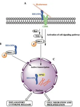
Figure 1.2. The interaction between cell surface RHAMM and HA activates the Ras-Mek-ERK pathway, and the phosphorylation of Ras-Mek-ERK1,2, which results in active transcription of mitogenic genes (A). Increased cytoplasmic RHAMM expression also results in
phosphorylation of ERK1,2, and therefore, the transcription of mitogenic genes (B). Increased nuclear RHAMM expression results in aberrant mitotic activity and genomic instability (C). All of these increases in RHAMM expression result in the release of
inflammatory cytokines and cellular migration and proliferation. Adapted from [29]. ... 3
Figure 1.3. RHAMM binds to microtubules and TPX2, and localizes at the centrosome, where it also binds to tubulin (A). TPX2 initiates the formation of microtubules at the kinetochore and activates AURKA (B). AURKA complexes with TPX 2 (C) and
accumulates at the centrosome (D). Adapted from [32]. ... 4
Figure 1.4. General scheme of solid-phase peptide synthesis ... 6
Figure 1.5. In an alpha-helix, the basic residues are often found along one exposed face of the protein, resulting in an amphipathic helical arrangement. ... 14
Figure 1.6. Analyte binding to the immobilized ligand at the sensor chip surface results in a change in resonance angle of reflected light. The Langmuir model with mass transport limitations describes the relationship between on (ka) and off (kd) rate constants, taking into
account the rate at which analyte is brought from the bulk solution to the sensor chip surface. Adapted from Nicoya Lifescience. ... 15
Figure 1.7. Analyte binding causes a shift in wavelength of absorbance position in localized SPR (LSPR). Adapted from Nicoya Lifesciences. ... 16
xiv
Figure 1.9. The plasma level of endogenous filtration markers depends on their generation (G) from cells and diet, elimination (E) by the liver and gut, and urinary excretion (U) by the kidneys. Urinary excretion includes filtration, tubular secretion and reabsorption. Under homeostatic conditions, U = G+E. Adapted from [98]. ... 22
Figure 2.1. 7 kDa RHAMM protein. (A) mRHAMM (706-767) sequence (B) Depiction of 7
kDa RHAMM, showing the protein’s two hyaluronan binding domains contained within two
helices, as proposed by [50, 51]. ... 38
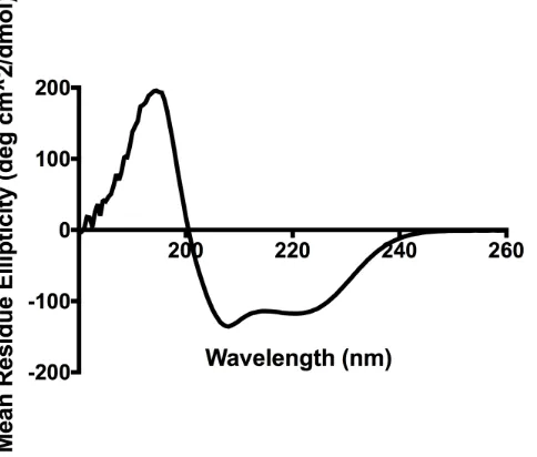
Figure 2.2. 7 kDa RHAMM is helical by CD spectroscopy. CD spectrum of 7 kDa RHAMM in water (0.5 mg/mL) showing alpha-helical character, similar to that of the full-length
protein’s predicted secondary structure [49, 50]... 40
Figure 2.3. 7 kDa RHAMM binds HA by SPR. SPR signals showing the interactions between immobilized 5-10 kDa HA and 7 kDa RHAMM (A) and a negative control mini-protein, which had no basic residues in the HA binding domains (B). Each signal corresponds to the responses of different HA concentrations, and solid lines indicate a linear 1:1 interaction curve fitting model for the ligand-analyte interaction. The dissociation constant was determined to be 9.0 nM for 7 kDa RHAMM and 3.2 M for the negative control mini-protein. ... 41
Figure 2.4. 7 kDa RHAMM binds HA by ELISA. ELISA was performed with HA-coated plates and varying concentrations of biotinylated 7 kDa RHAMM. ... 42
Figure 2.5. Excisional scratch wound assay performed on RHAMM-transfected 10T1/2 cells (LR21). LR21 cells treated with 7 kDa RHAMM (B) migrate slower over 24 hours than those not treated with the synthetic receptor (A). Statistical significance determined by students t-test (***p<0.005). Scale bar, 100 µM. ... 46
Figure 2.6. Biotinylated 7kDa RHAMM binds to the cell surface and occurs in intracellular perinuclear vesicles. The 7kDa RHAMM mini-protein was added to cultured RHAMM-transfected 10T1/2 fibroblasts, and its distribution was detected using a fluorescent
xv
is detected near or at the cell surface and that the majority of staining occurs in perinuclear vesicles, which is consistent with its endocytic uptake. Blue is Dapi to detect nuclei, and brightfield images are included to show intact cells. ... 47
Figure 2.7. SPR signals showing the interaction between immobilized 7 kDa RHAMM and previously reported tubulin-derived peptides, both binding and non-binding. Negative control (no peptide) graphs are also shown. Each signal corresponds to the responses of six peptide concentrations (1000 nM, 750 nM, 500 nM, 100 nM, 10 nM, and 1 nM). The solid lines indicate a global 1:1 interaction curve fitting model for each of the interactions. ... 49
Figure 3.1. Stapling RHAMM peptide mimetics increases helicity compared to the linear peptide by CD spectroscopy in both water (A) and 40% TFE solution (B) ... 82
Figure 3.2. SPR signals of linear and stapled peptides. Stapling and staple placement are important for HA-binding. Each signal corresponds to the response of 5 peptide
concentrations (750 nM, 1 M, 2.5 M, 5 M, and 10 M). The solid lines indicate a global 1:1 interaction curve fitting model for each of the interactions. ... 83
Figure 3.3. An inflammation protein array was performed on RAWBlue macrophages
stimulated with TLR agonist PAM3CSK4 in the presence or absence of 1 M of peptide ... 84
Figure 3.4. Preclinical evaluation of Peptide 3.1 was carried out in mice that were stimulated with lipopolysaccharide (LPS), a TLR agonist. Preliminary results demonstrate that a 54% decrease in TNF- concentration is observed in the presence of Peptide 3.1 compared to the LPS. ... 85
Figure 3.5. Stapling the peptide backbone (black) increases the half-life in human serum compared to the linear (red). Data was fit to nonlinear regression curves. ... 88
Figure 3.6. Modifications to lead compound, Peptide 3.1, affect helicity of peptides in water (A) by circular dichroism spectroscopy. Despite the modification, peptides all exhibit similar helical character in 40% TFE solution (B). ... 91
Figure 3.7. SPR experiments between modified variants of Peptide 3.1 and 5-10 kDa HA.
xvi
M, 5 M, and 10 M). The solid lines indicate a global 1:1 interaction curve fitting model for each of the interactions. ... 93
Figure 4.1. FT-IR spectrum showing shift in peaks as inulin is modified to CMI and Cy7.5-inulin conjugate and new functional groups are added ... 126
Figure 4.2. MALDI-MS of Inulin (A) and carboxymethyl inulin (CMI) (B) using DHB as matrix, acquired in reflectron mode. Masses were observed as sodiated adducts. A mass difference of 162 was observed for both inulin and CMI, and a mass difference of 57 Da was observed between inulin and CMI (boxed) (B). ... 128
Figure 4.3. ESI-MS spectrum of inulin ... 128
Figure 4.4. Dynamic light scattering of inulin, CMI, Cy7.5-inulin conjugate ... 130
Figure 4.5. Cy7.5-inulin is stable in plasma from CKD patients over 2 hours. Each
measurement was done in triplicate. The eluent following centrifugation, which contained water and particles <3 kDa in size, had absorption values < 0.05 at all time points. ... 131
Figure 4.6. Plasma from patients with chronic kidney disease does not absorb significantly at 788 nm, where Cy7.5-inulin has its maximal absorption in plasma. ... 132
Figure 4.7. Transcutaneous pulse dye densitometry reading of Cy7.5-inulin at 805 nm in the plasma of a farm-raised pig after intravenous injection of the dye. GFR was calculated as the ratio of the amount of dye injected to the AUC, resulting in an estimated value of 120
xvii
List of Schemes
Scheme 1.1. Fmoc-based solid-phase peptide synthesis ... 7
Scheme 1.2. General synthetic protocol for the formation of a lactam bridge between
glutamic acid and lysine residues ... 19
xviii
List of Supplemental Information
Figure S 2.1. UHPLC trace of 7 kDa RHAMM ... 66
Figure S 2.2. UHPLC trace of Ala-7 kDa RHAMM ... 66
Figure S 2.3. UHPLC trace of 2b (H-VEGEGEEEGEEY-NH2) ... 67
Figure S 2.4. UHPLC trace of 3b (H-SVEAEAEEGEEY-NH2) ... 67
Figure S 2.5. UHPLC trace of 10b (H-EEDFGEEAEEEA-NH2) ... 68
Figure S 2.6. UHPLC trace of 11b (H-GEFEEEAEEEVA-NH2) ... 68
Figure S 2.7. UHPLC trace of 12b (H-EAFEDEEEEIDG-NH2)... 69
Figure S 2.8. UHPLC trace of 14b (H-FTEAESNMNDLV-NH2) ... 69
Figure S 2.9. UHPLC trace of 7a (H-GEFSEAREDMAA-NH2) ... 70
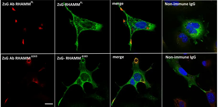
Figure S 2.10. 10T1/2 mesenchymal cells display RHAMM in cell processes and adhesion sites. 10T1/2 cells were transfected with full-length Zs-Green tagged RHAMM (green, top panel) and Zs-Green tagged RHAMM 163 (bottom panel). The tagged cell surface RHAMM was detected by co-localization of Zs-green antibody staining (red) with Zs-green fluorescence in non-permeabilized cells and is present in cell processes. Intracellular RHAMM, which is detected as green fluorescence only, is diffused in the cytoplasm. Scale bar, 20 µM... 70
Figure S 2.11. ESI+ Mass Spectrum for 7 kDa RHAMM ... 71
Figure S 2.12. ESI+ Mass Spectrum for Ala-7 kDa RHAMM... 71
Figure S 2.13. ESI+ Mass Spectrum for Peptide 2b (H-VEGEGEEEGEEY-NH2) ... 73
Figure S 2.14. ESI+ Mass Spectrum for Peptide 3b (H-SVEAEAEEGEEY-NH2) ... 74
xix
Figure S 2.16. ESI Mass Spectrum for Peptide 11b (H-GEFEEEAEEEVA-NH2) ... 75
Figure S 2.17 ESI+ Mass Spectrum for Peptide 12b (H-EAFEDEEEEIDG-NH2) ... 75
Figure S 2.18. ESI+ Mass Spectrum for Peptide 14b (H-FTEAESNMNDLV-NH2) ... 76
Figure S 2.19. ESI+ Mass Spectrum for Peptide 7a (H-GEFSEAREDMAA-NH2) ... 76
Figure S 3.1. HPLC trace of Linear peptide (Ac-KIKHVVKLKDENSQLKSEVSKL RSQLVKRK-NH2) ... 105
Figure S 3.2. HPLC trace of Peptide 3.1 (Ac-KIKHVVKLK [EENSK]-[EKSEK] SKLRSQLVKRK-NH2) ... 105
Figure S 3.3. HPLC trace of Peptide 3.2 (Ac-KIKHVVKLKD [ENSQK]-[ESEVK] KLRSQLVKRK-NH2) ... 106
Figure S 3.4. HPLC trace of Peptide 3.3 (Ac-KIKHVVKLK [EENSK]-[KKSEE] SKLRSQLVKRK-NH2) ... 106
Figure S 3.5. HPLC trace of Peptide 3.4 (Ac-KIKHVVKLK[KENSE]-[EKSEK] SKLRSQLVKRK-NH2) ... 107
Figure S 3.6. HPLC trace of Peptide 3.1-KIK (Ac-HVVKLK [EENSK]-[EKSEK] SKLRSQLVKRK-NH2) ... 107
Figure S 3.7. HPLC trace of Peptide 3.1_1st staple (Ac-KIKHVVKLK [EENSK]-LKSEV SKLRSQLVKRK-NH2) ... 108
Figure S 3.8. HPLC trace of Peptide 3.1_2nd staple (Ac-KIKHVVKLK DENSQ-[EKSEK] SKLRSQLVKRK-NH2) ... 108
Figure S 3.9. HPLC trace of Peptide 3.1+Gly (Ac-KIKHVVKLK [EENSK]-Gly-[EKSEK] SKLRSQLVKRK-NH2) ... 109
xx
Figure S 3.11. HPLC trace of Peptide3.1+GG (Ac-KIKHVVKLK [EENSK]-G-G-[EKSEK] SKLRSQLVKRK-NH2) ... 110
Figure S 3.12. HPLC trace of Peptide 3.1+AA (Ac-KIKHVVKLK [EENSK]-A-A-[EKSEK] SKLRSQLVKRK-NH2) ... 110
Figure S 3.13. HPLC trace of Peptide 3.1-KIK+AA (Ac-HVVKLK [EENSK]-A-A-[EKSEK] SKLRSQLVKRK-NH2) ... 111
Figure S 3.14. ESI+ Mass Spectrum for Linear peptide (Ac-KIKHVVKLKDENSQL
KSEVSKLRSQLVKRK-NH2) ... 113
Figure S 3.15. ESI+ Mass Spectrum for Peptide 3.1 (Ac-KIKHVVKLK [EENSK]-[EKSEK]
SKLRSQLVKRK-NH2) ... 113
Figure S 3.16. ESI+ Mass Spectrum for Peptide 3.2 (Ac-KIKHVVKLKD
[ENSQK]-[ESEVK] KLRSQLVKRK-NH2) ... 114
Figure S 3.17. ESI+ Mass Spectrum for Peptide 3.3 (Ac-KIKHVVKLK [EENSK]-[KKSEE] SKLRSQLVKRK-NH2) ... 114
Figure S 3.18. ESI+ Mass Spectrum for Peptide 3.4 (Ac-KIKHVVKLK[KENSE]-[EKSEK] SKLRSQLVKRK-NH2) ... 115
Figure S 3.19. ESI+ Mass Spectrum for Peptide 3.1-KIK (Ac-HVVKLK [EENSK]-[EKSEK]
SKLRSQLVKRK-NH2) ... 117
Figure S 3.20. ESI+ Mass Spectrum for Peptide 3.1_1st staple (Ac-KIKHVVKLK
[EENSK]-LKSEV SKLRSQLVKRK-NH2) ... 117
Figure S 3.21. ESI+ Mass Spectrum for Peptide 3.1_2nd staple (Ac-KIKHVVKLK
DENSQ-[EKSEK] SKLRSQLVKRK-NH2) ... 118
Figure S 3.22. ESI+ Mass Spectrum for Peptide 3.1+Gly (Ac-KIKHVVKLK
xxi
Figure S 3.23. ESI Mass Spectrum for Peptide 3.1+Amb (Ac-KIKHVVKLK [EENSK]-Amb-[EKSEK] SKLRSQLVKRK-NH2) ... 119
Figure S 3.24. ESI+ Mass Spectrum for Peptide3.1+GG (Ac-KIKHVVKLK
[EENSK]-G-G-[EKSEK] SKLRSQLVKRK-NH2) ... 119
Figure S 3.25. ESI+ Mass Spectrum for Peptide 3.1+AA (Ac-KIKHVVKLK
[EENSK]-A-A-[EKSEK] SKLRSQLVKRK-NH2) ... 120
Figure S 3.26. ESI+ Mass Spectrum for Peptide 3.1-KIK+AA (Ac-HVVKLK
[EENSK]-A-A-[EKSEK] SKLRSQLVKRK-NH2) ... 120
Figure S 3.27. CD spectra of double stapled Peptides 1-4 (B-E) and their linear counterpart (A) showing helicity in water and 40% TFE solution. Peptides were run at 0.25 mg/mL ... 121
Figure S 3.28. CD spectra of modified double stapled peptides, in which the linker region and N-terminal sequence of Peptide 3.1 were modified. All peptides were run at 0.25 mg/mL in water and 40% TFE solution... 122
Figure S 3.29. Serum stability of Peptides 3.1-3.4 and their linear counterpart ... 123
Figure S 4.1. Proton NMR spectra of Inulin (A), CMI (B), Conjugate (C). The signal at 5.15 ppm in inulin corresponds to the anomeric carbon. All samples were run in DMSO-d6. .... 141
Figure S 4.2. Carbon NMR of Inulin (A), CMI (B), Conjugate (C). All samples were run in DMSO-d6. ... 142
xxii
List of Abbreviations
CD: Circular dichroism
CKD: Chronic kidney disease
Cy7.5: Cyanine 7.5
DMEM: Dulbecco’s Modified Eagle Medium
DIPEA: N, N-Diisopropylethylamine
DMF: Dimethylformamide
DMSO: Dimethylsulfoxide
ECM: Extracellular matrix
eGFR: Estimated glomerular filtration rate
ERK1,2: Extracellular signal-regulated kinase 1,2
ELISA: Enzyme-linked immunosortbent assay
FBS: Fetal bovine serum
FL: Full-length
Fmoc: 9-fluorenylmethoxycarbonyl
GAG: Glycosomaminoglycan
GFR: Glomerular filtration rate
GPI: Glycosylphosphatidylinositol
GST: Glutathione S-transferase
xxiii
HABD: Hyaluronan binding domain
HATU: 1-[Bis(dimethylamino)methylene]-1H-1,2,3-triazolo[4,5-b]pyridinium 3-oxid hexafluorophosphate
HCTU: 2-(6-Chloro-1-H-benzotriazole-1-yl)-1,1,3,3-tetramethylaminium hexafluorophosphate
Hmb: 2-hydroxy-4-methoxybenzyl
HMW: High molecular weight
HPLC: High-performance liquid chromatography
HRP: Horseradish peroxidase
ka: rate constant of association
kd: rate constant of dissociation
km: rate constant of mass transport
LSPR: Localized surface plasmon resonance
MAP: Microtubule-associated protein
MAPK: Mitogen activated pathway kinase
MBHA: 4-methylbenzhydrylamine
MDRD: Modification of Diet in Renal Disease
MMP: Matrix metalloproteinase
m/z: mass over charge ratio
NTA: Nitrilotriacetic acid
xxiv
PBS: Phosphate buffered saline
PDB: Protein Data Bank
ROS/RNS: Reactive oxygen species/Reactive nitrogen species
RHAMM: Receptor for hyaluronan mediated motility
SAR: Structure-activity relationship
SPPS: Solid-phase peptide synthesis
SPR: Surface plasmon resonance
TBME: Tert-butyl methyl ether
TBS: Tris-buffered saline
TFA: Trifluoroacetic acid
TFE: Tetrafluoroethylene
TIPS: Triisopropylsilane
TMB: Tetramethylbenzidine
TPX2: Targeting protein for Xklp2
xxv
Ce qui embellit le désert, dit le petit prince, c’est qu’il cache un puits quelque part.
“What makes the desert beautiful,” said the little prince, “is that somewhere it hides a well.”
Chapter 1
1
Introduction
Hyaluronan
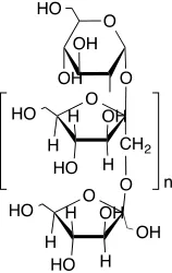
Hyaluronan (HA) is a simple linear extracellular matrix polysaccharide that belongs to the glycosaminoglycan (GAG) family of macromolecules and consists of dimeric repeats of N-acetylglucosamine followed by D-glucuronic acid (Figure 1.1). In homeostatic tissues, the majority of HA occurs in its high molecular weight (HMW) and native form (e.g. >500 kDa), which is organized into scaffolds and matrices of the extracellular matrix (ECM) [1, 2], making it a ubiquitous component of the ECM. The ECM environment is tightly regulated during normal physiological conditions, providing important structural and biochemical support to surrounding cells [3, 4]. Its functions are understudied, but at a minimum, it promotes tissue hydration, provides lubrication, protects against mechanical damage, reduces proliferation, modulates immune
complex interactions control a variety of signaling pathways that regulate cell adhesion/motility, mitotic spindle integrity and transcriptomes.
Figure 1.1. Structure of hyaluronan, depicting the dimeric repeat of D-glucuronic acid and N-acetyl glucosamine
Receptor for hyaluronan mediated motility
(RHAMM)
RHAMM (gene name HMMR) is a largely hydrophilic protein that was originally isolated from embryonic chicken heart explant cultures exhibiting high HA production and increased cell migration. It is one of a number of HA receptors that are expressed on immune cells that bind to complex mixtures of HA polymer sizes and activate pathways required for an inflammatory or tumorgenic response. RHAMM exists in two
Figure 1.2. The interaction between cell surface RHAMM and HA activates the Ras-Mek-ERK pathway, and the phosphorylation of ERK1,2, which results in active transcription of mitogenic genes (A). Increased cytoplasmic RHAMM expression also results in phosphorylation of ERK1,2, and therefore, the transcription of mitogenic genes (B). Increased nuclear RHAMM expression results in aberrant
mitotic activity and genomic instability (C). All of these increases in RHAMM expression result in the release of inflammatory cytokines and cellular migration
and proliferation. Adapted from [29].
RHAMM interacts with HA via two HA binding domains (HABDs) located near the
any basic residue and X represents any non-acidic residue [4]. These clusters of basic residues allow for ionic interactions with the carboxylate ions of HA [4].
In addition to being a cell surface receptor for HA, RHAMM binds to extracellular-regulated kinase (ERK) kinase, and regulates the expression of ERK [30]. ERK has two closely related isoforms (ERK1 and -2) that are required for cellular differentiation and proliferation [28, 30]. RHAMM is also a microtubule-associated protein (MAP) that binds directly to microtubules and interacts with TPX2, a different MAP that is required for microtubule formation [31]. RHAMM localizes to the centrosome, where it interacts with tubulin and helps to maintain mitotic spindle integrity and polarity [32] (Figure 1.3). TPX2 initiates the formation of microtubules at the kinetochore, and activates Aurora kinase A (AURKA), which accumulates at the centrosome from S phase to the end of mitosis and facilitates the formation of microtubules [33-35]. This results in centrosome maturation and spindle assembly [33]. High expression of AURKA results in aberrant mitotic spindle formation, and is, therefore, correlated with genetic instability and poor prognosis in human diseases [36].
Figure 1.3. RHAMM binds to microtubules and TPX2, and localizes at the centrosome, where it also binds to tubulin (A). TPX2 initiates the formation of microtubules at the kinetochore and activates AURKA (B). AURKA complexes with
While RHAMM expression is tightly regulated or absent in most tissues, it is important for a number of wound repair processes that require cell migration, invasion, and remodelling of the ECM. Its restricted expression makes it a potential target for cancer and wound repair therapy with low toxicity. RHAMM peptides are currently being tested in phase II clinical trials for multiple myeloma and myelodysplastic syndrome and are showing efficacy and low toxicity in patients [37, 38].
Peptide synthesis
Peptides can be synthesized chemically by two main techniques, solid phase peptide synthesis (SPPS) and solution phase peptide synthesis. Solution phase peptide synthesis can be more problematic than its solid phase counterpart, as dimerization and the formation of other unwanted by-products requires the purification after the addition of each amino acid.
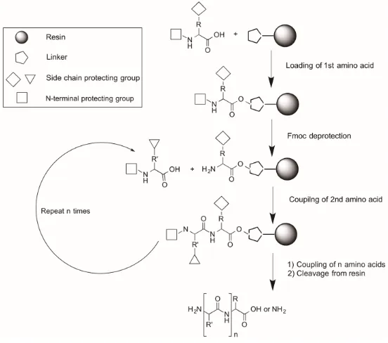
In the early 1960s, Merrifield pioneered the idea of SPPS, using a polystyrene-based solid support for peptide synthesis, which would deal with the difficulties associated with solution phase synthesis [39]. The technique of solid phase peptide synthesis covalently links the peptide chain to an insoluble resin, which acts as a solid support off of which the peptide chain can grow or elongate. The solid support facilitates the addition of excess soluble reagents in order to drive each reaction to completion, and the excess reagents can be easily removed by multiple washing and filtration steps, eliminating the need for tedious purification steps after the addition of each amino acid [39]. Following synthesis, the crude peptide is released from the solid support and purified by traditional HPLC methods, with higher yields often reported for SPPS than for solution phase methods [39]. SPPS can be performed either manually or using an automatic peptide synthesizer, which is convenient, and improves throughput.
achieved, it can be removed from the resin, yielding the free and linear peptide (Figure 1.4).Similarly, the synthesis can be monitored by LC-MS at any point during the synthesis by removing a small aliquot of resin and cleaving the peptide off of it.
Figure 1.4. General scheme of solid-phase peptide synthesis
Successful SPPS depends on the choice of resin used. Resins are insoluble polymers that are pre-loaded with varying linkers, which provide flexibility during synthesis, and allow for control over the final product by determining the functionalization of a cleaved peptide [40]. For example, Rink amide resins are functionalized with an amine group that covalently binds to the first amino acid in the sequence to obtain an amide bond at the carboxyl terminus of the peptide. In addition, resins can have different substitution levels, which when decreased, facilitate the synthesis of long peptides by avoiding interchain crowding, resulting in improved the synthetic yields [41].
basic conditions for the removal of the Fmoc group from each amino acid in order for the next amino acid to be added, and by the strong acidic conditions that are required to liberate the peptide from the resin, as well as remove all of the acid-labile side-chain protecting groups.
Fmoc-based SPPS is carried out as depicted in Scheme 1.1. A protected amino acid is added to the resin through standard coupling procedures to form an amide bond. The N-terminal Fmoc group of the amino acid is then deprotected with piperidine, resulting in a free amine. The remainder of the peptide sequence is built following the same procedure until the final peptide length is acquired. The free peptide is then obtained by treatment of the resin with acid in order to simultaneously deprotect amino acid side chains and
remove the peptide from the solid support.
Scheme 1.1. Fmoc-based solid-phase peptide synthesis
Rational design of peptides
Targeting a protein-polysaccharide or protein-carbohydrate interaction is often challenging, as there is a large surface area of interaction and a lack of well-defined binding pockets that prevent the use of small molecules for interfering with these binding interfaces and inhibiting the interaction. Similarly, a challenge exists in trying to develop
H N Fmoc H N OH O 20% Piperidine/DMF HCTU, DIPEA Fmoc 2. 1. N H H N O H2N
O n 20% Piperidine/DMF NH2 H N O H2N
O n 20% acetic anhydride/DMF
N H H N O N H O n NH2 H N O N H O n 95% TFA 2.5% TIPS 2.5% H2O
R R R R R R' R' R' R' Ac Ac N H H N O N H Fmoc
On R R'
n
mimics that contain the specific multivalences that will foster interaction with its target [43]. This is further complicated by the conformational changes that these HA receptor proteins undergo upon binding to HA [44]. Higher molecular weight entities such as peptides, proteins, and antibodies are more readily able to block protein-polysaccharide interactions due to their ability to interact over a larger surface area.
The discovery of peptides that modulate protein-carbohydrate interactions has focused largely on two approaches: unbiased or random peptide library screening and rational design based upon known structure or binding sites. Unbiased peptide library screening has used phage display, which is a biochemical approach to identify high affinity peptides displayed on a bacteriophage, and one-bead one-compound (OBOC) libraries, which is a chemical approach of screening peptides using polymer beads. These unbiased peptide libraries were primarily used for identifying peptide mimetics that scavenge HA and HA fragments. The discovery approaches used for finding HA binding peptides that mimic HA receptors have primarily been based upon rational design, with many structural leads
being derived from RHAMM’s known binding sites for HA.
In many cases, the rational design of targeting peptides can start with a known crystal
structure of the peptide, which provides important information on the compound’s
secondary and tertiary structure [45]. Based on the initial crystal structure, alanine scans, small focused libraries, and structure-activity relationship (SAR) studies are carried out in order to identify the essential amino acids and sites for possible modification. This
process allows for the identification of those amino acids that are unstable and
susceptible to events such as isomerization, glycosylation, or oxidation [46]. In addition
to optimizing the peptide’s sequence, rational design involves improving the
Peptides as therapeutics
Targeting a specific receptor or other class of target can be achieved with a number of different classes of molecules, including small molecules, peptides, or antibodies. There are advantages and disadvantages to using the different classes of compounds. Small molecules have relatively low molar mass, and therefore, can access many biological targets in the body. In addition, small molecules are not easily recognized or degraded by enzymes; however, they unfortunately suffer in lipophilicity, and because of their small surface area, suffer from nonspecific interactions, which can lead to toxicity [47]. Peptides, on the other hand, can be synthesized by standard synthetic protocols, and improvements in manufacturing technology have allowed for peptides of various length ranges up to 40 amino acids to be synthesized, as well as quick and easy synthesis of peptide analogues [48, 49].
Peptide-based drugs are generally shorter than 50 amino acids in length, with molecular weights that lie between those of small molecules and large biologics [50]. Peptides as a class of molecule are particularly attractive as drug candidates because they are
biocompatible with typically low toxicity, structurally diverse permitting high selectivity and potency, and have a predictable metabolism [51]. These features have enabled
peptides to have high affinity for their targets at nanomolar and picomolar concentrations. Unfortunately, peptides suffer from a relatively short circulating half-life because they are susceptible to the same digestive enzymes designed to break down amide bonds of ingested proteins, and can be cleaved by both endopeptidases and exopeptidases. As a result, they have poor oral bioavailability and are quickly eliminated [45]. In addition, they are prone to hydrolysis and oxidation, and have low membrane and tissue
permeability due to their polarity and relatively high molecular weight [45]. However, there has been an increasing number of commercially available unnatural amino acids and strategies that have helped improve the stability and other physical properties of peptides. For example, the identification of possible cleavage sites within the peptide sequence and the substitution of relevant amino acids is a preliminary approach to
unnatural amino acids, the conjugation of additional moieties that extend half-life or improve solubility, and cyclization of the peptide backbone with synthetic structural
constraints, which may stabilize the peptide’s secondary structure [48, 52].
Current peptide mimetics that inhibit RHAMM
-
HA
interactions
1.6.1 Rationally designed peptide mimetics
To date, several peptide mimetics have been rationally designed to interfere and block the HA-RHAMM interaction. RHAMM-sequence based peptide mimetics that bind to HA fragments and have been shown to have therapeutic effects in a number of processes, including inflammation, wound repair, and fibrosis/adipogenesis. One of the first rationally designed HA-binding peptides was based on the RHAMM BX7B HA binding
motif, but does not otherwise have any amino acid sequence homology with RHAMM. This peptide strongly reduced BAL macrophages in bleomycin-induced lung injury and blunted destruction of lung architecture [53], reduced surfactant protein A-induced
macrophage chemotaxis [54] and ozone induced lung hyper-responsiveness [55]. Another peptide, pep-35, has 70% homology with the RHAMM sequence and essentially joins four RHAMM HA binding sequences together. This peptide reduced Staphyloccus
aureus burden in infected surgical wounds and increased the production of CXCL1,2 by
inflammatory cells, which subsequently increased neutrophil influx into the wound [56]. Other peptides have been designed to mimic the three BX7B motifs of CD44 and were
shown to block tumor cell growth [57]. Finally, RHAMM sequence mimics (NPI-0102, NPI-0104), which disrupt HA binding to RHAMM, were reported to promote
adipogenesis and reduce tissue fibrosis [58] by increasing the production of adiponectin, an anti-fibrogenic adipokine [59, 60].
In another rationally designed approach, Esguerra et al. developed HA peptide mimics
those that contained a repeating amino acid motif of EEXEE, suggesting that both electrostatic forces and conformational effects may be important for the development of RHAMM-binding ligands.
Other RHAMM peptide-based therapies could reasonably be developed from varying the peptide backbone and/or altering the peptide structure, which may confer improved specificity and affinity towards its target. Such strategies include the development of stapled or cyclized peptides, resulting in more drug-like compounds.
1.6.2 Peptide library screening for HA- and HA receptor-binding
In addition to rational design, many peptides with therapeutic potential were discovered through peptide library screenings. The P15-1 peptide (STMMSRSHKTRSHHV) is a 15mer peptide, which was the first peptide mimetic that was reported to bind specifically to HA fragments of <10 kDa. It was identified by screening a recombinant phage display library with a complexity of approximately 1013 transformants for peptides that both bind
to HA fragments (MW range 5-200 kDa) linked to Sepharose beads and that block cell motility [62]. Two peptide sequences were recovered in the screen and of these, P15-1 exhibited the highest affinity for HA fragments (KD = 10-7 M), and most strongly blocked
cell motility. It has low homology with known HA receptors but contains a BX7B motif
Pep-1, a 12mer peptide (GAHWQFNALTVR) was identified as an HA binding sequence by screening an M13 phage display library expressing random 12mer peptides fused to gene 3 (pIII) minor coat proteins with a complexity of approximately 109 transformants
[67]. This peptide (Pep-1) was isolated by panning the library for sequences that bind to HA-coated plates. Pep-1 binds to HA with moderate affinity (KD = 1.4 µM), inhibits HA
binding to innate immune cells, and was shown to inhibit leukocyte attachment to HA substrates [67, 68]. The systemic, subcutaneous or topical administration of this peptide inhibited dinitrofluorobenzene/oxazolone induced-contact hypersensitivity by blocking both the in-trafficking of inflammatory cells and the migration of dendritic cells out of the epidermis [67]. Skin dendritic cells utilize HA as a motogenic stimulus for migrating from the epidermis to lymph nodes, where they function as antigen-presenting cells, a process that is required for generating protective pro-inflammatory and tolerogenic immune responses during tissue injury [69, 70]. Aberrant activation of these cells contributes to inflammatory disease processes [70]. These results provided early support for the development of HA inhibitors for inflammatory disorders. Pep-1 was later shown to inhibit the production of fragmented HA-promoted chemokine MIP-2 by bone marrow macrophages [71], reduce bronchial inflammation [72], reduce pro-inflammatory
cytokine production (TNF-α, IL-6, MMP13 and iNOS), and preserve cartilage
architecture in a mouse model of collagen-induced arthritis [69, 73]. In addition, Pep-1 dramatically inhibited interleukin-2 (IL-2)-induced vascular leak syndrome (VLS) [74], which may be linked to its anti-inflammatory effects. P15-1 and Pep-1 have been useful in dissecting the signaling pathways that are regulated by HA fragments, and their
efficacy in blocking inflammation in animal models propelled the development of peptide mimetics that block fragmented HA-induced activity that leads to disease.
Recombinant CD44 protein has also been used to screen peptide libraries [75]. In this study, a Ph.D.TM -12 phage display peptide library with a peptide complexity of 2.7x10⁻⁹
transformants was screened using recombinant CD44 as bait. The screen isolated several peptides, one of which exhibited a KD = 7.5 pM for recombinant CD44. However, none
peptides that bind to HA receptors is a viable approach for potentially developing novel inhibitors of HA receptor signaling.
Peptide-displaying phage and peptide library technology have also been used to identify peptides that mimic carbohydrates [76]. HA peptide mimics that bind with high affinity to recombinant RHAMM containing hyaluronan binding sequences, were originally identified by Ziebell, M. et al [77, 78].Two libraries of 8mer peptides were designed to target recombinant RHAMM fragments, with one library consisting of peptides made of entirely random sequences. The second library was biased, with alternating acidic residues incorporated in every other position of the sequence, with the intention of mimicking the placement of the glucuronic acid moieties of HA [77]. Peptides from the unbiased (random) library bound to recombinant RHAMM in an HA fragment-dependent manner with µM to nM affinity, and exhibited some similarities with respect to regions of hydrophobic residues (e.g. PVY), but contained very few negatively charged amino acids. These peptides were then computationally modeled to evaluate their binding to an NMR-based model of RHAMM, from which residues within RHAMM were identified and theorized to stabilize RHAMM-HA interactions [78]. However, these peptides have not yet been reported to affect cellular functions.
Protein
-
Carbohydrate interactions
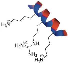
to exist on proteins, which facilitates the protein’s interaction with the negatively charged groups of the sugar. Heparin-binding proteins have been identified as having binding sites with sequences of XBBXBX or XBBBXXBX, where B is any basic residue, and X is any non-acidic residue [83]. Similar binding sequences have been observed in HA-binding proteins, such as RHAMM and CD44, both of which exhibit a BX7B binding
motif [4]. Depending on the secondary structure of the protein, it is possible that very few residues in these binding sequences actually contribute to or participate in binding. For example, when the binding domains are arranged in an alpha-helix, the basic residues are often found along one exposed face of the protein, resulting in an amphipathic helical arrangement [84]. Therefore, in order for an interaction to take place between linear carbohydrates, such as HA, and largely helical proteins, such as RHAMM, the positively charged residues arrange themselves so that they are on the same face of the protein segment (Figure 1.5).
Figure 1.5. In an alpha-helix, the basic residues are often found along one exposed face of the protein, resulting in an amphipathic helical arrangement.
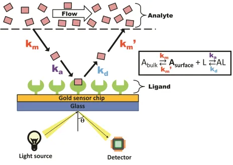
molecule, for signal readout. SPR, on the other hand, is a label-free method for evaluating biomolecular interactions, measuring single concentrations of compound at a single time, decreasing the throughput of measurement compared to ELISA. Despite this, label-free methods of evaluating the interaction between two compounds is especially important for small proteins or peptides, as secondary structure often influences binding, and
chemically modifying these molecules can potentially alter their secondary structure. In addition, unlike ELISA, SPR delivers a kinetic analysis of the interaction in addition to the binding affinity in real-time, providing information on the association (ka) and
dissociation (kd) phases of the interaction.
Figure 1.6. Analyte binding to the immobilized ligand at the sensor chip surface results in a change in resonance angle of reflected light. The Langmuir model with
mass transport limitations describes the relationship between on (ka) and off (kd)
The principle of SPR utilizes the immobilization of a compound of interest, known as the ligand, to a metal-coated sensor chip surface, and the measurement of changes in
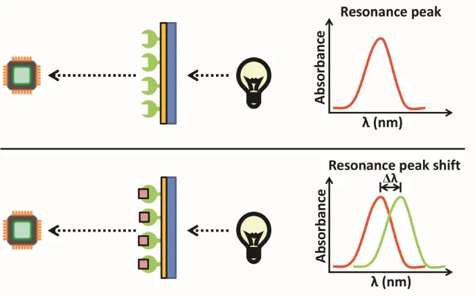
refractive index at the surface as another molecule, the analyte, binds. A light source shines light or a laser on the metal-coated film, reflecting with a resonance angle that shifts when a binding event occurs at the sensor chip surface, resulting from a change in refractive index (Figure 1.6). This shift depends on the mass and density of analyte at the chip surface. In this thesis, a variation of SPR, localized SPR (LSPR), was studied. LSPR differs from traditional SPR by using gold nanoparticles at the sensor chip surface instead of the continuous metal film used in traditional SPR. In addition, LSPR produces a strong resonance absorbance peak, with its position being highly sensitive to the refractive index localized around the nanoparticles. As a result, it measures changes in the wavelength of the absorbance position resulting from binding events rather than changes in angles (Figure 1.7), like is measured in traditional SPR.
Figure 1.7. Analyte binding causes a shift in wavelength of absorbance position in localized SPR (LSPR). Adapted from Nicoya Lifesciences.
associates with it, it accumulates on the chip surface, resulting in a change in molecular weight at the chip surface, which is denoted by a change in signal. The rate of association between the two compounds depends directly on the on-rate of the interaction (ka) and
the mass transport of the analyte from the bulk flow to the sensor chip surface (km). Once
the flow of analyte switches to buffer, the analyte dissociates from the chip surface, resulting in a decrease in signal (kd). A strong ligand-analyte interaction requires the use
of a regeneration buffer to completely remove analyte from the chip surface, while leaving the immobilized ligand in-place.
An important consideration in SPR is that tethering the ligand to the sensor chip surface will not disrupt its activity, prevent rotational freedom, or block any sites of binding. Therefore, the correct method of immobilizing one of the binding partners to the sensor chip surface is important. Biomolecule immobilization can be either direct, by covalent interaction with the sensor chip surface, or indirect, by binding to an immobilized capturing molecule [86]. Examples of direct immobilization include biotin-streptavidin interactions, gold-thiol interactions, and amine-carboxylic acid interactions. In many of these instances, orientation of these proteins is difficult to control, resulting in mixed orientations of protein on the surface, and the interaction can be so strong that
regeneration of the sensor chip surface is not possible, such as with biotin-streptavidin interactions [87]. An alternative approach is to indirectly immobilize a biomolecule by labeling it with an oligo histidine tag (His tag) at either its C- or N-terminus, which interacts with nitrilotriacetic acid (NTA) that is pre-immobilized on the sensor chip. This method of attachment involves the capture of the His-tag protein by Ni2+ NTA chelation
without altering secondary structure or blocking important analyte-binding residues, and allows for regeneration of the sensor chip. The exact immobilization chemistry depends on the specific interaction that is being evaluated.
Stapled peptides
1.8.1 Alpha-helices
62% alpha-helical proteins, suggesting that the alpha-helical conformation plays an important role in mediating a number of biological processes, including interactions with other proteins and macromolecules [52, 88].
There is a high energy requirement associated with organizing three consecutive amino acids into a helical conformation, which is referred to as the helix-coil transition theory [89]. Many short peptides lack a helical structure, as the energy required to organize them into well-defined helices is too great. However, pre-folding short peptides synthetically has the potential to overcome this energy barrier and can therefore facilitate the adoption of a stable secondary structure, such as an alpha-helix.
1.8.2 Stapled peptides
One complete turn of an alpha-helix is made up of 3.6 amino acids, resulting in the residues at positions i, i+4, and i+7 occurring on the same face of the helix [52].
Synthetically, the alpha-helical conformation of a peptide is carried out by introducing a
covalent bond, or “staple”, at these amino acids positions, stabilizing the structure and
inducing an alpha-helical conformation [90]. The positions of these staples can be important, however, and care must be taken in order to not replace or obstruct those residues that are important for binding to the target. Four methods for synthesizing stapled peptides have been extensively studied thus far, and include their formation by a lactam bridge, a hydrocarbon chain, a metal-ion clip, and a hydrogen bond surrogate [50, 52]. Staples formed by a hydrocarbon chain and lactam bridge are the preferred methods of peptide cyclization for the improvement in bioactivity and target recognition that they confer [91].
Staple formation in peptides depends on the linear peptide sequence and can only take place if the unfolded peptide sequence has the natural propensity to adopt an alpha-helical conformation. If the peptide sequence is unable to naturally adopt an alpha-alpha-helical conformation, it is possible that the side chains forming the staple will not arrange
themselves appropriately, and therefore, will not bridge. Staple length depends on the size of the ring that is created following bridging, with shorter staples between side
Amino acid side chains should produce a staple length or size that is large enough to not cause significant ring strain, and therefore, decrease the demand for activation energy for successful cyclization to be completed [92]. Side chains involved in creating the linker do not contribute to target recognition, and therefore, block at least one face of the helix once the staple is created.
A lactam bridge is created by means of an amide bond that forms between the side chains of two amino acids. Natural amino acids, glutamic acid or aspartic acid and lysine, are commonly applied to make the lactam bridge, but unnatural amino acids, such as ornithine and aminoadipic acid, can also be used. During synthesis,
orthogonally-protected glutamic acid and lysine residues replace the amino acids of the natural peptide sequence in the appropriate positions (i.e. positions i, i+4, i+7, i+11). The peptide chain is built on resin following standard Fmoc-based SPPS procedures, allowing for the selective deprotection of the two amino acid side chains forming the lactam bridge, and their subsequent coupling to staple the peptide backbone and yield the cyclized product. Specifically, allylester and alloxycarbonyl protecting groups on glutamic acid and lysine side chains, respectively, are deprotected with palladium (0) catalyst under basic
conditions, and the deprotected side chains are coupled together with a coupling agent, such as 1-[Bis(dimethylamino)methylene]-1H-1,2,3-triazolo[4,5-b]pyridinium 3-oxid hexafluorophosphate (HATU). The remainder of the peptide sequence can be modified as necessary, and eventually removed from the resin, with the staple intact (Scheme 1.2).
1.8.3 Circular Dichroism spectroscopy
The secondary structure of proteins and peptides can be analyzed and quantified by analytical techniques, such as circular dichroism (CD) spectroscopy, which measures the extent to which asymmetric molecules can absorb right- and left-handed circularly polarized light [93]. The amide bonds in the primary sequence of proteins and peptides contain chromophores that produce different excitation interactions, resulting in
identifiable characteristics on the spectra [94]. Each type of secondary structure produces a unique CD spectrum, which are blended together when different secondary structure elements exist within a single compound. The CD spectrum of a perfect alpha-helix (3.6 amino acids per turn) is characterized by two minima at 222 nm and 208 nm, and a maximum at 193 nm, but these signals may shift in wavelength or in intensity if the compound contains elements of -sheet or random coil within its structure [94]. Many large proteins are made up of multiple secondary structure elements, and many small stapled peptides lack a perfectly stable secondary structure beyond the stapled sequence. In both of these cases, the CD spectrum is unlikely to reflect a perfect single secondary structure.
Glomerular filtration rate
Glomerular filtration rate (GFR) measures renal clearance, and can therefore, be a valuable tool in assessing kidney function and identifying the presence of chronic kidney disease (CKD) or other diseases that may target the kidneys. GFR may also be used for monitoring kidney function following kidney transplant, and therefore, identifying threats associated with organ rejection and nephrotoxicity.
Figure 1.8. Structure of inulin
In the classic method for measuring clearance by Homer Smith, patients receive a continuous intravenous infusion of inulin following a period of fasting, multiple collections of blood and urine samples at precise times over 3 hours, and bladder
catheterization in order to ensure complete urine collection [95]. Because this method of determining GFR is time-consuming, impractical, and invasive for patients, the clinical practice of using inulin has decreased, and the common clinical practice has been replaced with measuring the clearance of endogenous markers, such as serum creatinine levels, which provide an estimated GFR (eGFR) value quickly for clinical decision making using the Modification of Diet in Renal Disease (MDRD) Study equation [96] or the Cockcroft-Gault formula [97].
Cockcroft-Gault formula:
𝐶𝑟𝑒𝑎𝑡𝑖𝑛𝑖𝑛𝑒 𝑐𝑙𝑒𝑎𝑟𝑎𝑛𝑐𝑒 =(140 − 𝑎𝑔𝑒 𝑖𝑛 𝑦𝑒𝑎𝑟𝑠) × (𝑏𝑜𝑑𝑦 𝑤𝑒𝑖𝑔ℎ𝑡 𝑖𝑛 𝑘𝑔) (72 × 𝑠𝑒𝑟𝑢𝑚 𝑐𝑟𝑒𝑎𝑡𝑖𝑛𝑖𝑛𝑒 𝑖𝑛 𝑚𝑔𝑑𝐿 )
The value is multiplied by 0.85 if the individual is female.
MDRD formula:
Plasma levels of endogenous filtration markers, such as creatinine, are determined by their generation from cells and diet, renal excretion, including filtration through the glomeruli, and tubular secretion and reabsorption, and elimination by the gut and liver (Figure 1.9) [98]. Unfortunately, equations that estimate GFR consider only 4 variables: age, gender, race, and body weight. Factors that influence muscle mass or diet, changes in diet or muscle mass due to illness or amputation that are not considered, and
differences in race other than those considered in the equation, result in inaccuracies that underestimate the normal physiologic GFR value.
Figure 1.9. The plasma level of endogenous filtration markers depends on their generation (G) from cells and diet, elimination (E) by the liver and gut, and urinary excretion (U) by the kidneys. Urinary excretion includes filtration, tubular secretion
and reabsorption. Under homeostatic conditions, U = G+E. Adapted from [98].
In order to improve the efficiency and accuracy of GFR measurements, several
alternative exogenous tracer agents [98] have been developed, all of which allow for real-time measurement of kidney function by monitoring the decay of a bolus infusion of the reporter molecule from the plasma or the extracellular space over a period of time [99]. These alternative techniques include the use of radioisotope-labeled probes, such as 125
I-iothalamate [100], 51Cr-ethylenediaminetetraacetic acid (EDTA) [101], and 99m
including iohexol [104], unlabeled iothalamate [105, 106], FITC-labeled sinistrin [107, 108], and FITC-labeled inulin [109, 110]. Methods for detecting these reporter molecules have included corometric assays, scintillation counting, X-ray fluorescence, HPLC, visible fluorescence, and two-photon intravital microscopy [99]. While many of these techniques are useful for developing and evaluating probes, they can be costly and are not easily translated to a clinical environment. In addition, these alternative probes have all been found to underestimate GFR, suggesting that they might be suffering from tubular reabsorption or plasma protein binding [98].
Rationale of thesis
This thesis will focus on two separate frameworks. The first framework encompasses chapters 2 and 3, studying the protein-carbohydrate interaction between RHAMM and HA and developing tools that inhibit it, while the second framework is discussed
exclusively in chapter 4, involving a different carbohydrate, inulin, and the development of an optical agent based on it. Chapter 2 describes the chemical synthesis and
optimization of a truncated RHAMM receptor, 7 kDa RHAMM, for use as the target receptor in screening and discovering novel peptide ligands. Chapter 3 describes the development of double stapled RHAMM peptide mimetics that bind HA with high affinity, and that are active in blocking inflammation both in vitro and in vivo. Chapter 4 describes the development of a dye-labeled inulin conjugate for measuring glomerular filtration rate by transcutaneous pulse dye densitometry and has been evaluated in vivo in farm-raised pigs.
References
1. Stern, R., Asari, A.A., and Sugahara, K.N., Hyaluronan fragments: An
information-rich system. Eur J Cell Bio, 2006. 85(8): p. 699-715.
2. Tolg, C., et al., Hyaluronan and RHAMM in wound repair and the
3. Misra, S., et al., Interactions between Hyaluronan and Its Receptors (CD44,
RHAMM) Regulate the Activities of Inflammation and Cancer. Front Immunol, 2015. 6:
p. 201.
4. Yang, B., et al., Identification of a common hyaluronan binding motif in the
hyaluronan binding proteins RHAMM, CD44 and link protein. Embo J, 1994. 13(2): p.
286-296.
5. Kavasi, R.M., et al., HA metabolism in skin homeostasis and inflammatory
disease. Food Chem Toxicol, 2017. 101: p. 128-138.
6. Schiraldi, C., et al., Hyaluronan viscosupplementation: state of the art and insight into the novel cooperative hybrid complexes based on high and low molecular weight HA
of potential interest in osteoarthritis treatment. Clin Cases Miner Bone Metab, 2016.
13(1): p. 36-37.
7. Litwiniuk, M., et al., Hyaluronic Acid in Inflammation and Tissue Regeneration.
Wounds, 2016. 28(3): p. 78-88.
8. Lauer, M.E., et al., The Rise and Fall of Hyaluronan in Respiratory Diseases. Int J Cell Biol, 2015. 2015: p. 712507-712522.
9. Hull, R.L., et al., Hyaluronan: A Mediator of Islet Dysfunction and Destruction in
Diabetes? J Histochem Cytochem, 2015. 63(8): p. 592-603.
10. Schwertfeger, K.L., et al., Hyaluronan, Inflammation, and Breast Cancer
Progression. Front Immunol, 2015. 6: p. 236.
11. Tolg, C., et al., Hyaluronan modulates growth factor induced mammary gland
branching in a size dependent manner. Matrix Biol, 2017. 63: p. 117-132.
12. Cowman, M.K., et al., Viscoelastic Properties of Hyaluronan in Physiological
13. Monslow, J., Govindaraju, P., and Pure, E., Hyaluronan - a functional and
structural sweet spot in the tissue microenvironment. Front Immunol, 2015. 6: p. 231.
14. De la Motte, C.A. and Kessler, S.P., The role of hyaluronan in innate defense
responses of the intestine. Int J Cell Biol, 2015. 2015: p. 481301-481306.
15. Wight, T.N., et al., Interplay of extracellular matrix and leukocytes in lung
inflammation. Cell Immunol, 2017. 312: p. 1-14.
16. Viola, M., et al., Extracellular Matrix in Atherosclerosis: Hyaluronan and
Proteoglycans Insights. Curr Med Chem, 2016. 23(26): p. 2958-2971.
17. Lee-Sayer, S.S., et al., The where, when, how, and why of hyaluronan binding by
immune cells. Front Immunol, 2015. 6: p. 150.
18. D'Agostino, A., et al., Is molecular size a discriminating factor in hyaluronan
interaction with human cells? Carbohydr Polym, 2017. 157: p. 21-30.
19. Dong, Y., et al., Endotoxin free hyaluronan and hyaluronan fragments do not stimulate TNF-alpha, interleukin-12 or upregulate co-stimulatory molecules in dendritic
cells or macrophages. Sci Rep, 2016. 6: p. 36928.
20. Schaefer, L., Complexity of danger: the diverse nature of damage-associated
molecular patterns. J Biol Chem, 2014. 289(51): p. 35237-35245.
21. Werb, Z. and Lu, P., The Role of Stroma in Tumor Development. Cancer J, 2015.
21(4): p. 250-253.
22. Avenoso, A., et al., Hyaluronan in experimental injured/inflamed cartilage: In
vivo studies. Life Sci, 2017. 193: p. 132-140.
23. Maytin, E.V., Hyaluronan: More than just a wrinkle filler. Glycobiology, 2016.
26(6): p. 553-559.
24. Garantziotis, S., et al., The role of hyaluronan in the pathobiology and treatment