Copyright © 2002, American Society for Microbiology. All Rights Reserved.
Detection of
Cryptococcus neoformans
DNA in Tissue Samples by
Nested and Real-Time PCR Assays
Ralf Bialek,
1* Michael Weiss,
2Kubrom Bekure-Nemariam,
1Laura K. Najvar,
3Maria B. Alberdi,
4John R. Graybill,
3and Udo Reischl
4Institute for Tropical Medicine, University Hospital Tübingen,1and Botanical Institute, University of Tübingen,2Tübingen, and Institute of Medical Microbiology, University of Regensburg, Regensburg,4Germany, and University of Texas
Health Science Center at San Antonio, San Antonio, Texas3
Received 10 September 2001/Returned for modification 3 December 2001/Accepted 3 January 2002
Two PCR protocols targeting the 18S rRNA gene ofCryptococcus neoformanswere established, compared, and evaluated in murine cryptococcal meningitis. One protocol was designed as a nested PCR to be performed in conventional block thermal cyclers. The other protocol was designed as a quantitative single-round PCR adapted to LightCycler technology. One hundred brain homogenates and dilutions originating from 20 ICR mice treated with different azoles were examined. A fungal burden of 3ⴛ101to 2.9ⴛ104CFU per mg of brain
tissue was determined by quantitative culture. Specific PCR products were amplified by the conventional and the LightCycler methods in 86 and 87 samples, respectively, with products identified by DNA sequencing and real-time fluorescence detection. An analytical sensitivity of 1 CFU ofC. neoformansper mg of brain tissue and less than 10 CFU per volume used for extraction was observed for both PCR protocols, while homogenates of 70 organs from mice infected with other fungi were PCR negative. Specificity testing was performed with genomic DNA from 31 hymenomycetous fungal species and from the ustilaginomycetous yeast Malassezia
furfur, which are phylogenetically related toC. neoformans. Twenty-four strains, including species of human
skin flora likeM. furfurandTrichosporonspp., were PCR negative. Amplification was observed with
Crypto-coccus amylolentus, Filobasidiella depauperata, Cryptococcus laurentii, and five species unrelated to clinical
specimens. LightCycler PCR products fromF. depauperataandTrichosporon faecalecould be clearly discrim-inated by melting curve analysis. The sensitive and specific nested PCR assay as well as the rapid and quantitative LightCycler PCR assay might be useful for the diagnosis and monitoring of human cryptococcal infections.
Cryptococcal disease, once rare, has become a major fungal infection in Europe, Northern America, and Africa in immu-nocompromised, mainly human immunodeficiency virus (HIV)-infected, patients (13). Diagnosis is established easily for cryp-tococcal meningoencephalitis by staining cerebrospinal fluid with india ink, by antigen detection, or by culture, but it can be more difficult for other clinical specimens such as bronchoal-veolar lavage fluids, lung biopsies, or blood (7). While antigen detection is a sensitive and specific diagnostic tool, false-posi-tive as well as false-negafalse-posi-tive assays have been described, and it is unreliable for monitoring the efficacy of antifungal treatment (3, 13, 18). Recently, molecular biological tools like PCR as-says have been introduced successfully to diagnose cryptococ-cal disease (5, 9, 11, 12, 17). The LightCycler (Roche Molec-ular Biochemicals, Mannheim, Germany) technology is capable of detecting and quantifying specific fungal DNA si-multaneously (8).
The LightCycler uses hot and cool air for rapid temperature cycling in the range of 20°C/s, and the PCR process is moni-tored either by fluorescence quantification of the DNA-bind-ing dye SYBR Green I for the general detection of double-stranded DNA or by hybridization probes. In contrast to other
real-time PCR platforms, LightCycler hybridization probes consist of a pair of oligonucleotides annealing next to each other on a given nucleic acid sequence and labeled with two different fluorescent dyes on their 3⬘and 5⬘ends, respectively. In the presence of the specific target sequence, the probes hybridize head-to-tail, bringing the two fluorescent dyes into close physical proximity. After each primer-annealing step, the fluorescein dye at the 3⬘end of probe 1 is excited. According to the principle of fluorescence resonance energy transfer, this emitted energy is transferred to the acceptor fluorophore lo-cated on the 5⬘ end of probe 2. The fluorescence intensity, which is directly proportional to the amount of specific target sequence present in the amplification mixture, is quantitatively measured during each PCR cycle. Moreover, this concept is capable of providing sequence confirmation for the amplified product by a simple melting curve analysis. After the PCR process is completed, melting curve analysis starts at 40°C and the temperature in the thermal chamber is slowly increased. When one of the probes melts off and the two fluorescent dyes are no longer in close proximity, fluorescence decreases and a characteristic melting point (Tm) is observed for the given target sequence. Every point mutation present in the hybrid-ization probe annealing region will lower the observed Tm. Since 18S ribosomal DNA (rDNA) sequences of different fun-gal species share a relatively high degree of homology, melting curve analysis could be a major advantage for the design of species-specific assays.
* Corresponding author. Mailing address: Institut für Tropenmedi-zin, Universitätsklinikum Tübingen, Keplerstrasse 15, 72074 Tübingen, Germany. Phone: 49 7071 298 2367. Fax: 49 7071 29 5267. E-mail: [email protected].
461
on August 17, 2020 by guest
http://cvi.asm.org/
Compared to block cycler technology, a rapid cycling real-time PCR protocol could simplify the diagnostic laboratory workflow and could reduce the possibility of product contam-inations frequently associated with amplicon manipulations. However, every modified or new diagnostic protocol has to be carefully examined before it is introduced into routine testing. Therefore, we evaluated and compared the sensitivities of a nested block cycler PCR protocol and the single-round Light-Cycler PCR protocol using tissue samples of known Cryptococ-cus neoformansburden. Specificity testing was performed with cultured strains of phylogenetically related fungal species, some of which might occur in clinical specimens either as pathogens or as contaminants.
MATERIALS AND METHODS
Animal model.Two strains ofC. neoformans(strains 8107 and 8108; M. G. Rinaldi, Fungus Testing Laboratory, University of Texas Health Science Center at San Antonio) isolated from an HIV-infected patient before and during flu-conazole treatment were tested with ICR mice. The mice were infected intracra-nially according to a protocol described by Nguyen et al. (10). After treatment with one of two doses of either fluconazole or posaconazole or without treat-ment, mice were sacrificed at day 11 postinfection. The complete brains were removed aseptically, weighed, and homogenized in 2 ml of sterile 0.9% saline supplemented with piperacillin and amikacin. Then, the homogenates were
se-rially diluted 10-fold up to 10⫺4, and 2⫻100l of each dilution was plated on
Sabouraud dextrose agar followed by incubation at 37°C for 72 h. The CFU on each plate were counted, and the number of CFU/milligram of brain tissue was calculated. The remaining homogenates and dilutions from the brains of 20 mice, in total 100 specimens representing a broad range of CFU/milliliter, as well as brain homogenates of 2 uninfected mice, were stored frozen until DNA extrac-tion was done.
Template DNA preparation.After thawing, 200l of each sample was pro-cessed with the QIAamp tissue kit (Qiagen, Hilden, Germany) according to the manufacturer’s instructions, with minor modifications. Briefly, incubation with
180l of ATL-buffer and proteinase K at a final concentration of 1 mg/ml was
performed in screw-cap reaction tubes for 3 h or overnight at 55°C. Then, three cycles of freezing in liquid nitrogen for 1 min and boiling for 2 min were applied to inactivate the proteolytic enzyme and to disrupt the rigid cell walls of the fungal organisms. After cooling to room temperature, AL-buffer and ethanol were added and the mixture was applied to the spin column. Following the
centrifugation and washing steps, total DNA was eluted with 100l of AE-buffer,
and 5- or 10-l aliquots (depending on the amplification platform used) were
directly transferred to a PCR mixture. The remainder was stored at⫺20°C for
further experiments.
Primer and LightCycler hybridization probe design.Oligonucleotide primers Fungus I and Fungus II are complementary to highly conserved regions within the nuclear gene coding for a small subunit of rRNA (18S rDNA) of several
pathogenic fungi, includingC. neoformans, and generate a 429-bp amplicon (2).
Oligonucleotides Cryp I and Cryp II are complementary toC. neoformans
-selective regions within the 18S rDNA target, spanning a 278-bp region, and can serve as nested primers for the Fungus I/II amplicon. The relatively high melting temperatures of Cryp I and Cryp II allowed for a two-temperature amplification profile, associated with an increased specificity and shorter effective cycle times.
For the real-time detection ofC. neoformans-specific amplicons, a pair of
Light-Cycler (Roche Diagnostics, Mannheim, Germany) hybridization probes was
de-signed. The fluorescence-labeled oligonucleotides Cryp-HP-1 and Cryp-HP-2
were designed to hybridize adjacent to each other in aC. neoformans-selective
region within the 278-bp amplicon, generated by primers Cryp I and Cryp II. The nucleotide sequences of PCR primers and LightCycler hybridization probes are listed in Table 1.
Block cycler PCR.Amplification mixtures of the first-round PCR contained 10
mM Tris-HCl (pH 8.3), 50 mM KCl, and 2.5 mM MgCl2(10⫻Perkin-Elmer
buffer II plus MgCl2solution [Roche Molecular Systems, Branchburg, N.J.]), 1.5
U of AmpliTaq DNA polymerase (Perkin-Elmer), 100M concentrations of
each deoxynucleoside triphosphate (Promega, Madison, Wis.), 1M
concentra-tions of each I and II primer oligonucleotides (Roth, Karlsruhe, Germany), and
10l of template DNA in a final volume of 50l.
Identical reaction mixtures were prepared for the nested PCR, except that
concentrations of 1M for each primer oligonucleotide Cryp I and II (Roth)
were used, the final concentration of deoxynucleoside triphosphates was 50M,
and 1l of the first-round amplification reaction mixture was used as a template.
Following an initial denaturation at 94°C for 5 min, the 35-cycle amplification profile of the first-round PCR consisted of 94°C for 30 s, 50°C for 30 s, and 72°C for 1 min. A final elongation step was applied at 72°C for 5 min. Following initial denaturation at 94°C for 5 min, the 30-cycle two-step amplification profile of the nested PCR consisted of 94°C for 30 s and 72°C for 1 min. A final elongation step was applied at 72°C for 5 min. Amplification was performed in a Primus Tc 9600 Thermocycler (MWG Biotech, Ebersberg, Germany). The PCR products were electrophoretically analyzed on 1.5% agarose gels, stained with ethidium bro-mide, and visualized on a UV transilluminator.
LightCycler PCR.Amplification mixtures contained 2l of 10⫻LightCycler FastStart DNA Master Hybridization Probes mix (Roche Diagnostics), 5 mM
MgCl2(total concentration), 0.5M concentrations of each Cryp I and II primer
oligonucleotide (Roth), 0.2M concentrations of each Cryp-HP-1/-II
hybridiza-tion probe oligonucleotide (TIB Molbiol, Berlin, Germany), and 5l of template
DNA in a final volume of 20l. Following an initial denaturation at 95°C for 10
min to activate the FastStartTaqDNA polymerase, the 50-cycle amplification
profile consisted of heating at 20°C/s to 95°C with a 10-s hold, cooling at 20°C/s to 50°C with a 10-s hold, and heating at 20°C/s to 72°C with a 20-s hold. The temperature transition rate was 20°C/s. Fluorescence values of individual Light-Cycler reaction capillaries were measured and recorded at 640 nm at the end of each annealing step and plotted against PCR cycle numbers. To minimize vari-ations in positioning or geometry of individual capillaries, the absolute signal of the reporter dye (LightCycler Red 640; channel F2) divided by the signal of the
donor dye (fluorescein; channel F1) is shown at theyaxis of the LightCycler
screen plots.
Following the amplification phase, a melting curve analysis was performed
with a temperature transition rate of 0.2°C/s to determine the melting point (Tm)
values for the sequences targeted by the hybridization probes. For melting curve
analysis, the first negative derivative of the fluorescence (⫺dF/dT) was plotted
versus temperature and theTmvalues were manually assigned from the turning
point of this curve.
Amplification controls.Starting with aC. neoformanscell suspension adjusted
to 1,000 CFU/ml, genomic DNA was extracted and 5- or 10-l aliquots were used
as positive controls in every series of PCR experiments. A water control (DNA extraction procedure performed on sterile water instead of on a clinical speci-men) was applied after every 10th sample of the nested PCR to monitor for crossover contamination. A no-template control (complete amplification mixture without addition of template DNA) was included in every first-round and nested PCR experiment series to monitor for contaminants. Moreover, organ
homog-enates of 70 ICR or BALB/c mice infected either withHistoplasma capsulatum
orParacoccidioides brasiliensisfrom earlier studies (1, 2) were examined by the
CryptococcusPCR to detect nonspecific amplification or other cross-reactions.
TABLE 1. Oligonucleotide primers and LightCycler hybridization probes used in the PCR assays
Oligonucleotide Sequence (5⬘-3⬘)a Target gene Nucleotide position GenBank
accession no. Reference
Fungus I GTT AAA AAG CTC GTA GTT G 18S rDNA 617–635 L05427 2
Fungus II TCC CTA GTC GGC ATA GTT TA 18S rDNA 1045–1026 L05427 2
Cryp I TCC TCA CGG AGT GCA CTG TCT TG 18S rDNA 661–683 L05427 This study
Cryp II CAG TTG TTG GTC TTC CGT CAA TCT A 18S rDNA 938–914 L05427 This study
Cryp-HP-1 TCC TGG TTC CCC TGC ACA C-[FL] 18S rDNA 743–725 L05427 This study
Cryp-HP-2 [Red640]-CAG TAA AGA GCA TAC AGG ACC ACC-Ph 18S rDNA 723–700 L05427 This study
a[FL], fluorescein; [Red 640], LightCycler-Red 640-N-hydroxy-succinimide ester; Ph, 3⬘-phosphate.
on August 17, 2020 by guest
http://cvi.asm.org/
Analytical sensitivity.DNA extracts ofC. neoformanssuspensions with fungal cell concentrations defined by quantitative culture were analyzed by nested PCR and LightCycler PCR. The minimal CFU count per sample resulting in a positive nested PCR or LightCycler PCR result was determined.
Analytical specificity.Total genomic DNA was prepared from axenic cultures
of basidiomycetous yeast-like species belonging toHymenomycetes(31 strains)
andUstilaginomycetes(Malassezia furfur) (6) using a sodium dodecyl sulfate
extraction protocol, as described by Weiet al. (19). Species names and strain
numbers of the hymenomycetous species are given in Fig. 1. All PCR assays were carried out blind: the examiner did not know the CFU concentration of the sample or the species examined for specificity testing. The numbered sample tubes were correlated after results were obtained.
DNA sequencing.Nested PCR amplification products were purified with the QIAquick PCR purification kit (Qiagen), based on size-selective DNA binding to a silica membrane. Sequencing reactions were performed with the BigDye ter-minator cycle sequencing kit (Perkin-Elmer) using the set of amplification prim-ers and analyzed on an ABI 373 automated DNA sequencer (Applied Biosystems Division, Perkin-Elmer Biosystems, Foster City, Calif.). Sequences of both strands were determined, edited, and aligned with the Sequence Navigator soft-ware (Applied Biosystems) and used in a BLAST search of the GenBank data-base (National Center for Biotechnology Information, Washington, D.C.).
Quantification of LightCycler PCR products.The quantitative interpretation of LightCycler results was assisted by the fit point method algorithm with the minimize error option (LightCycler software version 3.5; Roche Diagnostics). The LightCycler software performs all additional calculation steps necessary for generation of a standard curve (Absolute quantification with external standards, Technical Note LC 11/2000, p. 1–18, Roche Molecular Biochemicals). Briefly, a noise band (threshold) is automatically set at a fluorescence level at which the fluorescence signal development reflects that the PCR is in the log-linear phase. The software then calculates the logarithmic values by interpolating a straight line through three data points above the threshold value, and the points of intersection (crossing points) with the noise band are determined. Finally, the crossing points are plotted against the logarithm of the concentration. By using standard concentrations, a curve is generated to determine unknown concentra-tions of samples by comparison of their crossing points with the standard curve.
Phylogenetic analysis.To estimate the phylogenetic relationships of the hym-enomycetous species used in the tests for analytical specificity, a DNA alignment was constructed with sequences of the nuclear coded large ribosomal subunit (D1/D2 region) including the strains used in this study and also some sequences of additional species. PAUP* version 4.0b8 (15) was used to perform a boot-strapped maximum parsimony analysis (16) on this data set (1,000 rounds of heuristic search; TBR branch swapping on starting trees obtained by random addition; MULTREES and steepest descent options in effect, gaps treated as missing data; bootstrap analysis with 1,000 replicates, each employing 10 rounds of heuristic search with parameters as indicated above).
RESULTS
Tissue burden.The quantification by culture determined a concentration range of 3⫻101to 2.9 ⫻104CFU ofC. neo-formansper mg of brain tissue, depending on the fungal isolate and treatment of the 20 mice investigated in this study. The dilutions used for template DNA preparation had calculated concentrations of 1.1 ⫻ 10⫺1 to 1.1 ⫻ 106 CFU per 200-l
sample volume.
PCR results.Amplification products were obtained with all 20 brain homogenates by both PCR protocols but not with 2 brain homogenates from uninfected ICR mice kept under identical conditions. Both PCR assays gave rise to positive results in 63 samples containing more than 102 CFU of C. neoformansper processed sample volume of 200l, i.e., before DNA was extracted. Of 37 samples withC. neoformans con-centrations of less than 102CFU/200l, amplification products
were detected in 23 samples by nested PCR and in 24 samples by LightCycler PCR. Thus, of the 100 samples (total), 14 and 13 samples from 10 infected mice were found negative by nested PCR and by LightCycler PCR, respectively. Whereas 10 samples were concordantly negative in the two PCR assays, 3
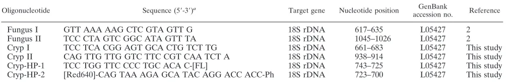
were negative by the LightCycler PCR assay alone and 4 were negative by the nested PCR assay alone. Since all water and no-template controls gave negative results, crossover or exter-nal-contamination events were ruled out. Visual examination of ethidium bromide-stained agarose gels or amplification screen plots generated by the LightCycler software (cycle num-ber versus fluorescence) allowed for a clear discrimination between positive and negative samples. The LightCycler re-sults of a representative panel of brain homogenates are de-picted in Fig. 2A. Post-PCR melting curve analysis demon-strated a consistent hybridization probe melting point of 66°C for all the C. neoformans-positive samples investigated (Fig. 2B). Specificity of nested PCR was further verified by sequenc-ing of characteristic PCR products. The amplicon sequences of the 86 positive samples were found to be identical to theC. neoformans18S rDNA sequence in GenBank (accession no. L05427) (data not shown).
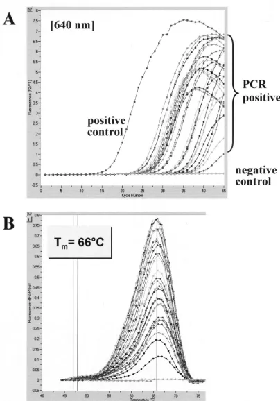
Analytical sensitivity. When nested PCR or LightCycler PCR was applied to dilution series of culturedC. neoformans
organisms, a detection limit of 1 to 10 CFU per processed sample volume, i.e., in 200l used for DNA extraction, was observed for both amplification protocols (Fig. 3).
With mouse brain homogenates and dilutions, 11 of 14 sam-ples negative by the nested PCR assay and 10 of 13 samsam-ples negative by the LightCycler PCR had a calculated concentra-tion of less than 4 CFU ofC. neoformansin 200-l samples used for DNA extraction. The remaining negative samples had concentrations of 10 to 30 CFU per processed sample volume. Thus, the detection limit determined by defined culture sus-pensions is in good correlation with the PCR results from tissue samples. Neither the fungal isolate used for infection nor the treatment regimen had significant influence on the PCR results (data not shown).
Specificities of the nested PCR and LightCycler PCR assays.
For specificity testing, strains of 32 fungal species, most of them belonging to the Tremellomycetidae (Hymenomycetes), and 70 homogenates of H. capsulatum- or P. brasiliensis -in-fected organs were examined. Of the Tremellomycetidae
strains, 12 revealed amplification products by the nested PCR. DNA sequencing identified the nested PCR products from one of two strains ofCryptococcus laurentii, fromFilobasidium cap-suligenum,Filobasidium floriforme,Filobasidium uniguttulatum,
Holtermannia corniformis, and from one of two strains of
Bullera variabilis, in total six products, as being identical with the 278-bp segment of C. neoformans 18S rDNA (Fig. 1). Additionally, the sequence gained fromFilobasidiella depaupe-ratashowed two different nucleotide positions but was identi-fied as being 99% homologous toC. neoformans. It could not be identified to the species level, because the 18S rDNA se-quence ofF. depauperatahas not yet been deposited in Gen-Bank. Amplicons from the remaining five strains, likeB. varia-bilis, Bullera globispora, or Cryptococcus amylolentus, were correctly differentiated down to species level by their nucleo-tide sequences.
Template DNA from these seven cross-reacting strains, fromTrichosporon faecaleandFibulobasidium inconspicuumas well as fromC. amylolentus, were positive by LightCycler PCR. However, T. faecale- and F. depauperata-derived amplicons could be clearly differentiated by melting point analysis (Tmof 66°C forC. neoformans;Tmof 56°C forT. faecale;Tmof 61°C
on August 17, 2020 by guest
http://cvi.asm.org/
FIG. 1. Estimate of phylogenetic relationships in theTremellomycetidae. Maximum parsimony analysis of an alignment of nuclear DNA sequences coding for the D1/D2 region of the ribosomal large subunit. Strict consensus of the three most parsimonious trees found in 1,000 rounds of heuristic search. Numbers on branches are bootstrap values from 1,000 replicates. The topology was rooted withCystofilobasidium capitatum. Strain numbers are given for the species used in the PCR experiments of this study. CBS, Centraalbureau voor Schimmelcultures; T superscript, type strain; open and solid circles, negative and positive reaction results, respectively, of nested-PCR and LightCycler hybridization experiments.
on August 17, 2020 by guest
http://cvi.asm.org/
forF. depauperata) (data not shown). As expected, six strains with 100% identity of the 278-bp fragment to the sequence of
C. neoformanscannot be distinguished by melting point anal-ysis. According to the search in GenBank, the 278-bp sequence ofF. depauperatais only 99% identical toC. neoformans. Since two different nucleotide positions are within the binding re-gions of the hybridization probes, it was recognized as being different fromC. neoformansby melting point analysis.
Compared to C. neoformans sequences, the 278-bp se-quences ofT. faecaleandF. inconspicuumare different in nine nucleotide positions within the binding sites of the primers. Therefore, the primers will not bind at the 72°C temperature used for the combined annealing and extension step in the second reaction of the nested PCR assay. Accordingly, the nested PCRs were negative. In contrast, the annealing temper-ature of 50°C used in the LightCycler PCR assay favors primer binding and amplification. Four nucleotide mismatch positions within the hybridization probe annealing sites revealed the product ofT. faecaleto be different from that ofC. neoformans
by subsequent melting point analysis. In contrast, the product ofF. inconspicuumwas not distinguished.
LightCycler PCR including discriminative melting point analysis cannot distinguishC. amylolentusandC. neoformans, because the characteristic nucleotide variations within the 278-bp PCR product are outside the annealing sites of primers and hybridization probes.
All 70 homogenates of organs infected withH. capsulatum
orP. brasiliensistested negative by both PCR protocols.
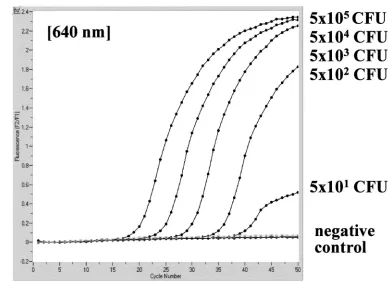
Quantification of PCR products.The quantitative capability of the LightCycler concept was obvious in the course of inves-tigating defined brain samples of mice which had been infected with various doses ofC. neoformansorganisms. Based on the results with a series of external standards (5⫻101to 5⫻104
CFU ofC. neoformans), a standard curve was generated with the help of the LightCycler software. The calculated quantity of template DNA extracted from the tissue samples correlated well with the results of quantitative culture. The original am-plification curves obtained on a representative set of four tissue sections containing 2.6⫻101to 2.6⫻104CFU ofC. neofor-mansper processed sample volume are depicted in Fig. 4. This figure also shows the calculated standard curve and the indi-vidual CFU concentrations determined on the basis of crossing points.
Single-round block cycler PCR. A protocol for the block cycler PCR using the primer oligonucleotides Cryp I and Cryp II, and consisting of 45 cycles with an annealing temperature of 50°C, was unsuccessful. A significant reduction with respect to sensitivity and specificity of the PCR assay was observed. All brain samples with fewer than 102CFU per processed sample
volume became negative, and products were amplified from 24 of 32 related fungal species. As could be expected theoretically, the second round of amplification increases sensitivity, and the highly stringent annealing step at 72°C reduces nonspecific amplification significantly.
When LightCycler PCR mixtures were analyzed by agarose gel electrophoresis, amplification products of the expected sizes were detected for 30 of the 32 related fungal species investigated. This demonstrates the value of the LightCycler hybridization probe concept for total assay specificity.
Phylogenetic analysis.Heuristic maximum parsimony anal-ysis of the ribosomal D1/D2 DNA data set yielded three equally most parsimonious trees; a strict consensus of these is shown in Fig. 1. The phylogenetic hypothesis obtained is con-sistent with analyses on data sets including more species (6).
DISCUSSION
The excellent sensitivity of a novel nested PCR protocol and a LightCycler PCR protocol for the detection ofC. neoformans
DNA was demonstrated in a murine model of cryptococcal meningitis. When the two are compared, the LightCycler PCR assay appears more sensitive, because only 5l instead of 10l of DNA extract is used, and 45 instead of a total of 65 cycles are necessary to detect a tissue burden of less than 1 CFU of
C. neoformans per mg of brain. In addition, real-time PCR using the hybridization probe detection format and the use of external standards offers quantitative results without addi-tional work or costs. This may prove beneficial for monitoring antifungal treatment in humans in a clinical setting.
PCR assays for the detection of C. neoformans DNA in clinical specimens have been described, targeting 18S, 28S, or the ITS and 5.8S ribosomal DNA (rDNA) (5, 9, 11, 12). The detection limits of our novel PCR assays compare favorably with the reported detection limits of 1 to 10 cells ml⫺1or per
volume used for DNA extraction (5, 11, 12). A number of DNA extraction procedures have been published for efficient disruption of cryptococcal cells, including enzyme digestion or glass beads (17). After identifying fungal DNA contaminants in enzyme preparations, we included instead three cycles of freez-ing and boilfreez-ing in our DNA extraction protocol for dimorphic fungi (1, 2). As demonstrated in this study, the simple freeze-and-boil procedure is sufficient to liberate cryptococcal DNA from tissue homogenates as well.
The PCR assays showed cross-reactions with some fungal species related toC. neoformans(Fig 1). Some of these can be pathogenic, and their detection is beneficial. The presence of the remaining cross-reacting species is most unlikely in clinical samples but might occur as contaminants in mycological labo-ratories. In general, this demonstrates the specificity limits of PCR assays targeting conserved regions of the rDNA. Target-ing multicopy rDNA genes will undoubtedly raise the analyti-cal sensitivity for a given fungal pathogen, but specificity can decrease due to possible cross-reactions with phylogenetically related fungi.
Prariyachatigul et al. showed that their primers and hybrid-ization probe used for an 18S rDNA PCR assay for cryptococ-cal disease did not cross-react with phylogeneticryptococ-cally distant pathogens like Candida spp. (11). According to GenBank, their probe will exclusively hybridize withC. neoformans18S rDNA. However, the 18S rDNA gene sequences from most of the related fungal species used in this study have not been submitted to GenBank. Therefore, it is not surprising that although we used GenBank alignments for the selection of two unique LightCycler hybridization probes and a unique pair of nested primers for block cycler PCR within Cryptococcus -specific 18S rDNA regions, cross-reactions were observed. Whereas two of the cross-reacting species can be differentiated by LightCycler melting point analysis, others disclose a 100% sequence identity withC. neoformansin the partial sequence
on August 17, 2020 by guest
http://cvi.asm.org/
FIG. 2. Evaluation of the LightCycler PCR assay with brain tissue sections ofC. neoformans-infected mice. A representative set of 28 samples with variousC. neoformansconcentrations was tested for the presence ofC. neoformans18S rDNA. Amplicon curves representingC. neoformans
PCR-positive isolates are indicated by braces (A), and the corresponding results in melting curve analysis are depicted (B).
on August 17, 2020 by guest
http://cvi.asm.org/
FIG. 3. Analytical sensitivity of nested PCR (A) and LightCycler PCR (B) for theC. neoformanssmall-subunit rRNA gene (18S rDNA), determined with serial dilutions of a culturedC. neoformans strain. Detection limits in CFU per milliliter, determined by standard plating procedure, are given at the corresponding lanes in agarose gel electrophoresis or next to the LightCycler amplicon curves.
on August 17, 2020 by guest
http://cvi.asm.org/
targeted by the PCR protocols. To minimize the event of false-positive results in the nested PCR protocol, a high an-nealing temperature was selected to discriminate between sim-ilar but not identical primer binding regions, and amplification products were subjected to DNA sequencing.
According to our findings, hybridization with probes com-plementary to segments of the rDNA may be unreliable for absolute species confirmation, and amplicons should be se-quenced if an unambiguous species identification is desired.
Including a variety of other pathogens, specificity testing should include phylogenetically closely related fungal species
and potential contaminants of clinical specimens, especially fungi residing on normal skin or mucous membranes. From a clinical point of view, false-positive PCR results with other fungal pathogens are acceptable, because antifungal treatment is indicated in that situation as well. But any cross-reaction of the PCR assay with harmless contaminants will falsely indicate the necessity of therapy, which might be harmful to the patient.
C. laurentii, for example, was shown to cross-react with the presented PCR assays. Since this is a potential pathogen (4), its detection is beneficial. Due to strain variation, which is known to occur (14), not all strains will be detected. Potential skin
FIG. 4. Quantitative analysis of LightCycler results obtained on a representative set of four tissue sections containing 2.6⫻101to 2.6⫻104
CFU ofC. neoformansper processed sample volume. Based on the slope of the original amplification curves during the log-linear phase determined by three data points, artificial crossing points with the noise band (horizontal line) were determined by the LightCycler software (A). The standard curve represents the linear regression line through the data points on a plot of crossing points (threshold cycle) versus logarithm of standard sample concentration (B). Slope,y-intercept, mean squared error, and regression coefficient of the standard curve are given.
on August 17, 2020 by guest
http://cvi.asm.org/
contaminants such as Trichosporon spp. or Malassezia furfur
either will not be detected or will be distinguished by Light-Cycler melting point analysis.Bulleraspp.,F. depauperata,F. inconspicuum, someFilobasidiumspecies,C. amylolentus, and
H. corniformiswill be incorrectly identified asC. neoformans. However, these species have not been described to cause hu-man disease, nor is a contamination of clinical specimens likely to occur.
Since procedures for sequence-specific detection of ampli-cons evolved as the “gold standard” in the field of diagnostic PCR, LightCycler technology avoids the application of time-consuming and laborious postamplification procedures like Southern blotting or DNA sequencing. Enhancing the reliabil-ity of the results by sequence-specific probes and simplifying the PCR workflow by a completely automated amplification and online detection procedure, the LightCycler system proved to be a valuable tool for rapid and sensitive identification ofC. neoformans. Once DNA is extracted from suitable specimens and reaction mixtures are completed, the results of sensitive and quantitative PCR are available within 60 min. A current limitation of the LightCycler system is the total reaction vol-ume of 20l, which limits the input of template DNA and, as a consequence, total assay sensitivity.
In conclusion, we have successfully evaluated sensitive con-ventional nested PCR and LightCycler PCR protocols for the detection ofC. neoformansin tissue samples. The LightCycler real-time PCR is more rapid and offers quantitative results, whereas the block cycler PCR is broadly applicable. Although we have identified some cross-reacting fungal species, the true diagnostic value of these assays will be further evaluated in prospective studies on human cryptococcal disease.
ACKNOWLEDGMENTS
We thank Hans Wolf, Ulrike Zelck, Jürgen Knobloch, José Paulo Sampaio, and Dominik Begerow for their active support and Jeffrey Emch for critical comments. We gratefully acknowledge the technical assistance of Markus Bollwein and Birgit Leppmeier during the study.
REFERENCES
1.Bialek, R., A. Ibricevic, C. Aepinus, L. K. Najvar, A. W. Fothergill, J. Knobloch, and J. R. Graybill.2000. Detection ofParacoccidioides brasiliensis
in tissue samples by a nested PCR assay. J. Clin. Microbiol.38:2940–2942.
2.Bialek, R., J. Fischer, A. Feucht, L. K. Najvar, K. Dietz, J. Knobloch, and
J. R. Graybill.2001. Diagnosis and monitoring of murine histoplasmosis by
a nested PCR assay. J. Clin. Microbiol.39:1506–1509.
3.Blevins, L. B., J. Fenn, H. Segal, P. Newcomb-Gayman, and K. C. Carroll.
1995. False-positive cryptococcal antigen latex agglutination caused by
dis-infectants and soaps. J. Clin. Microbiol.33:1674–1675.
4.Cheng, M.-F., C. C. Chiou, Y.-C. Liu, H.-Z. Wang, and K.-S. Hsieh.2001.
Cryptococcus laurentiifungemia in a premature neonate. J. Clin. Microbiol.
39:1608–1611.
5.Evertsson, U., H. J. Monstein, and A. G. Johansson.2000. Detection and identification of fungi in blood using broad-range 28S rDNA PCR
amplifi-cation and species-specific hybridisation. APMIS108:385–392.
6.Fell, J. W., T. Boekhout, A. Fonseca, G. Scorzetti, and A. Statzell-Tallman.
2000. Biodiversity and systematics of basidiomycetous yeasts as determined by large-subunit rDNA D1/D2 domain sequence analysis. Int. J. Syst. Evol.
Microbiol.50:1351–1371.
7.Kralovic, S. M., and J. C. Rhodes.1998. Utility of routine testing of
bron-choalveolar lavage fluid for cryptococcal antigen. J. Clin. Microbiol.36:3088–
3089.
8.Loeffler, J., N. Henke, H. Hebart, D. Schmidt, L. Hagmeyer, U. Schumacher, and H. Einsele.2000. Quantification of fungal DNA by using fluorescence resonance energy transfer and the Light Cycler system. J. Clin. Microbiol.
38:586–590.
9.Mitchell, T. G., E. Z. Freedman, T. J. White, and J. W. Taylor.1994. Unique
oligonucleotide primers in PCR for identification ofCryptococcus
neofor-mans. J. Clin. Microbiol.32:253–255.
10.Nguyen, M. H., L. K. Najvar, C. Y. Yu, and J. R. Graybill.1997. Combination therapy with fluconazole and flucytosine in the murine model of cryptococcal
meningitis. Antimicrob. Agents Chemother.41:1120–1123.
11.Prariyachatigul, C., A. Chaiprasert, V. Meevootisom, and S. Pattanakit-sakul.1996. Assessment of a PCR technique for the detection and
identifi-cation ofCryptococcus neoformans. J. Med. Vet. Mycol.34:251–258.
12.Rappelli, P., R. Are, G. Casu, P. L. Fiori, P. Cappuccinelli, and A. Aceti.
1998. Development of a nested PCR for detection ofCryptococcus
neofor-mansin cerebrospinal fluid. J. Clin. Microbiol.36:3438–3440.
13.Saag, M. S., J. R. Graybill, R. A. Larsen, P. G. Pappas, J. R. Perfect, W. G. Powderly, J. D. Sobel, and W. E. Dismukes, for the Mycoses Study Group cryptococcal subproject.2000. Practical guidelines for the management of
cryptococcal disease. Clin. Infect. Dis.30:710–718.
14.Sugita, T., M. Takashima, R. Ikeda, T. Nakase, and T. Shinoda.2000.
Intraspecies diversity ofCryptococcus laurentiias revealed by sequences of
internal transcribed spacer regions and 28S rRNA gene and taxonomic
position ofC. laurentiiclinical isolates. J. Clin. Microbiol.38:1468–1471.
15.Swofford, D. L.1998. PAUP*. Phylogenetic analysis using parsimony (*and other methods). Sinauer Associates, Sunderland, Mass.
16.Swofford, D. L., G. J. Olsen, P. J. Waddell, and D. M. Hillis.1996.
Phylo-genetic inference, p. 407–415.InD. M. Hillis, C. Moritz, and B. K. Mable
(ed.), Molecular systematics. Sinauer Associates, Inc., Sunderland, Mass. 17.Tanaka, K.-I., T. Miyazaki, S. Maesaki, K. Mitsutake, H. Kakeya, Y.
Yamamoto, K. Yanagihara, M. A. Hossain, T. Tashiro, and S. Kohno.1996.
Detection ofCryptococcus neoformansgene in patients with pulmonary
cryp-tococcosis. J. Clin. Microbiol.34:2826–2828.
18.Tintelnot, K., S. Adler, F. Bergmann, K. Schoenherr, and M. Seibold.2000. Case reports. Disseminated cryptococcoses without cryptococcal antigen
de-tection. Mycoses43:203–207.
19.Wei, M., Z.-L. Yang, and F. Oberwinkler.1998. Molecular phylogenetic
studies in the genusAmanita. Can. J. Bot.76:1170–1179.