1
Human in situ study of the effect of bis(2-methacryloyloxyethyl) dimethylammonium
bromide immobilized in dental composite on controlling mature cariogenic biofilm
Mary Anne S. Melo1, Michael D. Weir1, Vanara F. Passos2, Juliana P. M. Rolim3, Christopher D. Lynch4, Lidiany K. A. Rodrigues2, Hockin H. K. Xu1,5,6
1Department of Advanced Oral Sciences and Therapeutics, University of Maryland School of
Dentistry, Baltimore, MD 21201, USA;
2Postgraduate Program in Dentistry, Faculty of Pharmacy, Dentistry and Nursing, Federal
University of Ceara, Fortaleza, Ceara, Brazil;
3Faculty of Dentistry UniChristus, Fortaleza, Ceara, Brazil;
4Restorative Dentistry, University Dental School and Hospital, University College Cork, Wilton,
Cork, Ireland;
5Center for Stem Cell Biology & Regenerative Medicine, University of Maryland School of
Medicine, Baltimore, MD 21201, USA;
6Marlene and Stewart Greenebaum Cancer Center, University of Maryland School of Medicine,
Baltimore, MD 21201, USA
Correspondence:
Mary Anne Melo, 650 West Baltimore St, Baltimore, MD, 21201, USA,
Email: [email protected];
Lidiany K. A. Rodrigues, Rua Monsenhor Furtado S/N, Bairro Rodolfo Teófilo, 60430-325
Fortaleza, Brazil, Email: [email protected];
Hockin H. K. Xu, 650 West Baltimore St, Baltimore, MD, USA 21201, Email:
2
Abstract
Cariogenic oral biofilms cause a considerable amount of recurrent dental caries around
composite restorations every year, resulting in unprosperous oral health status and expensive
restorative treatment for many patients. Quaternary ammonium monomers that can be
copolymerized with the current dental resin systems have been increasingly explored for
modulation of dental plaque biofilm growth over dental composite surfaces. Here, we
investigated the effect of bis(2-methacryloyloxyethyl) dimethylammonium bromide (QADM),
against human overlying mature oral biofilms grown intra-orally in human participants for 7 and
14 days, for the first time. Seventeen volunteers wore palatal devices containing composite
specimens containing 10% by mass of QADM or a control composite without QADM. After 7
and 14 days, the adherent biofilms were collected for determination of bacterial counts via
colony-forming unit (CFU) counts. The biofilm viability, chronological changes, and percentage
coverage were also determined by live/dead staining. QADM composites caused a significant
inhibition of S. mutans biofilm formation for up to seven days. No difference in the CFU values were found for the 14-day period. Our findings suggest that (1) QADM composite was
successful in inhibiting 1-3 day biofilms in the oral environment in vivo; (2) QADM significantly reduced the portion of S.mutans group in a time course where patients at high risk of caries would develop initial enamel carious lesions; and (3) stronger antibiofilm activity is required for
the control of mature long-term cariogenic biofilms. These results provide a perspective on the
value of integrating bioactive restorative materials with traditional caries management
approaches into clinical practice. Contact-killing strategies via dental materials aiming to prevent
or at least reduce high numbers of cariogenic bacteria seem to be a promising approach in
patients at high risk of recurrence of dental caries around composites.
Keywords: Antibacterial, Biofilm, Caries, Dental composite, Quaternary ammonium monomers,
3
INTRODUCTION
In the last decade, dental materials research has intensified attempts to reduce or
modulate dental plaque biofilm growth over dental composite surfaces [1]. This is because
recurrent caries or caries around restorations (CARS) are identified as one of the major reasons
for the failure of composite restorations [2,3]. Replacement rates of failed restorations have been
reported to be 37% to 70% with consequences that can seriously compromise the oral health
status [4,5].CARS are frequently located at the gingival margins of the proximal restorations,
which are common areas for food impaction [6]. Despite the favorable outcomes of mechanical
biofilm removal by brushing; patients consider the cleaning of dental biofilm at the proximal
space challenging and very often fail to control biofilm build-up over time.
As dental caries is characterized by continuous mineral loss promoted by organic acids
released by bacteria after sugar metabolization, [7] the presence of cariogenic bacteria, mainly
streptococci from the mutans group, is a key factor to initiate carious lesions. Cariogenic bacteria
are characterized as pathologically shifted species with the ability of generate of large amounts
of acid and being able to survive in acidic microenvironments [8]. Hence, the introduction of
novel treatment approaches, supplementary to the conventional therapeutic strategies, is
considered crucial for the efficient control of CARS. The cariogenic oral biofilm influences the
initiation and progression of carious lesions, not just in its primary development but also its
recurrence [9]. Reports in the literature have stated a temporal relationship between changes in
biofilm composition and enamel demineralization following exposure to sucrose[10,11] An
undisturbed dental biofilm exposed to frequent sucrose leads to enamel demineralization after
seven days of biofilm accumulation, although changes in the biofilm composition can be
observed earlier [10]. As the cariogenic biofilm becomes more mature, some acidogenic and
aciduric bacteria become dominant in the biofilm [12].
Another factor of microbiological relevance is that the resin composites facilitate
cariogenic biofilm growth [13]. The suggested rationale is based on degradation products from
dental monomers such as bisphenol A glycidyl dimethacrylate (BisGMA) and triethylene glycol
dimethacrylate (TEGMA), which may help alter the metabolism and promote the proliferation of
4
ammonium groups in their chemical structures paved the way for a compelling, noninvasive,
biofilm-targeted, method that can be used against oral biofilms [15]. Reactive and easily miscible
quaternary ammonium monomers have the advantage of copolymerization with the current
dental resin systems by covalenty bonding with the polymer network. These polymers are
referred to as non-leaching antimicrobial or contact-killing agents. The antibacterial action
results from the direct contact of the polymer with the microorganisms, with no release of active
molecules. Although the exact antimicrobial mechanism of action has not been fully elucidated,
it is generally stated that the predominant mode of action is disruption of the cell membrane [16].
This method promises to impart a durable and permanent antibacterial capability to dental
composites.
Along with a similar line, studies have been carried out on the synthesis of quaternary
ammonium monomers for dental applications. Studies have presented different positions of the
functional groups and alkyl chain length for improved balance between mechanical properties,
anti-bacterial effect, and biocompatibility [17]. The majority of the synthetic quaternary
ammonium monomers have only one methacrylate group and are classified as mono
methacrylates. Incorporating a high content of mono methacrylate could compromise the overall
cross-linked polymer matrix and, consequently, the mechanical properties [18].
Over the years, several in-vitro studies investigated the antibacterial performance bis(2-methacryloyloxyethyl) dimethylammonium bromide, a quaternary ammonium monomer
containing two methacrylate groups (QADM)[18]. In these studies, QADM was loaded at 10wt%
in different parental formulations such as commercial and experimental adhesive systems [19,20]
as well as nanocomposite,[21]rendering great reductions in S. mutans and total microorganisms. Overall, these studies achieved a significant reduction of biofilm viability, metabolic activity,
lactic acid, and bacterial counts using a 48-h human saliva microcosm biofilm model [22]. The
incorporation of QADM also did not compromise the mechanical or bonding performance of the
parental materials, and its antibacterial and mechanical properties were long-term and maintained
after o one-year follow-up [23].
Although encouraging results were found in vitro [19-22], only a few studies used native
in situ dental plaque to study the effects of quaternary ammonium methacrylate [24,25]. In these studies, bacterial colonization over a short period (hours to 3 days) was assessed. However,
5
biofilm formed over seven days has not been studied to date. There has been no report of in situ
studies for longer than three days. Such a longer-term in situ study would give meaningful insights in the in vivo antibacterial performance of this material in challenging conditions that mimic the clinical scenario of retentive proximal areas where the biofilm could not be removed
in high caries-risk patients. Moreover, over a 7-day period, dysbiosis is present due to the
proliferation or overgrowth of cariogenic bacteria in a low pH eco-niche and enamel is prone to
demineralization.
In light of the evidence available to support quaternary ammonium monomers on initial
oral biofilm, the present study evaluated the antibacterial performance of QADM by challenging
the effectiveness of the material against in situ-formed mature oral biofilms in a relatively long-term study beyond 3 days for the first time. Intact oral biofilms were grown under a cariogenic
challenge in situ on composites within the oral cavity for 7 and 14 days, respectively. In addition to the determination of bacterial counts, the chronological changes in the biofilm were also
visualized by live/dead staining, and the percentages were measured.
RESULTS
All 17 volunteers completed the study, and no protocol deviation was identified.
Treatment compliance was satisfactory. The mean and standard deviation values of colony
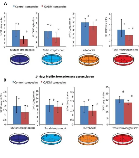
forming unit (CFU) of biofilms collected at 7 and 14-day are plotted in Fig. 2A-B. The QADM
composite had a significant effect on the viability of S. mutans at the 7-day period (p=0.0303). This effect corresponds to a 43% reduction of both solutions compared with the control. The
effect of QADM composite on the viability of total streptococci, lactobacilli and total
microorganisms of the in situ biofilms showed a slight reduction in CFU counts in the same period. However, there was no statistical significance between the groups (p> 0.5). At the
14-day period, the microbiological composition of biofilms formed on restoration was statistically
similar for all evaluated conditions (p>0.5).
Figure 3 displays the variable percentage of mutans streptococci related to total
streptococci (MS/TS) showed the statistically significant difference between the tested groups
6
to total microorganisms was similar at the same period. These variables have shown no different
at the 14-day period (Fig. 3B).
Figure 4 shows (A-D and F-I) live/dead staining images of biofilms grown on the QADM
and control composites at the 1st, 3rd, 7th and 14th days. Biofilms grown on the control composite at the 1st and 3rd days were primarily alive with continuous green staining (4A-B). Widespread bacterial cell killing was more pronounced on the 1st and 3rd day-biofilm accumulation for
QADM composite (4E-F). At the 7th and 14th day, the overly mature biofilm structure was
compact with innumerous layers presenting a mushroom-like configuration with channels in the
outer layer (Fig. 4C-D for control and Fig. 4G-H for QADM). Complete coverage of the
composite surface is observed after seven days in the oral cavity. No significant difference
between control and QADM can be observed.
In Fig. 5, image analyses indicated that the living cells grown over control composites
accounted for 93 ± 3% (±SD) and 93 ± 7% (±SD) of the total biofilm cells for the 1st and 3rd day-biofilm, respectively. In contrast, lower percentages of living cells (46 ± 8% for the 1st day and
37± 3% for the 3rd day) were determined on QADM composite for the same period where the
remaining cells were inactive and dead.
DISCUSSION
Bacterial attachment to dental composite surfaces and subsequent cariogenic biofilm
formation are a complex process [26]. It is controlled by the interplay among biological factors,
such as the bacterial ability to rapidly convert dietary sugars to acid, lower the pH, and
demineralize the tooth structure, patient-related factors, and physicochemical factors such as
surface topography, surface charge and surface energy of the dental materials [27,28].Typical
treatment methods for biofilm-mediated recurrence of caries lesions around the composite
restorations involves operative replacement of the composites, which incurs additional health
care costs and additional loss of tooth structures.
To avoid the formation of biofilms over dental materials, an attractive alternative and
complementary method to dental caries management is the use of biomaterials that possess
antibacterial surfaces [27]. Resin-based materials that today are widely used clinically present no
antibacterial activity. The new contact-active antibacterial material is effective to prevent biofilm
7
surrounding microenvironment thereby significanty the potential for extending the material’s
service life.
The performance of quaternary ammonium monomers for dental applications has been
intensely investigated in vitro in the past years with the overall positive outcome. Investigations have shown immediate and robust antibacterial effect (more than 3 log reductions) against oral
microorganisms [21,23,25]. QADM presents a greater affinity for polymerization due to its
di-functional monomers and effectiveness in producing active surfaces with higher densities of
immobilized antimicrobial agents [30,31].For the specific quaternary ammonium used in our
study, the previous in vitro studies have pointed out the reduction of 68% and 79% in biofilm CFU counts and lactic acid production, respectively, on cured primers specimens [20].
The present study represents the first study on a human in vivo effect of antibacterial dental resins for relatively long periods of biofilms growing more than three days. For this in situ
study, we challenged the antibacterial performance of a dual methacrylate-group-QADM
incorporated in composites against overly mature oral biofilms formed inside the oral cavity for
up to 14 days for the first time. The rationale for it was to investigate the effect of this type of
monomer in an acidogenic biofilm structure capable of causing substantial mineral loss and deep
lesions on the enamel surface. This approach has high clinical relevance since anti-caries
therapies aimed at controlling the assembly of cariogenic biofilms should contribute with its
bioactivity for prevention of the onset of early carious lesions clinically known as white spots.
The data revealed that QADM has compromised the S. mutans group biofilm accumulation at seven days period formation. This result is particularly interesting since S. mutans group presents the two most common species constantly linked to caries formation,
S.mutans and S. sobrinus.[32]. The cariogenic potential of S. mutans is determined genetically being accentuated when sucrose is available [33].Sucrose-mediated biofilm formation created
spatial organizations as expressed by a complex network of microcolonies, which modulate the
development of compartmentalized acidic microenvironments across the 3D biofilm architecture
[34]. Furthermore, within the 3D biofilm, S. mutans displays properties that are dramatically distinct from their planktonic counterparts, including much higher resistance to antibacterial
approaches, which make the biofilm much more difficult to kill than planktonic bacteria [32].
8
the contact-killing mechanism of quaternization of the amino groups of QADM available on the
bottom layer of the biofilm adjacent to composite. The negatively charged counterions that
stabilize the bacterial membrane are displaced by the positively charged cationic N+ sites in the
chemical structure of the quaternary ammonium-based resin. Indeed, the live/dead images
obtained at the initial period of biofilm formation showed the presence of a higher proportion of
nonviable bacteria (red-orange color areas). Accordingly, previous studies have highlighted
similar viability of 3D biofilms growing on resin-based materials containing quaternary
ammonium monomers [35,36]. Furthermore, Beyth and co-workers have suggested an
intracellularly mediated death program, in which, the bacterial lysis promoted by the presence of
quaternary ammonium on the resin surface may function as a stressful condition triggering
programmed cell death to the bacteria further away in the biofilm [25,37].
Subsequently, no expressive microbial reduction results on CFU values or microorganism
proportions were observed for S. mutans, and the others evaluated culture media after the 14-day period. These results point out the challenge faced by anti-caries approaches against mature
biofilms. The bacterial adhesion processes under in vivo and in vitro conditions differ considerably [30]. Bacteria in biofilms are far less sensitive to antibacterial agents because of
the exopolymeric matrix, extracellular polysaccharides, specific gene expression, and metabolic
activity; factors that protect antibacterial therapies to reach target bacteria [38].The live-dead
images of biofilms show a well-developed dense and compact EPS-matrix, and the presence of
bacterial cell clusters or microcolonies (Fig. 4C-D for control and Fig. 4H-I for QADM). The
relative alteration of the proportion dead/life found at 7-14-day images are related to the uneven
spatial distribution of vital and dead microorganisms found in matured and thick dental biofilms
with decreased vitality towards the outer layers [38]. Tawakoli et al. [39]also supported the high
variability of the live/dead distribution, and the CFU counts as challenges found in situ biofilm models. Recently, new investigations have started to emerge using nanoparticles to improve
penetration of therapeutics agents into the biofilm matrix of oral cariogenic biofilm [40]
Another aspect to consider is the spatial arrangement, charge density and counter anion of
the quaternary ammonium monomers and their antibacterial activity [29]. Previous studies have
designed antibacterial monomers containing an eight-carbon or longer chain and have correlated
9
These monomers need be investigated for in vitro to a human in vivo translation of their antibacterial performance [42]. Future studies are warranted to investigate this further, especially
whether the charge density can be reflected as a relevant factor for the antibacterial effect of
these new quaternary ammonium monomers.
In summary, the findings in this paper demonstrate that QADM composite at 10% can
promote a substantial and valuble bacterial reduction of S. mutans biofilm. This was achieved not only in the initial days of contact but also reaching a 7d-period, a time course where patients at
high risk of caries would develop initial enamel carious lesions. However, dental caries results
from interactions over time. An undisturbed cariogenic biofilm well-established on the
composite surface over long periods is extremely hard to eradicate. Its inhibition should not rely
only on contact with an antibacterial surface. Removing/disturbing biofilm from all tooth and
composite surfaces and reducing sugar intake within three days is expected to control carious
lesion development.
Concomitant and multitargeting strategies are needed against mature long-term
cariogenic biofilms. The results of the present study provide a perspective on the value of
integrating bioactive restorative materials with traditional caries management approaches into
clinical practice. Contact-killing strategies via dental materials aiming to prevent or at least
reduce high numbers of cariogenic bacteria seem to be a promising approach to help patients at
high risk of recurrence of dental caries around composites.
MATERIALS AND METHODS
Study design and participants
This study involved a prospective, randomized, single-blind, split-mouth in-situ design conducted according to the code of ethics of the World Medical Association (Declaration of
Helsinki) for experiments involving humans. The region’s ethical committee (protocol #
1232012) has also approved it. Seventeen healthy volunteers of both genders, aged from 21 to 36
years, accepted to participate in this study fulfilling the required criteria. Inclusion criteria were
normal salivary flow rate, a good general and oral health with no active caries lesions or
periodontal treatment needs, ability to comply with the experimental protocol, not have used
10
devices. Exclusion criteria were failing to use the device according to the established protocol,
taking medication interfering with saliva flow rate or containing antimicrobial agents. The
sample size was determined by a power analysis and based on previous data [25].Seventeen
volunteers were recruited: 16 for the study and one volunteer to allow temporal visualization of
the biofilm formation by live/dead staining. After being screened, the volunteers were verbally
informed about the study aim and procedures and received written information and the informed
consent form. At the next appointment, maxillary alginate impressions were made to the
fabrication of the palatal devices.
During the experimental period, each volunteer used a removable acrylic custom-made
palatal device containing the tested materials as shown in Fig. 1A. Seven days before the
experiment beginning (washout period) and during the whole experiment, the volunteers were
asked to use a standard toothbrush and non-fluoridated paste. Each acrylic palatal device
enclosing four composite specimens (5 x 5 x 2 mm); two specimens for control composite and
two specimens to QADM composite. To promote plaque accumulation and to protect it from
disarrangement, recessions were created by placing the surface of the composite specimens about
1 mm below the covered plastic mesh (Fig. 1A-detail) [43].
To divert any possible carry-across effect, the sequence in which the experimental units
were assigned in the palatal device took into consideration that antibacterial dental materials
should be placed in one side of the palatal appliance and, consequently, control materials on the
opposite side (Fig. 1A). The split-mouth experimental design is a practical approach for testing
the effects of various agents on the composition of dental plaque [44]. Within each side of the
palatal device, the positions of the specimens were randomly determined, according to a
computer-generated randomization list [45]. The outcome variables evaluated were colony
forming unit counts for total microorganisms, total streptococci, mutans streptococci, and
lactobacilli on the specimens.
Specimen preparation
The light-curable composite was made by blending a monomer resin consisting of
BisGMA (bisphenol-glycidyl dimethacrylate) and TEGDMA (triethylene glycol dimethacrylate)
at 1:1 ratio (all by mass) with 0.2% camphorquinone and 0.8% ethyl 4-N,
11
with a median diameter of 1.4 µm (Caulk/ Dentsply, Milford, DE, USA) were silanized with 4%
3-methacryloxypropyltrimethoxysilane and 2% n-propylamine [21]. The fillers were mixed with
the resin at a total filler mass fraction of 60% to form a cohesive paste.
The synthesis of bis(2-methacryloyloxyethyl) dimethylammonium bromide via a
modified Menshutkin reaction was previously described [18], and it is summarized in Fig. 1B.
Briefly, 10 mmol of 2-(N, N-dimethylamino)ethyl methacrylate (DMAEMA; Sigma-Aldrich, St
Louis, MO, USA) and 10 mmol of 2-bromoethyl methacrylate (BEMA; Monomer-Polymer
Labs, Trevose, PA, USA) were combined with 3 g of ethanol in a closed vial. After stirring at 60
°C for 24 h for the reaction to complete, the solvent was removed via evaporation under vacuum.
This process yielded QADM as a clear and viscous liquid. QADM was mixed with the
BisGMA-TEGDMA resin at a QADM mass fraction of 10%. A preliminary study has shown that this mass
fraction yielded strong antibacterial properties without compromising the resin’s mechanical
properties [21].
Thirty-six light-curable composite specimens were fabricated for the experimental
composition (QADM at 10wt.%) each, and a further 36 specimens without antimicrobial
monomer served as control. The composite was inserted and light-activated for 20 sec using a
Light Emitting Diode (Radii-cal, SDI Limited Victoria, Australia; standard curing mode,
irradiance output provided of 689 mW/cm2). The specimens were mounted in standardized sample chambers inside the device with an anterior-posterior position using body impression
material (Aquasil Ultra, Dentsply DeTray GmbH, Konstanz, Germany) as demonstrated in Fig.
1A.
Clinical phase
Audiovisual orientation and written instructions of the in situ protocol were given to the volunteers to assure their adhesion and avoid protocol deviation during the study. To provide a
cariogenic challenge during the clinical phase, the application of a 20% sucrose solution
extra-orally on the restored specimens was performed by the volunteers, eight times per day at
predetermined times. According to previous studies, the sucrose was gently dried after 5 min,
and the device was reinserted into the mouth [42,44]. No restriction was made about the
volunteer's diet, but they were instructed to avoid F-rich food containing bioavailable F, such as
12
Microbiological and biochemical analysis
On the 7th and 14th examination days, the subjects refrained from eating, drinking, and tooth cleaning 12 h after the last application of the sucrose solution and dentifrice before
presenting at the clinic. On the 7th day, the device was removed from the mouth, and the biofilm
and one enamel disc, respectively, from each side, were carefully removed and collected (Fig.
1B). Then, the device with the remaining discs was reinserted into the mouth. On the 14th day, a similar process was performed to collect the two residual biofilms from the specimens. After the
collection, the biofilm was processed for analysis. Firstly, it was weighed (± 1 mg) in
pre-weighed microcentrifuge tubes and agitated during a 2 min period in a Disrupter Genie Cell
Disruptor (Precision Solutions, Rice Lake, WI, USA). An aliquot of 50 µL of the sonicated
suspension was diluted in 0.9% NaCl and serial decimal dilutions were inoculated in triplicate by
the drop-counting technique in the following culture media: (1) Mitis salivarius agar containing
20% sucrose, to determine TS, and in mitis salivarius agar plus 0.2 bacitracin/mL, to determine
MS; (2) Rogosa agar supplemented with 0.13% glacial acetic acid to assess the number of CFU
of lactobacilli (LB); and (3) brain heart infusion enhanced with 5% sterile defibrinated sheep
blood agar plates were used to determine total microorganisms (TM). The plates were incubated
in 10% CO2 at 37 °C for 48 h. The CFU were counted, and the results were expressed as
CFU/mg biofilm wet weight, the percentage of MS in relation to TM, and the percentage of MS
in relation to TS.
Live/dead assay
To visualize the microorganisms during the initial phase of formation as well during the
experimental periods, one volunteer used a palatal device containing eight composite specimens:
four specimens for control composite and four specimens for QADM composite. One specimen
of each group was removed at the 1st, 3rd, 7th and 14th days. The specimens were immediately washed with phosphate buffered saline (PBS) and stained using the LIVE/DEAD BacLight
Bacterial Viability Kit (Molecular Probes, USA) to qualify the bacterial cell viability. This assay
13
propidium iodide stain [39]. These stains differ in their ability to penetrate healthy bacterial cells.
When used alone, SYTO 9 stain labels both live and dead bacteria.
In contrast, propidium iodide penetrates only bacteria with damaged membranes,
reducing SYTO 9 fluorescence when both dyes are present. Thus, live bacteria with intact
membranes fluoresce green, while dead bacteria with damaged membranes fluoresce red. A
volume of 100 μL of the previously described fluorescence dyes was pipetted onto the specimens
and incubated in a dark chamber for 15 min. The biofilms grown over the specimens were then
examined using an epifluorescence microscope (TE2000-U, Nikon, Melville, NY, USA) at a
magnification of 100x. Images (n=4) were acquired and analyzed (NIS Elements software, Nikon
Instruments Inc, Melville, NY, USA) for quantification of live (green fluorescence) and dead
(red fluorescence) bacteria.
Statistical Analysis
The assumptions of equality of variances and normal distribution of errors were checked
for all the response variables tested and those that did not satisfy these assumptions were
transformed using Box-Cox power transformation [42]. To determine the differences between
test and control values in the in situ experiment, the viable bacteria counts, percent MS/TS and percent MS/TM were submitted to a two sample independent Student’s t-test. The significance
level was set at α = 0.05. The statistical appraisal was computed with SPSS for Windows XP
17.0 (SPSS Inc., Chicago, IL, USA).
Acknowledgments
The authors thank the volunteers for their valuable participation and Dr. Agostinho Soares de
Alcântara Neto (Faculdade de Veterinária, Universidade Estadual do Ceará) for his technical
assistance and laboratory facilities for the cell viability assay. This study was supported by
CNPq/Brazil (141791/2010-1), CAPES/ Fulbright Doctoral Program (BEX 0574/06-6),
University of Maryland Baltimore Seed grant (HX), and University of Maryland School of
14
Conflict of interest statement
The authors declare that there is no conflict of interest pertaining to the data presented in this
article.
References
1. Hwang G, Koltisko B, Jin X, Koo H. Nonleachable Imidazolium-Incorporated Composite
for Disruption of Bacterial Clustering, Exopolysaccharide-Matrix Assembly, and
Enhanced Biofilm Removal. ACS Appl Mater Interfaces. 2017, 8, 44, 38270-38280. 2. Hollanders ACC, Kuper NK, Maske T, Huysmans MDNJM. Secondary Caries in situ
Models: A Systematic Review. Caries Res. 2018 Apr 5;52(6):454-462.
3. Bernardo M, Luis H, Martin MD, Leroux BG, Rue T, Leitao J. Survival and reasons for
failure of amalgam versus composite posterior restorations placed in a randomized
clinical trial. J Am Dent Assoc 2007,138, 775–83.
4. Owen B, Guevara PH, Greenwood W. Placement and replacement rates of amalgam and
composite restorations on posterior teeth in a military population.US Army Med Dep J. 2017, 2, 88-94.
5. Roumanas ED. The frequency of replacement of dental restorations may vary based on a
number of variables, including type of material, size of the restoration, and caries risk of
the patient. J Evid Based Dent Pract. 2010, 1, 23-4.
6. Mjör IA. The location of clinically diagnosed secondary caries. Quintes Int. 1998, 29, 313-7.
7. Fejerskov O. Changing paradigms in concepts on dental caries: consequences for oral
health care. Caries Res. 2004, 38, 182-91.
8. Marsh P.D. Are dental diseases examples of ecological catastrophes? Microbiol 2003, 279-294.
9. Paes Leme AF, Koo H, Bellato CM, Bedi G, Cury JA. The role of sucrose in cariogenic
dental biofilm formation--new insight. J Dent Res. 2006, 85, 878-87.
10. Vale GC, Tabchoury CP, Arthur RA, Del Bel Cury AA, Paes Leme AF, Cury
JA.Temporal relationship between sucrose-associated changes in dental biofilm
15
11. Cury, J. A., & Tenuta, L. M. A. Enamel remineralization: Controlling the caries disease
or treating early caries lesions? Braz Oral Res 2009, 23, 23–30.
12. N. Takahashi, B. Nyvad. Caries ecology revisited: microbial dynamics and the caries
process. Caries Res 2008; 42, 409-418.
13. Singh J, Khalichi P, Cvitkovitch DG, Santerre JP. Composite resin degradation products
from BisGMA monomer modulate the expression of genes associated with biofilm
formation and other virulence factors in Streptococcus mutans. J Biomed Mater Res A
2009, 88, 551-60.
14. Sadeghinejad L, Cvitkovitch DG, Siqueira WL, Santerre JP, Finer Y. Triethylene Glycol
Up-Regulates Virulence-Associated Genes and Proteins in Streptococcus mutans. PLoS One 2016: 7,11, 665-760.
15. Melo MA; Weir MD; Li F.; Cheng L.; Zhang K, Xu HHK. Control of Biofilm at the
Tooth-Restoration Bonding Interface: A Question for Antibacterial Monomers? A
Critical Review. Rev. Adhesion Adhesives 2017, 3,287-305.
16. Yang Jiaoa, Li-na Niua, Sai Ma, Jing Li , Franklin R. Tay , Ji-hua Chen. Quaternary
ammonium-based biomedical materials: State-of-the-art,toxicological aspects and
antimicrobial resistance. Prog Pol Sci 2017, 71, 53–90.
17. Zhang N, Ma Y, Weir M, Xu HHK, Bai Y, Melo MA. Current Insights into the
Modulation of Oral Bacterial Degradation of Dental Polymeric Restorative Materials,
Mater (Basel) 2017,10, 507-514.
18. Joseph M. Antonucci, Diana N. Zeiger, Kathy Tang, Sheng Lin-Gibson, Bruce O.
Fowler, Nancy J. Lin. Synthesis and characterization of dimethacrylates containing
quaternary ammonium functionalities for dental applications. Dent Mater. 2012, 28, 219– 228.
19. Melo MAS, Cheng L,Weir MD, Hsia R, Rodrigues, LKA. Xu HHK. Novel dental
bonding agents containing antibacterial agents and calcium phosphate nanoparticles. J Biomed Mater Res B Appl Biomater. 2013,101, 620-9.
20. Cheng L, Zhang K, Melo MAS, Weir MD, Zhou X, Xu HHK. Anti-biofilm dentin primer
16
21. Cheng L, Weir MD, Xu HH, Antonucci JM, Kraigsley AM, Lin NJ, Lin-Gibson S, Zhou
X. Antibacterial amorphous calcium phosphate nanocomposites with a quaternary
ammonium dimethacrylate and silver nanoparticles. Dent Mater. 2012,28, 561-72. 22. Zhang K, Melo MAS, Cheng L, Weir MD, Bai Y, Xu HHK. Effect of quaternary
ammonium and silver nanoparticle-containing adhesives on dentin bond strength and
dental plaque microcosm biofilms. Dent Mater. 2012, 28, 842-52.
23. Cheng L, Zhang K, Zhou CC, Weir MD, Zhou XD, Xu HH. One-year water-ageing of
calcium phosphate composite containing nano-silver and quaternary ammonium to inhibit
biofilms. Int J Oral Sci. 2016, 29, 172-81.
24. Feng J, Cheng L, Zhou X, Xu HH, Weir MD, Meyer M, Maurer H, Li Q, Hannig M,
Rupf S. In situ antibiofilm effect of glass-ionomer cement containing
dimethylaminododecyl methacrylate. Dent Mater 2015, 31,992-1002.
25. Beyth N, Yudovin-Farber I, Perez-Davidi M, Domb AJ, Weiss EI. Polyethyleneimine
nanoparticles incorporated into resin composite cause cell death and trigger biofilm stress
in vivo. Proc Natl Acad Sci U S A 2010,107, 22038–22043.
26. Marsh PD. Microbiology of dental plaque biofilms and their role in oral health and caries.
Dent Clin North Am. 2010, 54,441-54.
27. Chen C, Weir MD; Wang L.; Zhou X.; Xu HHK, Melo MA. Dental Composite
Formulation Design with Bioactivity on Protein Adsorption Combined with
Crack-Healing Capability. J. Funct. Biomater. 2017, 8, 40-50.
28. Melo MA, Codes BM, Passos VF, Lima JPM, Rodrigues LK. In Situ Response of
Nanostructured Hybrid Fluoridated Restorative Composites on Enamel Demineralization,
Surface Roughness, and Ion Release. Eur J Prosthodont Restor Dent. 2014, 22,185-190. 29. Chatzistavrou X, Lefkelidou A, Papadopoulou L, Pavlidou E, Paraskevopoulos KM,
Fenno JC, Flannagan S, González-Cabezas C, Kotsanos N, Papagerakis P. Bactericidal
and Bioactive Dental Composites. Front Physiol. 2018 ,16, 99-103.
30. Sjollema J, Zaat SAJ, Fontaine V, Ramstedt M, Luginbuehl R, Thevissen K, Li J, van der
Mei HC, Busscher HJ. In vitro methods for the evaluation of antimicrobial surface
17
31. Hu X, Lin X, Zhao H, Chen Z, Yang J, Li F, Liu C, Tian F. Surface Functionalization of
Polyethersulfone Membrane with Quaternary Ammonium Salts for Contact-Active
Antibacterial and Anti-Biofouling Properties. Mater (Basel). 2016,17, 9, 35-45.
32. Bowen WH, Koo H. Biology of Streptococcus mutans-derived glucosyltransferases: role
in extracellular matrix formation of cariogenic biofilms. Caries Res. 2011, 45, 69-86. 33. Hanada N, Kuramitsu HK. Isolation and characterization of the Streptococcus mutans
gtfD gene, coding for primer-dependent soluble glucan synthesis. Infect Immun 1989, 57, 2079–2085
34. Xiao J, Hara AT, Kim D, Zero DT, Koo H, Hwang G. Biofilm three-dimensional
architecture influences in situ pH distribution pattern on the human enamel surface. Int J Oral Sci. 2017, 9, 74-79.
35. Han Zhou, Michael D Weir,1 Joseph M Antonucci, Gary E Schumacher, Xue-Dong
Zhou, and Hockin HK Xu. Evaluation of three-dimensional biofilms on antibacterial
bonding agents containing novel quaternary ammonium methacrylates. Int J Oral Sci. 2014 , 6, 77–86.
36. Zhou H, Liu H, Weir MD, Reynolds MA, Zhang K, Xu HH. Three-dimensional biofilm
properties on dental bonding agent with varying quaternary ammonium charge densities.
J Dent. 2016,53,73-81.
37. Beyth N, Yudovin-Farber I, Bahir R et al. Antibacterial activity of dental composites
containing quaternary ammonium polyethylenimine nanoparticles against Streptococcus
mutans. Biomate 2006; 27, 995–4002.
38. Auschill TM, Arweiler NB, Netuschil L, Brecx M, Reich E, Sculean A. Spatial
distribution of vital and dead microorganisms in dental biofilms. Arch Oral Biol. 2001,46, 471-6.
39. Tawakoli PN, Al-Ahmad A, Hoth-Hannig W, Hannig M, Hannig C. Comparison of
different live/dead stainings for detection and quantification of adherent microorganisms
in the initial oral biofilm. Clin Oral Investig. 2013, 17,841-50.
40. Liu Y, Naha PC, Hwang G, Kim D, Huang Y, Simon-Soro A, Jung HI, Ren Z, Li Y,
Gubara S, Alawi F, Zero D, Hara AT, Cormode DP, Koo H. Topical ferumoxytol
nanoparticles disrupt biofilms and prevent tooth decay in vivo via intrinsic catalytic
18
41. F. Li, M.D. Weir, and H.H.K. Xu. Effects of Quaternary Ammonium Chain Length on
Antibacterial Bonding Agents. J Dent Res. 2013, 92, 932–938.
42. Fang Li, Michael D. Weir, Jihua Chen, and Hockin H. K. Xu. Effect of charge density of
bonding agent containing a new quaternary ammonium methacrylate on antibacterial and
bonding properties. Dent Mater. 2014, 30, 433–441.
43. Melo MAS, Weir MD, Rodrigues, LKA. Xu HHK. Novel calcium phosphate
nanocomposite with caries-inhibition in a human in situ model. Dent Mater 2013, 29, 231-40.
44. Hara AT, Turssi CP, Ando M, González-Cabezas C, Zero DT, Rodrigues AL Jr, Serra
MC, Cury JA. Influence of fluoride-releasing restorative material on root dentine
secondary caries in situ. Caries Res. 2006, 40, 435-9.
45. Melo MA, Morais WA, Passos VF, Lima JPM, Rodrigues LK. Fluoride releasing and
enamel demineralization around orthodontic brackets by fluoride-releasing composite
19
Legends
Figure 1. A. In situ palatal devices used by 16 volunteers. Each device containing four slabs: 2 filled with QADM composite on one side, and two filled with control composite on the other
side in this in situ study. The biofilm is collected from the surface of specimens on 7th and 14th day, respectively. Top left is a magnified description showing details of biofilm formation over
the composite specimens inside the device. B. Synthesis route of Bis(2-methacryloyloxyethyl)
dimethylammonium bromide monomer via Menshutkin reaction. Details for dual polymerizable
groups and bacterial terminal.
Figure 2. A. Colony-forming unit (CFU) counts for the viability of mutans streptococci, total
streptococci, lactobacilli and total microorganisms present in biofilms formed in situ after 7-day
and B. after the 14-day period. Error bars represent the standard deviation of mean and data
followed by different letters differ statistically (p <0.05). The reduction in CFU counts from
biofilms adherent on the QADM composites was significantly different from control for
Streptococcus mutans after 7 days of growth. After 14 days, no further reduction was observed for S. mutans.
Figure 3. A. The percentage of mutans streptococci related to total streptococci (MS/TS) and
percentage of mutans streptococci related to total microorganisms (MS/TM) present in biofilms
formed in situ after 7-day and After B. 14-day period. The MS/TS was greatly reduced for
biofilms adherent on the QADM composite in relation to control at the 7-day period. Error bars
represent SD and data followed by different letters differ statistically (p <0.05).
Figure 4. Fig. 4 shows (A-H) live/dead staining images of biofilms grown on the QADM and
control composites at 1st, 3rd, 7th and 14th day period. Biofilms grown on the control composite at 1st and 3rd day were primarily alive with continuous green staining (4-A-B). Widespread cell
killing of bacteria was more pronounced on the 1st and 3rd day-biofilm accumulation for QADM composite (4-F-G). At 7th and 14th day, the overly mature biofilm structure is compact with in
numerous layers presenting a mushroom-like configuration with channels in the outer layer (Fig.
20
Figure 5. Fig. 5 expresses the relative percentage of live/dead bacterial cell found for the biofilm
grown over the control (5A) and QADM (5B) composite, respectively. An increase in the dead
percentage is observed for the biofilm grown over the QADM composite during the 1st and 3rd
21
22
23
filled with QADM composite on one side, and two filled with control composite on the other
side in this in situ study. The biofilm is collected from the surface of specimens on 7th and 14th
day, respectively. Top left is a magnified description showing details of biofilm formation over
the composite specimens inside the device. B. Synthesis route of Bis(2-methacryloyloxyethyl)
dimethylammonium bromide monomer via Menshutkin reaction. Details for dual polymerizable
24
25
streptococci, lactobacilli and total microorganisms present in biofilms formed in situ after 7-day
and B. after the 14-day period. Error bars represent the standard deviation of mean and data
followed by different letters differ statistically (p <0.05). The reduction in CFU counts from
biofilms adherent on the QADM composites was significantly different from control for
26
27
percentage of mutans streptococci related to total microorganisms (MS/TM) present in biofilms
formed in situ after 7-day and After B. 14-day period. The MS/TS was greatly reduced for
biofilms adherent on the QADM composite in relation to control at the 7-day period. Error bars
28
29
control composites at 1st, 3rd, 7th and 14th day period. Biofilms grown on the control composite at
1st and 3rd day were primarily alive with continuous green staining (4-A-B). Widespread cell
killing of bacteria was more pronounced on the 1st and 3rd day-biofilm accumulation for QADM
composite (4-F-G). At 7th and 14th day, the overly mature biofilm structure is compact with in numerous layers presenting a mushroom-like configuration with channels in the outer layer (Fig.
30
Figure 5. Fig. 5 expresses the relative percentage of live/dead bacterial cell found for the biofilm
grown over the control (5A) and QADM (5B) composite, respectively. An increase in the dead
percentage is observed for the biofilm grown over the QADM composite during the 1st and 3rd