DOI: 10.1534/genetics.108.093450
Alternative Processing of Sterol Regulatory Element Binding Protein
During Larval Development in
Drosophila melanogaster
Krista A. Matthews, Amit S. Kunte,
1Edward Tambe-Ebot and Robert B. Rawson
2Department of Molecular Genetics, University of Texas Southwestern Medical Center, Dallas, Texas 75390-9046 Manuscript received July 3, 2008
Accepted for publication November 12, 2008
ABSTRACT
Sterol regulatory element binding protein (SREBP) is a major transcriptional regulator of lipid metab-olism. Nuclear Drosophila SREBP (dSREBP) is essential for larval development inDrosophila melanogasterbut dispensable in adults. dSREBPlarvae die at second instar owing to loss of dSREBP-mediated transcription but survive to adulthood when fed fatty acids. Activation of SREBP requires two separate cleavages. Site-1 protease (S1P) cleaves in the luminal loop of the membrane-bound SREBP precursor, cutting it in two. The NH2- and COOH-terminal domains remain membrane bound owing to their single membrane-spanning helices. The NH2-terminal cleavage product is the substrate for site-2 protease (S2P), which cleaves within its membrane-spanning helix to release the transcription factor. In mice, loss of S1P is lethal but the consequences of loss of S2P in animals remain undefined. All known functions of SREBP require its cleavage by S2P. We isolated Drosophila mutants that eliminate all dS2P function (dS2P). Unexpectedly, larvae lackingdS2Pare viable. They are deficient in transcription of some dSREBP target genes but less so than larvae lackingdSREBP. Despite loss of dS2P, dSREBP is processed in mutant larvae. Therefore, larvae have an alternative cleavage mechanism for producing transcriptionally active dSREBP, and this permits survival ofdS2Pmutants.
I
NCREASED transcription of genes targeted by sterol regulatory element binding protein (SREBP) requires proteolytic release of the NH2-terminal transcriptionfactor domain from the membrane-bound precursor. This process, which involves two separate cleavages by two different proteases, is an example of regulated intra-membrane proteolysis (Brownet al.2000). Once SREBP
is cleaved in the luminal loop of the precursor at site 1, the second cleavage occurs at site 2, which lies within the first membrane-spanning helix of SREBP (Duncanet al.
1998). This cleavage requires a highly hydrophobic integral membrane protein that contains a metallopro-tease active site motif (Rawsonet al.1997). This protein is
thus designated site-2 protease (S2P) and its homologs occur throughout all kingdoms of life. Recent structural analysis of an archaebacterial S2P shows that its active site is highly similar to that of thermolysin (Fenget al.2007).
Importantly, all currently known functions of SREBP require its cleavage by S2P (Bengoechea-Alonso and
Ericsson2007).
S2P is absolutely required for the survival of mamma-lian cells under standard culture conditions (Rawson
et al.1997). Cells lacking S2P cannot process SREBPs and are deficient in the transcription of many genes
needed for synthesis and uptake of lipid (e.g., genes of the biosynthetic pathways for cholesterol and unsatu-rated fatty acids and the low-density lipoprotein re-ceptor gene). Mutant cells survive when the ultimate products of SREBP activation, cholesterol and unsatu-rated fatty acids, are added to the medium (Limanek
et al. 1978; Goldsteinet al.2002), demonstrating that
the essential role for S2P in cultured mammalian cells is to process SREBPs and thereby enable them to mediate the transcriptional upregulation of the genes of lipid metabolism.
The SREBP pathway is also found in insects (Seegmiller
et al. 2002), even though they cannot make cholesterol from acetyl–coenzyme A and must get sterols from their diet (Clarkand Bloch1959). Accordingly, cleavage of
their single isoform of SREBP (dSREBP, also called
HLH106; Theopoldet al.1996) is regulated by
phospho-lipids rather than by sterols (Dobrosotskayaet al.2002).
We have shown that dS2P is required for release of dSREBP from the membranes in Drosophila S2 cells (Seegmiller et al. 2002). An asparagine–proline (NP)
motif found in the first membrane-spanning helix of all SREBP homologs is necessary for cleavage by S2P (Yeet al.
2000a). When N462P is mutated to phenylalanine–leucine
(FL), dSREBP is still correctly inserted into the mem-brane but no longer serves as a substrate for dS2P (Seegmilleret al.2002).
In Drosophila larvae, dSREBP itself is an essential gene. Without it, larvae raised on standard cornmeal– molasses–agar culture medium die at second instar (Kunteet al.2006). Supplementing the culture medium
1Present address:Department of Internal Medicine, Residency Training
Programs, Yale School of Medicine, P.O. Box 208030, New Haven, CT 06520-8030.
2Corresponding author:Department of Molecular Genetics, University of
Texas Southwestern Medical Center, 5323 Harry Hines Blvd., Dallas, TX 75390-9046. E-mail: [email protected]
with fatty acids affords substantial rescue of dSREBP
mutant flies. Expressing a truncated form of dSREBP that ends before the first membrane-spanning helix (and therefore bypasses proteolytic regulation) also rescues mutant flies. Rescued larvae show restored transcription of dSREBP target genes. The remaining portions of dSREBP neither rescue mutants nor are required for their rescue (Kunteet al.2006). OncedSREBPmutants
reach adulthood, dSREBP is dispensable (Cherryet al.
2006). These data indicate that the essential role for dSREBP in larvae requires it to reach the nucleus and mediate the increased transcription of target genes required for fatty acid synthesis and uptake.
There are currently no animal models lacking S2P and the consequences of its loss in whole animals are unknown. To address the role of S2P in the SREBP pathwayin vivo, we isolated mutant Drosophila lacking
dS2Powing to deletion of the locus. We also obtained mutants harboring a transposon insertion in exon 3. These mutations eliminate dS2P function. Unexpectedly, we found that flies lacking dS2P function are viable.
dS2P mutant larvae show modest transcriptional
deficits in some dSREBP target genes but the deficits are less severe than those observed indSREBPmutants. The present data indicate that some protease(s) other than dS2P can release the transcriptionally active NH2
-terminal domain of dSREBP from the membrane, free-ing it to go to the nucleus. This alternative cleavage thus supports larval development in the absence of dS2P.
MATERIALS AND METHODS
Plasmids:pUAS-dSREBP is described elsewhere (Kunteet al. 2006). pUAS-dSREBP(NP–FL) was constructed by subjecting pUAS-dSREBP toin vitromutagenesis using the Quickchange-XL kit (Stratagene). The primers used for mutagenesis were 59-GCCATCCTGGCCGTCTTTCTCTTCAAGACCTTTCTCC-39
and 59-GGAGAAAGGTCTTGAAGAGAAAGACGGCCAGGAT GGC-39. The mutant dSREBP cDNA fragment was then excised and recloned into the original pUAST vector and the open reading frame was completely sequenced. P{GAL4-dSREBPg} and P{UAS-GFP} are described in Kunteet al.(2006).
Genetic strains: All marker mutations and balancer chro-mosomes are described in and referenced by FlyBase Consortium(2003). Crosses were maintained at 25°in vials containing freshly yeasted cornmeal–molasses–agar (Kunte et al.2006) except where noted. Oregon-R flies served as wild type.P-element transposon insertion lines EP(2)2245 (1 kb upstream ofdS2P) and KG08356 (in exon 3 of dS2P) were obtained from the Bloomington Drosophila Stock Center. Transposon alleles were allowed to recombine freely with wild type for three generations prior to being formally isogenized and tested for lethal and sterile phenotypes. Deletion mutants were obtained as described (Kunteet al.2006).dSREBP189is a deletion extending into the open reading frame ofdSREBP isolated in a screen for imprecise excisants of a nearby P element (Kunte et al. 2006). The dSREBP and UAS-dSREBP(NP / FL) transgenes used are inserted on the second chromosome. These stocks were created as described (Kunteet al.2006). The 6487 GAL4 driver line is a P{GAWB} enhancer trap insertion (P{w[1mW.hs]¼GawB}OK376)
ob-tained from the Bloomington Stock Center. The P{ GAL4-dSREBPg} and P{UAS-GFP} transgenes were recombined onto a single third chromosome.
Characterization of alleles: The following primers were used in PCR analysis and sequencing of mutant alleles (F, forward; R, reverse; number indicates the nucleotide position relative to the predicted start site of transcription): 59-GGA ATTCCATGGATCCCTTCGTGTTCTTCATA-39(F, 285), 39-GTG TAAACACCTACTTAAATTTGGC-39(F,2381), 59-CTAGTCT AGATTCTTAAAGCAGGGGTCGCAG-39(R, 1915), 59-CTCAG TTAAGGTGAACTTGGTGGTGG-39(F,1041), 59-CATATAAG ACTTTTGCCGACTTGC-39(R,256), 59-GTATTTTAAGTCAC TTAACACAATGG-39 (F,202), 59-GGTGAGGTCTCAAGATG TCATTGG-39 (R, 258), 59-CGACGACTCAGGGTCAAGAGCG AGG-39(F,3977), 59-GTGCATAGGTTTAACCAGCGTTGGG-39
(R, 3338), 59-CCCAACGCTGGTTAAACCTATGCAC-39 (F, 3338, 59-GTTGGCAATTCTATCAAGAAACCCGG-39(R, 3441). Immunoblot analysis of dSREBP cleavage:On day 0, 24–48 hr old male flies were collected and fed for 1 day in freshly yeasted vials. On day 1, flies were distributed 60–70 flies/vial. Flies were harvested on day 4. After dead flies were discarded, the remaining flies were anesthetized under CO2 and the sample was placed on ice. Flies were homogenized in buffer A (Kunteet al.2006) supplemented with a mixture of protease inhibitors in 1.5 ml microcentrifuge tubes using pellet pestles (Kontes) for 15 strokes by hand followed by 30 sec with a motorized pestle (Fisher). Homogenates were filtered through 100 mm2 Nitex mesh by centrifuging twice at 1000 3 g for
1 min. The filtrates were passed through a 2212gauge needle 20 times and centrifuged at 10003gfor 7 min. The resulting pellets, designated as the nuclear fraction, were resuspended in an equal volume of buffer C (Huaet al.1996) supplemented with protease inhibitors and agitated for 1 hr at 4°. Nuclear fractions were then centrifuged at 100,0003gfor 30 min at 4°. The supernatant from this spin was designated the nuclear extract. The supernatant from the 10003gspin was further centrifuged at 100,0003gfor 30 min. The resultant pellets, designated membrane fraction, were then resuspended in SDS lysis buffer (1% SDS) and boiled in 13 SDS sample buffer immediately after resuspension. Nuclear extract (35mg) and 50 mg of solubilized membrane were then subjected to electrophoresis on 8% SDS-polyacrylamide gels and trans-ferred to nitrocellulose membranes for immunodetection with the IgG 3B2.
cDNA rescue experiments:The 6487 GAL4 driver was first crossed into a dSREBP189background to generate w;P[w1
, GAL4]/P[w1
, GAL4]; dSREBP189/TM6B, Tb Hu eflies. Similarly, the responder transgenes were crossed into the dSREBP189 background to generate w;P[w1
, UAS-dSREBP]/P[w1 ; UAS-dSREBP]; dSREBP189/TM6B, Tb Hu e. This was also done for P[w1
, UAS-dSREBP(NP-FL)] stocks. For rescue experiments, the driver and responder lines described above were crossed and the emergence of various classes of adults was scored using theHuandCymarkers. The genotype of sampled individuals was verified by PCR analysis.
Quantitative analysis of transcripts:Transcript abundance was determined by real-time PCR as described (Kunte2006, no. 3121). Briefly, total RNA was prepared from100 larvae for each genotype and time point examined using the RNA-Stat 60 reagent (Tel-Test). Real-time PCR was performed on an ABI 7900HT instrument, using SYBR green fluorescent probe and the primers described (Dobrosotskaya et al. 2002; Kunteet al.2006). The relative abundance of all mRNAs was calculated using the comparative CT method as described in User Bulletin No. 2 (PE Applied Biosystems).
the indicated crosses were collected overnight at 25°. Embryos (2 mg) were added to vials containing 9 ml supplemented medium. Adults were scored as they emerged and scoring was repeated multiple times daily through day 21 after egg laying (AEL) so that no mature adults remained in the cultures to produce F1 offspring. Percentage rescue was calculated by dividing the observed ratio of homozygotes to heterozygotes by the expected ratio (the expected ratio is 1 for crosses of heterozygotes with homozygotes and 0.5 for crosses of hetero-zygotes with heterohetero-zygotes owing to embryonic lethality of balancer chromosome homozygotes). The day of median eclosion is that day at which$50% of adults had emerged from each culture.
Mass of flies: Mass was determined by placing 3–10 flies/ tube into 8–10 preweighed tubes for each sex and genotype. These were then reweighed on a Mettler/Toledo XS105 dual range balance and the initial mass was subtracted from the subsequent mass to determine the mass of flies in each tube. This value was divided by the number of flies to determine mass/fly.
RESULTS
Flies lacking dS2P are viable: We used a P-element excision approach (Robertson et al. 1988) to isolate
events that removed transposon EP(2)2245 and extended into thedS2Plocus. The extent of each candidate deletion was determined using Southern blotting, PCR, and se-quencing. Excision line 74 harbors a deletion that removes allP-element sequences and encompasses the entiredS2P locus (Figure 1A). We designate this allele
dS2P1. We also obtained aP-element insertion in thedS2P
locus from the Bloomington Stock Center (KG08356). We designate this alleledS2P2.
We determined the site of transposon insertion in
dS2P2
to be 255 bp into exon 3 (Figure 1A) by se-quencing multiple PCR products generated from mu-tant genomic DNA using primers specific fordS2Pand for P-element sequences. This insertion disrupts the open reading frame of the transcript at codon 261 of the 508-amino-acid coding sequence (Figure 1B). We detected no dS2P transcripts from dS2P1homozygous
larvae by Northern blot analysis and only a truncated, 1.4-kb transcript fromdS2P2homozygous larvae (not
shown).
Sequencing of PCR-amplified cDNAs from dS2P2
mutants revealed an in-frame stop codon arising from
P-element sequences four codons after the insertion site and no additionaldS2P-derived sequence thereafter. A putative protein produced from this transcript could comprise only the first half of dS2P, plus three amino acids encoded by P-element sequences. The final 247 amino acids of dS2P include an aspartate residue at position 453 that is the third coordinating ligand for the active site metal atom (Kinchet al.2006; Fenget al.
2007) and is essential for S2P function. In all S2P homologs tested, alteration of this aspartate renders S2P inactive (Rudneret al.1999; Zelenskiet al.1999).
Thus, any protein product of thedS2P2allele cannot be
proteolytically active.
ThedS2P1deletion also removes a recently predicted
gene (CG34229, annotation of release 5.2 of the Drosoph-ila melanogastergenome). Two independent transposon insertions within CG34229 exhibit no associated pheno-types (FlyBaseConsortium2003). To eliminate
possi-ble phenotypic effects due to loss of this putative gene in
dS2P1
mutants, we performed experiments with mutants
trans-heterozygous fordS2P1and
dS2P2.
Figure 1C shows the results of a real-time PCR analysis of dS2P transcript abundance in first instar mutant larvae relative to wild-type larvae. The primers used here are specific for exon 1, which is present in the aberrant
dS2P2transcript. No transcript is detectable fromdS2P1
mutants; only low levels of the aberrant transcript in
dS2P2 mutants and levels intermediate to these are
apparent intrans-heterozygotes.
We also examined cleavage of dSREBP in adult flies. dSREBP is the only confirmed substrate for dS2P in flies (Seegmilleret al.2002). Both in wild-type and in
heterozygous adult flies, cleaved dSREBP is clearly detected in nuclear extracts (Figure 1D). No nuclear dSREBP is seen in homozygotes (Figure 1D, lanes 3 and 5). Instead, the intermediate form, which is the product of the cleavage of dSREBP at site 1 (and which is the substrate for dS2P), accumulates in membranes (Figure 1D, lanes 3 and 5, top). Thus, in adults, both alleles ofdS2Pare profoundly deficient for cleavage of dSREBP.
Mammalian cells lacking S2P die unless grown in medium supplemented with cholesterol and unsaturated fatty acids (Rawson et al.1997). This is owing to their
inability to cleave SREBPs at site 2 and the consequent loss of transcriptional upregulation of target genes.dSREBPis itself essential in flies (Kunteet al.2006). We expected
that loss ofdS2Pwould phenocopy loss ofdSREBPdue to inability of dS2P mutants to cleave dSREBP. It was therefore surprising that Drosophila mutants lacking
dS2P survive well enough to be easily maintained as homozygous stocks.
We have maintained both homozygous and balanced heterozygous stocks of dS2P1for.200 generations (and
of dS2P2 for .100 generations) without intentional
selection. Maintenance of homozygous stocks demon-strates that, in flies,dS2Pis not essential for viability. By contrast, maintenance of the lethal-allele-carrying bal-ancer chromosome at high frequency in the heterozy-gous stocks for so many generations indicates that loss ofdS2Pputs homozygotes at a substantial competitive disadvantage relative to their heterozygous culture mates (see below).
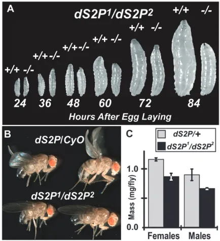
dS2P mutants grow more slowly than heterozygotes: We compared the growth ofdS2P1/
dS2P2mutants (from
crosses of dS2P2
/dS2P2 virgin females with
dS2P1
/CyO
males) to wild-type larvae raised in parallel cultures. Up to 48 hr AEL, there is no difference in size between
dS2P1/dS2P2mutants and wild-type larvae (Figure 2A).
By 60 hr AEL, dS2P1/dS2P2 mutants are distinctly
smaller than wild type. Disparity in size persists through 84 hr. By the time that mutants reach adulthood, they display a greater variability of body size than do their heterozygous siblings (males shown; Figure 2B) and are somewhat less massive, on average (Figure 2C). Morpho-logically, mutants are normal. These data show that the homozygotes grow more slowly than wild type or hetero-zygotes, taking longer to approach normal size.
Figure 3A shows typical emergence data from a het-erozygous cross ofdS2P1/dS2P1virgin females withdS2P2/
CyOmales. While the majority of heterozygotes emerge by day 11 AEL, the bulk of their dS2P1/dS2P2 siblings
emerge 2 days later. In multiple experiments, we consis-tently observe this2-day delay irrespective of the alleles used or the direction of thetrans-heterozygous cross. This delay becomes more pronounced with crowding (Figure 3B). We set up cultures with the indicated masses of embryos on standard medium and scored adults as they emerged. The delay is shown as the day AEL of median eclosion for homozygotes minus the day of median eclo-sion for heterozygotes. At 10 mg of embryos per culture, the delay fordS2P1
/dS2P1flies was 2 days. Doubling the
mass of embryos in the culture increased the delay to 5 days. At 40 or 70 mg of embryos, the delay extends to2 weeks. Results from flies lacking dSREBP (dSREBP189;
Kunteet al.2006) are shown for comparison.
Maternally supplied dS2P functions in dS2Pmutant larvae:We conducted extensive fertility, fecundity, and viability studies ondS2Pmutant stocks. In the course of these studies, we noted that the frequency of emergence of homozygotes was strongly affected by the maternal genotype. In experimental cultures, the homozygous offspring of heterozygous mothers emerged at about
the expected frequencies (Figure 4A, left, open bars). By contrast, the homozygous offspring of homozygous mothers survived markedly less well on unsupplemented medium, emerging at less than half the expected fre-quency (Figure 4A, middle, open bars). To determine if reduced viability resulted from disruption of fatty acid metabolism subsequent to deficient processing of dSREBP, we tested sibling cultures on medium supple-mented with fatty acids (Kunteet al.2006).
Supplemen-tation with fatty acids permitted near-expected survival of the homozygous offspring of homozygous mothers (Figure 4A, middle, shaded bars).dSREBP189flies served
as a control for rescue by fatty acid supplementation (Figure 4A, right).
Differential survival of homozygotes depending on the maternal genotype indicates that maternally sup-plied dS2P ameliorates the effects of the lack of dS2P
in the zygotic genome. We tested the hypothesis that at least some maternal dS2P activity is supplied via mRNA. Figure 4B shows real-time PCR analysis of transcript abundance in 0- to 2.5-hr AEL embryos and 36-hr AEL larvae. At 0–2.5 hr AEL, before the onset of most zygotic
transcription, the offspring of heterozygous mothers show significant levels of dS2P transcript, about one-third of wild-type levels, while no dS2P transcript is detectable in the offspring of homozygous mothers. By 36 hr AEL, no dS2P transcript is detectable in dS2P
mutant larvae irrespective of the maternal genotype. Transcript abundance of CG6295, a highly transcrip-tionally responsive target of dSREBP (Kunte et al.
2006), is shown as an indicator of dSREBP activity in these larvae. We found reduced transcript abundance in the homozygous offspring of heterozygous mothers and a much more substantial deficit in offspring of homo-zygous mothers (Figure 4C). Interestingly, these later
Figure 3.—dS2Pmutants develop more slowly than wild type. (A) Plot of the number of adults emergingvs. days after egg laying. On day 0, 3 mg of embryos was introduced into vials of standard cornmeal–molasses–agar medium. Begin-ning on day 9, and each day thereafter, adults were cleared from the culture and counted. (B) Crowding substantially ex-acerbates the developmental delay. The indicated mass of embryos was introduced into flasks of standard cornmeal– molasses–agar medium (80 ml/flask) on day 0. Beginning on day 9, and each day thereafter, adults were cleared from the culture, scored, and counted. ‘‘Days delayed’’ was calcu-lated as the day of median eclosion for homozygotes minus that of heterozygotes.
animals show greater abundance of CG6295 transcript than dodSREBP189larvae, even in the complete absence
of detectable dS2P transcripts (see below).
dSREBP mutated at site 2 rescues dSREBP null flies: The NH2-terminal transcription factor domain of dSREBP,
which is the product of cleavage by dS2P, is needed to rescue dSREBP mutants (Kunteet al.2006). Cleavage of
dSREBP by dS2P requires an asp462pro motif in the first
membrane-spanning helix of dSREBP (Yeet al.2000a).
When N462P is mutated to phenylalanine–leucine, dSREBP
cleavage is abolished (Seegmiller et al. 2002). Since
flies entirely lacking dS2P can survive, cleavage of dSREBP by dS2P is not essential for survival. Accord-ingly, an N462P/FL mutant dSREBP that cannot be
cleaved by dS2P should be able to rescue flies otherwise lacking dSREBP.
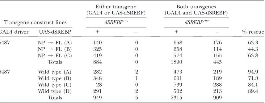
To test this hypothesis, we prepared transgenic flies expressing either wild-type or N462P / FL mutant
dSREBP cDNAs under the control of the yeast GAL4 upstream activating sequence. Expression was driven by the 4687 GAL4 enhancer trap line, which we have
pre-viously show is able to rescue dSREBP null mutant
animals to adulthood when driving expression of dSREBP (Kunteet al.2006). These transgenes were tested in a
dSREBP null background. Samples of emerging flies were analyzed by sequencing PCR products to confirm the presence of the indicateddSREBPtransgenes. Table 1 shows that both wild-type and mutant SREBPs can substantially rescue dSREBP null flies to adulthood,
although the NP/FL mutant does so less efficiently than wild-type dSREBP.
Alternative cleavage of dSREBP in flies: Nuclear dSREBP is essential for larval survival but cleavage of dSREBP by dS2P is not. This implies that transcription-ally active dSREBP must be present in the nuclei of
dS2P1
/dS2P2
larvae owing to a mechanism that does not require dS2P. To test this hypothesis, we used the
pre-viously described P{GAL4-dSREBPg} and P{UAS-GFP}
binary reporter system (Kunte et al. 2006) to assess
dSREBP processing indS2Pmutants (Figure 5A). Virgin females homozygous for either dS2P1 or
dS2P2 on the
second chromosome and homozygous for both the P{GAL4-dSREBPg} and P{UAS-GFP} transgenes on the third chromosome were crossed to males of the same genotype heterozygous on the second chromosome. Embryos were raised on standard medium until third instar when they were examined by fluorescence microscopy. Fluorescence owing to GFP expression is readily detectable in dS2P
mutants (Figure 5B, top and middle), although at levels lower than seen in heterozygous siblings (Figure 5B, bottom). Thus, release of the amino-terminal transcrip-tion factor domain from dSREBP occurs even in the absence of dS2P.
We noted above (cf.Figure 4) that thedS2P1
/dS2P2
homozygous offspring of homozygous mothers showed transcription of CG6295 that was greater than indSREBP
mutants. This is consistent with the presence of the dSREBP transcription factor domain in the nuclei of
TABLE 1
Rescue ofdSREBPlethality by wild-type and mutantdSREBPcDNA
Either transgene (GAL4 or UAS-dSREBP)
Both transgenes (GAL4 and UAS-dSREBP)
Transgene construct lines dSREBP189 dSREBP189
GAL4 driver UAS-dSREBP 1 1 % rescue
6487 NP/FL (A) 140 0 658 176 63.3
NP/FL (B) 325 0 658 114 44.3
NP/FL (C) 419 0 574 155 63.8
Totals 884 0 1890 445
6487 Wild type (A) 282 2 473 219 94.9
Wild type (B) 348 1 601 189 71.8
Wild type (C) 28 0 739 288 84.1
Wild type (D) 291 2 502 213 89.4
Totals 949 5 2315 909
We prepared P-element-based germline transformation constructs that encode either wild-type dSREBP cDNA or cDNA carrying the mutation N462P/FL, which abolishes cleavage by dS2P. Independent second chromosome insertions of each transgene were isolated (designated A, B, C, and D) and used to generate stocks of the genotypesw;P{w1
, UAS-dSREBP}/P{w1
,UAS-dSREBP}; dSREBP189/TM6B, Tb Hu e(for homozygous viable transgene insertions) andw; UAS-dSREBP/CyO; dSREBP189/TM6B, Tb Hu e(for homozygous lethal transgene insertions). These were crossed to flies carrying the 6487 GAL4 driver (expressed predominantly in anterior gut, fat body, and oenocytes) of the genotypew;P{w1
, GAL4}/P{w1
dS2Plarvae. To determine if this pattern held true for other target genes, we performed real-time PCR analy-sis. Figure 6 shows mRNA abundance at 36, 48, and 60 hr AEL for the indicated dSREBP target genes (Dobrosotskayaet al.2002; Kunteet al.2006). At 36
hr,dS2P1
/dS2P2
and wild-type larvae show similar abun-dance of transcripts for acetyl–coenzyme A carboxylase, synthase, and fatty acid synthase. These transcripts are less abundant indSREBP189
larvae. This pattern contin-ues through 60 hr. By contrast, transcripts for CG6295 are much less abundant indS2P1
/dS2P2
than in wild type, more closely matching their abundance in dSREBP189
larvae. We consistently observe the small increase in
abundance in dS2P1
/dS2P2
larvae vs. dSREBP189
larvae. Thus,dS2P1
/dS2P2
larvae have less severe transcriptional deficits than dodSREBP189
larvae.
Figure 5.—Larvae lacking dS2P nevertheless process dSREBP. (A) A binary reporter system for dSREBP activity (Kunte et al. 2006). The transcription factor domain of pP{dSREBPg} was replaced by a GAL4-VP16 transcription fac-tor to generate pP{GAL4-dSREBPg}. (B) Animals homozygous for both P{GAL4-dSREBPg} and P{UAS-GFP} transgenes in the indicateddS2Pbackground were examined for spatial locali-zation of GFP fluorescence. In larvae homozygous for either dS2Pallele, fluorescence is detectable in fat body but levels are decreased relative to heterozygous siblings. No fluores-cence is detectable in the midgut of dS2Phomozygotes, in contrast to heterozygotes. Although not clearly visible in pho-tographs, we detect a faint fluorescence in the oenocytes of manydS2Phomozygotes. All larvae are the offspring of moth-ers homozygous for the indicated dS2P allele. Images are 1-sec exposures taken using a Leica MZ16FA fluorescence microscope equipped with an Evolution MP digital camera (Media Cybernetics) and In Focus software (Meyer Instru-ments, Houston). GFP fluorescence was visualized using a GFP2(1) filter set for MZ16 FA, 480/40, 510 nm, and images were captured using ImagePro software.
DISCUSSION
We isolated mutant D. melanogaster harboring a de-ficiency that removes the entiredS2Ptranscription unit (Figure 1A). No dS2P mRNA is detectable in these animals and no dSREBP processing is observed in mutant adults under conditions where it is readily observed in wild-type flies. Instead, the substrate for dS2P cleavage, the intermediate form of dSREBP, accumulates in mem-branes (Figure 1D). Therefore, thedS2P1deletion is a
null allele ofdS2P.
Phenotypes of the P-element insertion allele,dS2P2,
are indistinguishable fromdS2P1and are no more severe
in trans to the deletion allele. Transcripts from dS2P2
cannot yield catalytically active dS2P (Figure 1B). Thus,
dS2P2
is a null allele by genetic and molecular criteria. Surprisingly, animals harboring either allele are viable and can be readily maintained as homozygous stocks. Reciprocally,dSREBP189flies can be rescued by expressing
a dSREBP cDNA harboring an N462P/FL mutation that
renders dSREBP refractory to cleavage by dS2P (Table 1). Thus, the site-2 protease is not essential for the de-velopment and growth ofD. melanogaster.
The dS2P1
allele must also be null for the predicted gene CG34229 (Figure 1A) that encodes a putative component of the higher eukaryotic NADH complex. The predicted sequence of the encoded polypeptide is highly conserved, supporting the case for this gene.
Are there consequences of the loss of CG34229 that influence the phenotypes that we report? We cannot absolutely exclude the possibility that some phenotypes could result, in part, from haplo-insufficiency for CG34229 indS2P trans-heterozygotes. However, CG34229 cannot be an essential gene; dS2P1
homozygotes are viable. We performed most of the experiments presented here with mutantstrans-heterozygous fordS2P1anddS2P2. In parallel
experiments, we found indistinguishable results with flies homozygous for eitherdS2P1or d
S2P2(not shown), which
indicates that the phenotypes that we observe are not the result of the loss of CG34229. Further, the reduced survival of dS2P mutants is rescued by feeding fatty acids, a treatment that also rescues lethality in animals lacking
dSREBP. This indicates that reduced survival is a conse-quence of reduced dSREBP activity.
The phenotype informative for the most important finding described here is cleavage of dSREBP in the absence of dS2P (Figure 5B). Whether or not insuffi-ciency for CG34229 (or any gene yet to be identified in this region) contributes in some way to reduced viability, smaller-average-size, or delayed development in dS2P
homozygotes, dS2P is absent and dSREBP does reach the nucleus without cleavage by dS2P (Figures 5 and 6). In mammals, S2P is needed to process other mem-brane-bound transcription factors, ATF-6aand -b, that play a crucial role in the endoplasmic reticulum (ER)-stress response [also known as the unfolded protein response or UPR (Ye et al. 2000b)]. The Drosophila
genome encodes a protein highly similar to mammalian ATF-6, CG3136. In mammals, ATF6 is required to transcribe XBP1 mRNA, and mutant cells lacking S2P are deficient in the induction of the spliced form of XBP1 mRNA (Yoshidaet al.2006). WhendS2Plarvae
are challenged with dithiothreitol or tunicamycin, treat-ments that elicit the UPR, we see no difference in XBP1 splicing compared to wild-type larvae (supplemental Figure 1). If the Drosophila UPR is closely similar to the mammalian UPR, these data suggest that ATF6 processing is relatively unimpaired indS2Plarvae. It might be that
the Drosophila homolog of ATF6 is not required for the fly UPR or that its activity does not require cleavage by dS2P. If dS2Pisrequired to activate this homolog in flies, the observed developmental delay ofdS2Plarvae may result
from defects in ATF6 activation. Nevertheless, while these putative additional functions of dS2P may be important, the crucial function of dS2P in flies is to process dSREBP. In striking contrast to dS2P adults, which lack
nuclear dSREBP under conditions where it is readily detected in wild type, dSREBP can reach the nucleus and activate transcription of target genes in dS2P mutant larvae (Figure 5B). Thus, Drosophila larvae lackingdS2P
have an alternative means of releasing the nuclear tran-scription factor domain of dSREBP from the membrane-bound precursor. This explains the greater abundance of dSREBP target transcripts indS2P1
/dS2P2
mutants com-pared withdSREBP189
mutants (Figures 4 and 6).
What is the role of this alternative mechanism for producing nuclear dSREBP? The current data show only that it occurs in the absence of dS2P. We do not yet know if it is a normal, physiologically relevant mecha-nism or whether it happens fortuitously in the absence of normal dSREBP processing. It is, however, sufficient to afford the survival, over many generations, of flies completely lacking dS2P.
How is the transcription factor domain of dSREBP
produced in dS2P mutants? A possible mechanism is
production of alternative transcripts that encode only the dSREBP transcription factor domain without the membrane-spanning helices. These might arise from different promoter usage or from differential splicing. Arguing against these possibilities is the fact that only a single transcript is detected for dSREBP in flies from embryogenesis through adulthood and in various
tis-sues examined (Theopold et al. 1996). We likewise
observe a single band on Northern blots for dSREBP (not shown). Any putative alternative transcripts or splice forms would have to be present at levels too low to be detected in these assays, while the activity of nuclear dSREBP in dS2P1/dS2P2 larvae is readily
de-tected (Figures 5 and 6). Moreover, a cDNA construct
harboring the N462P/ FL mutation and under
con-trol of a single, heterologous promoter rescuesdSREBP
We favor the hypothesis that in larvae lacking dS2P, dSREBP is released from the membrane by some other protease(s). This posited protease is unlikely to cleave within the first membrane-spanning helix of dSREBP: flies have no other S2P homologs, and other intra-membrane-cleaving proteases display different sub-strate preferences (cf. Hooperand Lendeckel2007).
The signal peptide peptidase (SPP) is an intramem-brane protease of the ER (Weihofenet al.2002). SPPs
are unlikely candidates for cleavage of SREBPs, how-ever. Like S2P, the SPPs require prior cleavage of the substrate by a separate protease. Chinese hamster ovary (CHO) cells express active SPP (Devet al. 2006), but
multiple, independently isolated lines of CHO cells lacking S2P show no processing of SREBPs (Sakaiet al.
1996). If SPPs could cleave SREBPs, one would expect
some evidence of SREBP processing in S2P cells.
Cleavage of dSREBP following its first
membrane-spanning helix cannot release the NH2-terminal
do-main. It is most probable that the alternative cleavage occurs in the cytoplasm, between the transcription factor domain and the first membrane-spanning helix of dSREBP. We term this portion of dSREBP the ‘‘stalk.’’
Cleavage of SREBPs within the stalk has been reported previously. Wang et al. showed that caspases 3 and 7
could each cleave mammalian SREBPs (Wang et al.
1995; Paiet al.1996) and that this cleavage was
detect-able during apoptosis (Wanget al.1996). The
physio-logical significance of this cleavage is presently unclear. The caspase cleavage sites identified by Wang et al.
(1995) are highly conserved among vertebrate SREBP isoforms, however, and all metazoan SREBPs (except those from Nematoda) contain potential caspase cleav-age sites within their stalk regions (R. B. Rawson,
un-published observations). Using reporter constructs, Higgins and Ioannou showed that SREBP cleaved during apoptosis by caspases can be transcriptionally active (Higginsand Ioannou2001). There is precedent
for caspase cleavage of SREBPs releasing the functional transcription factor.
Current data do not suggest that the production of nuclear dSREBP in dS2P mutants has any involvement with apoptosis. However, nonapoptotic roles of caspases have been found in Drosophila (Huhet al.2004) and
other systems (e.g., reviewed in Algeciras-Schimnich
et al.2002). Cleavage of dSREBP in the absence of dS2P may be an example of a nonapoptotic caspase function. We are currently testing the hypothesis that dSREBP is cleaved by caspases to produce transcriptionally active dSREBP indS2Plarvae.
We are grateful to Kaori Tanaka, Denise Parker, Phuong Pham, Praja Lakireddy, and Therese Schindler for excellent technical support and to Jeff Cormier for sequencing and real-time PCR analysis. This work was supported by grants from the National Institutes of Health (R01 GM07145701A1) and the Perot Family Foundation.
LITERATURE CITED
Algeciras-Schimnich, A., B. C. Barnhart and M. E. Peter, 2002 Apoptosis-independent functions of killer caspases. Curr. Opin. Cell Biol.14:721–726.
Bengoechea-Alonso, M. T., and J. Ericsson, 2007 SREBP in signal transduction: cholesterol metabolism and beyond. Curr. Opin. Cell Biol.19:215–222.
Brown, M. S., J. Ye, R. B. Rawson and J. L. Goldstein, 2000 Regulated intramembrane proteolysis: a control mecha-nism conserved from bacteria to humans. Cell100:391–398. Cherry, S., A. Kunte, H. Wang, C. Coyne, R. B. Rawsonet al.,
2006 COPI activity coupled with fatty acid biosynthesis is re-quired for viral replication. PLoS Pathog.2:e2.
Clark, A. J., and K. Bloch, 1959 Absence of sterol biosynthesis in insects. J. Biol. Chem.234:2578–2588.
Dev, K. K., S. Chatterjee, M. Osinde, D. Stauffer, H. Morganet al., 2006 Signal peptide peptidase dependent cleavage of type II transmembrane substrates releases intracellular and extracellu-lar signals. Eur. J. Pharmacol.540:10–17.
Dobrosotskaya, I. Y., A. C. Seegmiller, M. S. Brown, J. L. Goldstein and R. B. Rawson, 2002 Regulation of SREBP processing and membrane lipid production by phospholipids in Drosophila. Science296:879–883.
Duncan, E. A., U. P. Dave, J. Sakai, J. L. Goldsteinand M. S. Brown, 1998 Second-site cleavage in sterol regulatory element-binding protein occurs at transmembrane junction as determined by cys-teine panning. J. Biol. Chem.273:17801–17809.
Feng, L., H. Yan, Z. Wu, N. Yan, Z. Wanget al., 2007 Structure of a site-2 protease family intramembrane metalloprotease. Science
318:1608–1612.
FlyBaseConsortium, 2003 The FlyBase database of the Drosoph-ila genome projects and community literature. Nucleic Acids Res.
31:172–175.
Goldstein, J. L., R. B. Rawson and M. S. Brown, 2002 Mutant mammalian cells as tools to delineate the sterol regulatory ele-ment-binding protein pathway for feedback regulation of lipid synthesis. Arch. Biochem. Biophys.397:139–148.
Higgins, M. E., and Y. A. Ioannou, 2001 Apoptosis-induced release of mature sterol regulatory element-binding proteins activates sterol-responsive genes. J. Lipid Res.42:1939–1946.
Hooper, N. M., and U. Lendeckel(Editors), 2007
Intramembrane-Cleaving Proteases (I-CLiPs).Springer-Verlag, Berlin/Heidelberg, Germany/New York.
Hua, X., J. Sakai, M. S. Brownand J. L. Goldstein, 1996 Reg-ulated cleavage of sterol regulatory element binding proteins re-quires sequences on both sides of the endoplasmic reticulum membrane. J. Biol. Chem.271:10379–10384.
Huh, J. R., S. Y. Vernooy, H. Yu, N. Yan, Y. Shiet al., 2004 Multiple apoptotic caspase cascades are required in nonapoptotic roles for Drosophila spermatid individualization. PLoS Biol.2:E15. Kinch, L. N., K. Ginalskiand N. V. Grishin, 2006 Site-2 protease
regulated intramembrane proteolysis: sequence homologs sug-gest an ancient signaling cascade. Protein Sci.15:84–93. Kunte, A. S., K. A. Matthewsand R. B. Rawson, 2006 Fatty acid
auxotrophy in Drosophila larvae lacking SREBP. Cell Metab.3:
439–448.
Limanek, J. S., J. Chinand T. Y. Chang, 1978 Mammalian cell mu-tant requiring cholesterol and unsaturated fatty acid for growth. Proc. Natl. Acad. Sci. USA75:5452–5456.
Pai, J. T., M. S. Brownand J. L. Goldstein, 1996 Purification and cDNA cloning of a second apoptosis-related cysteine protease that cleaves and activates sterol regulatory element binding pro-teins. Proc. Natl. Acad. Sci. USA93:5437–5442.
Rawson, R. B., N. G. Zelenski, D. Nijhawan, J. Ye, J. Sakaiet al., 1997 Complementation cloning of S2P, a gene encoding a pu-tative metalloprotease required for intramembrane cleavage of SREBPs. Mol. Cell1:47–57.
Robertson, H. M., C. R. Preston, R. W. Phillis, D. M. Johnson -Schlitz, W. K. Benz et al., 1988 A stable genomic source of
Pelement transposase inDrosophila melanogaster. Genetics118:
461–470.
Sakai, J., E. A. Duncan, R. B. Rawson, X. Hua, M. S. Brownet al., 1996 Sterol-regulated release of SREBP-2 from cell membranes requires two sequential cleavages, one within a transmembrane segment. Cell85:1037–1046.
Seegmiller, A. C., I. Dobrosotskaya, J. L. Goldstein, Y. K. Ho, M. S. Brownet al., 2002 The SREBP pathway in Drosophila: regula-tion by palmitate, not sterols. Dev. Cell2:229–238.
Theopold, U., S. Ekengrenand D. Hultmark, 1996 HLH106, a Drosophila transcription factor with similarity to the vertebrate sterol responsive element binding protein. Proc. Natl. Acad. Sci. USA93:1195–1199.
Wang, X., J. T. Pai, E. A. Wiedenfeld, J. C. Medina, C. A. Slaughter
et al., 1995 Purification of an interleukin-1 beta converting enzyme-related cysteine protease that cleaves sterol regulatory element-binding proteins between the leucine zipper and trans-membrane domains. J. Biol. Chem.270:18044–18050. Wang, X., N. G. Zelenski, J. Yang, J. Sakai, M. S. Brown et al.,
1996 Cleavage of sterol regulatory element binding proteins (SREBPs) by CPP32 during apoptosis. EMBO J.15:1012–1020. Weihofen, A., K. Binns, M. K. Lemberg, K. Ashmanand B. Martoglio, 2002 Identification of signal peptide peptidase, a presenilin-type aspartic protease. Science296:2215–2218.
Ye, J., U. P. Dave, N. V. Grishin, J. L. Goldsteinand M. S. Brown, 2000a Asparagine-proline sequence within membrane-span-ning segment of SREBP triggers intramembrane cleavage by site-2 protease. Proc. Natl. Acad. Sci. USA97:5123–5128. Ye, J., R. B. Rawson, R. Komuro, X. Chen, U. P. Daveet al., 2000b ER
stress induces cleavage of membrane-bound ATF6 by the same pro-teases that process SREBPs. Mol. Cell6:1355–1364.
Yoshida, H., S. Nadanaka, R. Satoand K. Mori, 2006 XBP1 is crit-ical to protect cells from endoplasmic reticulum stress: evidence from site-2 protease-deficient Chinese hamster ovary cells. Cell Struct. Funct.31:117–125.
Zelenski, N. G., R. B. Rawson, M. S. Brownand J. L. Goldstein, 1999 Membrane topology of S2P, a protein required for intra-membranous cleavage of sterol regulatory element-binding pro-teins. J. Biol. Chem.274:21973–21980.