DOI: 10.1534/genetics.106.059709
Maternal Gametophytic
baseless1
Is Required for Development of the
Central Cell and Early Endosperm Patterning in Maize (
Zea mays
)
Jose´ F. Gutie´rrez-Marcos,*
,1Liliana M. Costa*
,1and Matthew M. S. Evans
†,2*Department of Plant Sciences, University of Oxford, Oxford, OX1 3RB, United Kingdom and†Department of Plant Biology, Carnegie Institution of Washington, Stanford, California 94025
Manuscript received April 19, 2006 Accepted for publication July 6, 2006
ABSTRACT
In angiosperms, double fertilization of an egg cell and a central cell with two sperm cells results in the formation of a seed containing a diploid embryo and a triploid endosperm. The extent to which the embryo sac controls postfertilization events in the seed is unknown. The novel gametophytic maternal-effect maize mutation,baseless1(bsl1) affects central cell development within the embryo sac, frequently by altering the position of the two polar nuclei. Despite this irregularity, fertilization is as efficient as in wild type. The spatial expression of basal endosperm-specific transcripts is altered in free-nuclear and cellular mutant endosperms. At later stages of seed development,bsl1predominantly affects development of the basal en-dosperm transfer layer (BETL). Whenbsl1/1diploid plants were pollinated by wild-type tetraploid plants, the BETL abnormalities observed inbsl1/bsl1/1/1tetraploid endosperms were diverse and of variable severity. Moreover, the frequency of kernels with severely perturbed BETL development correlated with the percentage of severely affectedbsl1central cells. Therefore, BSL1 is likely required in the central cell before fertilization for correct BETL patterning to occur. These findings provide new genetic evidence that a maternal gametophytic component is necessary for correct endosperm patterning.
M
OST flowering plants are sexually dimorphic, in that they possess both male and female reproduc-tive organs. The male gametophyte (pollen) typically contains a vegetative nucleus, which contributes to pol-len tube growth, and two male gametes (sperm cells) that participate in fertilization. The female gameto-phyte is usually of the Polygonum type, consisting of two synergid cells, some antipodal cells, and two female gametes—the egg cell and the larger central cell, which contains two polar nuclei (Drewsand Yadegari2002;Yadegariand Drews2004).
The process of double fertilization is unique to the flowering plants and results in formation of the seed. For this to occur, the pollen tube must first grow toward the ovule, where it is guided to the micropyle and enters the female gametophyte through one of the two syner-gids. Recent studies have shown that pollen tube growth and guidance are largely controlled by the female gameto-phyte and in particular by the synergids (Higashiyama
et al.2001; Hucket al.2003; Rotmanet al.2003; Marton
et al.2005). Under wild-type conditions, the pollen tube penetrates the female gametophyte through a degener-ated synergid, after which the pollen tube tip ruptures, releasing the two sperm cells. The sperm cells
subse-quently fuse individually with the egg and central cell to typically form a diploid (1 maternal:1 paternal) embryo and a triploid (2 maternal:1 paternal) endosperm, re-spectively. Although both seed components are typically identical genetically, their developmental fates diverge significantly. While the life cycle of the embryo is ex-tended to the next generation—where it forms the ma-ture plant—the endosperm life cycle is confined to the seed stage, where it provides nutritional and structural support to the growing embryo (Walbot and Evans
2003; Costaet al.2004).
In many flowering plants, the endosperm undergoes a nuclear-type mode of development (Olsen 2004).
Strikingly similar to early Drosophila embryogenesis, the fertilized proendosperm triploid nucleus divides through a series of mitoses that are uncoupled to cell wall formation, to form a free-nuclear structure or syn-cytium with a single layer of cortical nuclei suspended in the peripheral cytoplasm (Olsen 2001). Following
syncytial development, cellularization occurs and vari-ous cell types eventually differentiate within the fully cellular endosperm (Costaet al.2004; Olsen2004).
To date, relatively little is known about the mecha-nisms regulating early development of the endosperm. On the basis of current genetic evidence, endosperm patterning occurs in two main phases (Costa et al.
2004). The first, pivotal phase leads to the establishment of the proximal–distal axis, which is reflected by the polarized localization of transcripts (Doanet al.1996;
1These authors contributed equally to this work.
2Corresponding author:Department of Plant Biology, Carnegie
Institu-tion of Washington, 260 Panama St., Stanford, CA 94025. E-mail: [email protected]
Gomezet al.2002; Dreaet al.2005; Ingouffet al.2005a),
and, in Arabidopsis, the cytological organization of syn-cytial endosperm domains (Boisnard-Loriget al.2001;
Brown et al.2003). These early events are most likely
regulated by several maternally required genes (for ex-ample, Springeret al.2000; Griniet al.2002; Holding
and Springer2002), including members of thePolycomb
Group complex (Pc-G). The identification and charac-terization of the ArabidopsisPc-G medea (mea) mutant first demonstrated the principle of gametophytic mater-nal effects on seed development in plants (Grossniklaus
et al.1998). Mutations in MEAand in two other Arabi-dopsisPc-G genes,Fertilization Independent Endosperm(FIE) (Ohadet al.1996) andFertilization Independent Seed2(FIS2)
(Chaudhuryet al.1997), cause irregular nuclear
prolifer-ation in the unfertilized central cell as well as a delay in the developmental progression of the fertilized endo-sperm (Grossniklauset al.1998; Ingouffet al.2005b).
As a consequence of the latter, ectopic chalazal (poste-rior) endosperm-specific gene expression becomes man-ifest in inappropriately anterior positions (Sørensenet al.
2001; Guittonet al.2004). Similarly, development of the
chalazal endosperm is perturbed in two other Arabidop-sisPc-G mutants,borgiaandmedicis/multicopy suppressor of IRA1(Guittonet al.2004). Parallel studies have
demon-strated that alterations in the 2 maternal:1 paternal ge-nomic balance in the endosperm also strongly influence early development of the Arabidopsis chalazal endosperm and the maize basal endosperm transfer layer (BETL). In both species, development of these domains is more dramatically perturbed in 2 maternal (m):2 paternal (p) endosperms derived from crosses between diploid fe-males and tetraploid fe-males (reviewed in Costa et al.
2004).
Here we report the comprehensive characterization ofbaseless1(bsl1)—a novel gametophytic maternal-effect mutant in maize. Before fertilization,bsl1mutants dis-played defects in the central cell within the mature female gametophyte. The two polar nuclei were fre-quently displaced inbsl1central cells, yet, despite this irregularity fertilization was achieved, suggesting that sperm were able to efficiently locate and fuse with the displaced polar nuclei. Resulting mutant seeds dis-played irregular distributions of transcripts specific to the basal region of syncytial and cellular endosperms. Mutant cellular endosperms also exhibited aberrant BETL development. BETL abnormalities were dramati-cally enhanced in seeds resulting from crosses between
bsl1/1 females and wild-type tetraploid males. Our analysis ofbsl1mutants provides new genetic evidence in plants that essential endosperm patterning compo-nent(s) are present in the central cell before fertilization.
MATERIALS AND METHODS
Plant material and growth conditions:Thebaseless1mutant was originally isolated from a self-pollination of a W22 inbred maize (Zea mays) plant carrying a novel mutable allele,
r1-m-Bolivia, of theR1locus and was given the provisional desig-nationdex-4299* (Kermicle1978). The mutant was typically propagated as heterozygous by transmission through the fe-male and selection for viablebsl1defective kernels. Plants were grown in summer field conditions or in greenhouses under 16 hr light:8 hr dark cycles.bsl1heterozygous (bsl1/1) and wild-type (1/1) plants used for controls were grown under the same conditions for each experiment. For characterization of the effects ofbsl1on seed development,bsl1/1plants were pollinated with wild-type pollen. Segregating wild-type and mutant siblings were then taken from the same middle-third portion of the ear, as standard. With the exception of the mapping, all characterizations were performed in a standard W22 inbred background. Male transmission (Mt) ofbsl1was calculated on the basis of both linkage to and transmission of a linked genetic marker. Mtlinked markerequals the transmission of nonrecombinantbsl1pollen grains and recombinantBsl11
pollen grains. This was calculated as Mtbsl1times the frequency
of nonrecombinants (1R) betweenbsl1and the marker plus the frequency of transmission ofBsl11
(1Mtbsl1) times the
frequency of recombinants betweenbsl1and the marker (R). Solving for Mtbsl1gives Mtbsl1¼(Mtlinked markerR)/(1.02R), where R equals the recombination frequency between bsl1 and the linked marker, and Mtlinked marker equals the trans-mission of the allele linked tobsl1.
Mapping ofbsl1was performed in crosses betweenbsl1/1, W22/W23 hybrid females and W23 wild-type males. Onlybsl1 defective kernels were used for the mapping population, as incomplete penetrance causes some normal kernels to carry bsl1, necessitating progeny testing to ensure that they were wild type. DNA was extracted from seedlings from these defective kernels and maize simple sequence repeat (SSR) markers were analyzed as described previously (Evansand Kermicle2001). Pollen tube growth analysis:Pollen was analyzed from five bsl1/1heterozygotes and five homozygous wild-type plants. Old anthers were removed from plants the day before pollen collection. The following day, one newly extruded anther was picked from each plant prior to dehiscence, and anthers were quickly dissected to liberate pollen onto germination plates.In vitro pollen tube growth measurements were performed as described previously (Evansand Kermicle2001).
Histology: For examination of pollen morphology, pollen grains were collected in the same manner as for pollen tube growth measurements, except that anthers were dissected di-rectly on slides. Pollen was mounted under coverslips in iodide potassium iodine to visualize starch or stained with hematox-ylin/ferric ammonium sulfate to visualize nuclei (Kindiger 1994). Pollen grains were analyzed with a Nikon Eclipse E600 microscope.
For analysis of embryo sacs, samples collected from homo-zygous wild-type and heterohomo-zygousbsl1/1plants were fixed in 5% formaldehyde, 5% acetic acid, 45% ethanol under vacuum for 15 min, followed by overnight fixation in fresh fixing buffer at 4°. Samples were rinsed and stored in 70% ethanol. Ovules and developing kernels were bisected along the longitudinal axis of the ear, dehydrated through a standard ethanol series, and cleared in methyl salicylate. Samples were mounted in methyl salicylate and visualized on a Bio-Rad (Hercules, CA) laser scanning confocal microscope. Excitation was performed at 488 nm and emission was collected at both 522- and 585-nm wavelengths and combined. Image files were opened with NIH Image v. 1.62 and handled in Adobe Photoshop v. 5.0.
counting photometer attached to a side port of a Nikon Diaphot TMD inverted microscope. Samples were viewed with a Zeiss 1003oil immersion objective using an ultraviolet filter set with a excitation filter (360 nm, bandpass 10 nm), a dichroic mirror (400 nm), and a barrier filter (420 nm). For each mea-surement, individual calibration curves were prepared on the basis of a linear regression analysis as the relative fluorescence units (RFU) of DAPI-stained nuclei in proportion to DNA con-tent (Colemanand Goff1985).
mRNA in situhybridization: In situhybridization was per-formed on kernels as described previously (Costaet al.2003). Probes for MRP1 (Gomezet al.2002) and the maize homolog of barley END1 (Doanet al.1996) were generated by reverse transcriptase amplification (RT–PCR) with gene-specific oli-gonucleotides: MRP1.FOR 59-ATGAATCCCAACTTCAAC AGTGTGTG-39, MRP1.REV 59-TATCGGTTA TATATCTGGCT CTCC-39, END1.FOR 59-ATGAAACGAGAGTGCAAGCAGTT TGAG-39, and END1.REV 59-CATACTAAGAGGAACTGTAT AACTCC-39. Slides were viewed with a Zeiss Axiophot micro-scope under bright field optics and images were digitally re-corded with a coolpix Nikon camera.
Transgenic reporter gene analysis:To analyze BETL-specific reporter gene expression in mutant and wild-type seeds,bsl1/1
plants were crossed as females by males carrying one of two BETL transgenic reporters:ProBet1:GUS(Hueroset al.1999a)
orProMeg1:GUS(Gutierrez-Marcoset al.2004). Kernels were
cut along the longitudinal axis and stained for GUS activity as previously described (Costaet al.2003; Gutierrez-Marcos et al.2004). To analyze the combined effects ofbsl1and paren-tal genomic imbalance in the tetraploid endosperm on BETL-specific reporter gene expression, plants heterozygous forbsl1 and homozygous forProMeg1:GUS transgene were crossed by
wild-type (W22) tetraploid plants.
RESULTS
baseless1is a novel maternal-effect mutation in maize: Thebaseless1mutant arose spontaneously as a sector on a female inflorescence (ear) of a wild-type W22 inbred maize plant. One side of the ear had 70 normal seeds; the other side of the ear had 40 defective seeds and 58 normal seeds. Fortuitously, the viability of some abnor-mal seeds allowed recovery and propagation of the mutant when transmitted maternally. bsl1 was initially
mapped to chromosome 5 using waxy1 (wx1)-marked reciprocal translocations. This location was confirmed by linkage to the purple aleurone1 (pr1) gene (49/226 ¼
21.7%;P,0.0001). Mapping with SSR markers placed
bsl1on the short arm of chromosome 5 in proximity to the centromere—betweenumc1110andbnlg603.
After pollinatingbsl1/1plants with wild-type pollen, 45.762.6% of the seeds were visibly defective (Figure 1 and Table 1). Progeny testing of phenotypically normal seeds revealed that 8.4 62.3% were also carrying the
bsl1 defective allele, so that the percentage of bsl1/1
kernels was always50% (Table 1). Progeny testing of the defective seeds revealed that they almost exclusively (238/241 individuals) segregated defective kernels in the next generation (i.e., due tobsl1inheritance). Fur-ther, the penetrance and severity of the bsl1/1 seed phenotype varied from cross to cross within the same genetic background (Figure 1, B and C), suggesting that the mutant phenotype is most likely influenced by ex-trinsic environmental factors. Mature mutant seeds fell into different phenotypic classes, including reduced en-dosperm, loose pericarp, empty pericarp, and germless (Figure 1, D–H). Consequently, the most severely af-fected seeds failed to germinate (27/98), while those that did displayed root or shoot defects (18/71), in-cluding fasciations and/or absence of the root and shoot axes.
When wild-type females were crossed withbsl1/1 het-erozygous males, the frequency of defective kernels did not significantly differ from that observed in wild-type crosses (Table 1). These findings indicate thatbsl1
male transmission alone has no detrimental effect on seed development and thatbsl1is not dominant. More-over, progeny testing of the offspring ofbsl1/1 males revealed that the bsl1 mutant allele was transmitted through pollen less efficiently than the wild-type allele. The rate of bsl1 male transmission was calculated as 12.062.3%, both by progeny testingbsl1/1males and by segregation distortion of the linked markerpr1or a Figure1.—Thebsl1seed phenotype. (A) Ear from a homozygous wild-type plant pollinated with pollen from a bsl1/1 heterozygous plant, exhibiting normal kernels. (B) Ear from absl1/1
waxy1(wx1) marked reciprocal translocation, T5-9cwx1
(Table 1). These data indicate that the deficit ofbsl1
heterozygotes after male transmission was caused by reduced transmission of thebsl1allele rather than by frequent reversion ofbsl1to wild type.
We self-pollinatedbsl1/1heterozygotes in an attempt to generatebsl1/bsl1 homozygous progeny. If bsl1 ho-mozygous seeds were viable, they would be expected to occur at an average frequency of 6% (i.e., on the basis of the probability of 12% male transmission and 50% female transmission ofbsl1alleles). Furthermore, 12% of the resulting defective kernels (i.e., those with ma-ternalbsl1) are predicted to have inherited paternalbsl1
and be homozygous. With this in mind, all resulting defective kernels were selected, and plants from these seeds were grown to maturity. These plants did not ex-hibit any morphological abnormalities, and they were subsequently progeny tested for homozygosity ofbsl1on the basis of the frequency of defective seed production (i.e., from 50% defective seeds to 100% defective seeds). However, of 103 plants tested, none were
ho-mozygous for bsl1, indicating that the homozygotes were seed lethal (likelihood that bsl1/bsl1plants were missed by random chance,P,0.0001). No novel kernel phenotypes were visible in self-pollinations of bsl1/1
heterozygotes compared to pollinations of bsl1/1 fe-males by wild type, suggesting that thebsl1/bsl1 individ-uals are not phenotypically distinct from heterozygous individuals carrying maternal bsl1 alleles. To confirm this finding we carried out test crosses and self-pollina-tions ofbsl1 Wx11
/Bsl11
T5-9cwx1plants, which enabled us to distinguish kernels carrying wild-type or bsl1 al-leles. Progeny derived from self-pollinations ofbsl1/1
plants exhibited a higher proportion of lethal pheno-typic classes (18.9%) compared to those derived from test crosses (7%) (Table 2). Therefore the rise in seed lethality following self-pollinations can be accounted for by the presence of bsl1 homozygotes in these populations.
Taken together, these genetic data indicate thatbsl1
confers a maternal effect on seed development. That only half of the seeds are affected is consistent with
TABLE 2
Phenotypic classes of kernels produced in test crosses and self-crosses ofbsl1/1plants marked with T5-9cwx1in repulsion tobsl1
bsl1/1andbsl1/bsl1b 1/1:
Ear parent Pollen parent
Empty pericarpa
(%)
Loose pericarpa
(%)
Germlessa
(%)
Reduced endosperm
(%)
Normal morphology
(%)
Normal morphology
(%)
bsl1 Wx111T5-9c
wx1 (199 kernels/3 ears)
wx1 1.9 3.5 1.6 33.6 12.8 46.6
bsl1 Wx111
T5-9cwx1 (352 kernels/5 ears)
bsl1 Wx111
T5-9cwx1 3.2 10.3 5.4 28.1 8.7 43.9
bsl1phenotypic classes are listed from most to least severe (empty pericarp.loose pericarp.germless. reduced endo-sperm.normal).
a
Lethal phenotypic classes.
b
Include1/1defective kernels.
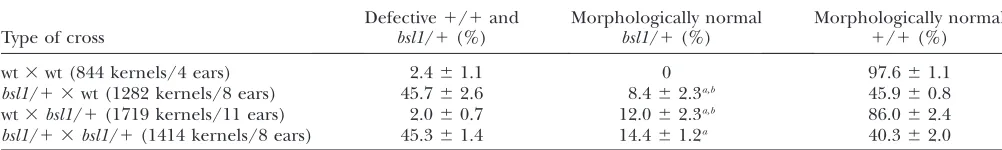
TABLE 1
Phenotypic classes of kernel progeny from pollinations with wild-type andbsl1/1plants
Type of cross
Defective1/1and bsl1/1(%)
Morphologically normal bsl1/1(%)
Morphologically normal
1/1(%)
wt3wt (844 kernels/4 ears) 2.461.1 0 97.661.1
bsl1/1 3wt (1282 kernels/8 ears) 45.762.6 8.462.3a,b 45.960.8
wt3bsl1/1(1719 kernels/11 ears) 2.060.7 12.062.3a,b 86.062.4
bsl1/1 3bsl1/1(1414 kernels/8 ears) 45.361.4 14.461.2a 40.362.0
Percentages given are the average of each kernel class per cross6the standard error of the mean.
aIn some crosses the transmission ofbsl1was calculated from the transmission and linkage of the genetic markerspurple
aleu-rone1(pr1), or awaxy1(wx1) marked reciprocal translocation T5-9c, linked in repulsion phase tobsl1. Male transmission rates of bsl1were calculated as Mtbsl1¼(Mtlinked markerR)/(12R) (seematerials and methods).
b
either a gametophytic maternal effect or an incom-pletely penetrant sporophytic maternal effect. The cor-relation between kernel phenotype and the inheritance ofbsl1in the progeny of these crosses demonstrates that the presence of the mutation in the embryo sac rather than the diploid parent is critical, thus supporting the view that thebsl1maternal effect is gametophytic.
baseless1 affects development of the central cell within the female gametophyte:The reduced male trans-mission ofbsl1and linked markers frombsl1/1 hetero-zygotes suggests that mutantbsl1pollen grains are at a competitive disadvantage to wild-type pollen. Thus it is possible thatbsl1causes either an increased frequency of aborted and abnormal pollen or more subtle defects, such as in pollen tube growth rate, its ability to target embryo sacs, or the fertilization process, any of which could result inbsl1pollen grains achieving less efficient fertilization than wild type. We therefore examined pollen from heterozygousbsl1/1plants and compared them to pollen from wild-type plants (see materials and methods). No abnormalities in pollen
morphol-ogy, starch filling, or nuclear number were apparent in pollen frombsl1/1heterozygous plants (Table 3), sug-gesting that bsl1 pollen grains develop normally to maturity. Further, germination and tube growth rates of pollen frombsl1/1heterozygous plants and wild-type plants were compared in vitro (see materials and methods) and were found to be identical (Table 3),
indicating that pollen tube growth is not perturbed by
bsl1.
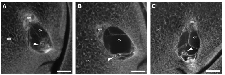
We also investigated the possible effects of the bsl1
mutation on female gametophyte development. Cell number, position, and morphology within embryo sacs ofbsl1/1plants were as in wild type, with discrepancies observed only in the central cell. In wild-type plants, the two polar nuclei of the central cell were always posi-tioned adjacent to the egg cell, along the central longi-tudinal axis of the embryo sac (Figure 2A). However, in
bsl1/1plants we found that the two polar nuclei were not properly positioned within the central cell in 38% (n¼142) of embryo sacs examined (Table 4). In 16.9% of embryo sacs, the two polar nuclei were found ap-posed to the abgerminal wall of the central cell, whereas the polar nuclei were situated proximal to the adgermi-nal wall in only 0.7% of embryo sacs (Figure 2B, Table 4). In contrast, the two polar nuclei were situated off center, in that they were abgerminal to the central longitudinal axis of the central cell, in 20.4% of embryo sacs from bsl1/1 heterozygotes—a phenotype rarely seen in wild-type plants (Figure 2C). In all cases, the central cytoplasmic strands that extend from the antip-odal cells to the polar nuclei, and from the polar nuclei to the egg, were also displaced in mutant embryo sacs compared to wild type. Because the observed frequency of abnormal embryo sacs in bsl1/1 heterozygotes was significantly lower than the expected 50% (x2¼8.14,
TABLE 3
Phenotypes of pollen from wild-type andbsl1/1plants
Plant genotype
Pollen morphology In vitropollen analysis
Aborted (%)
Normal (%)
Germination frequency (after 30 min) (%)
Pollen tube length (after 3.5 hr) (mm) Wild type 8.5 (n¼398) 91.5 (n¼398) 65 (n¼834) 463613a(n¼168)
bsl1/1 8.0 (n¼387) 92.0 (n¼387) 67 (n¼397) 451612a(n¼212)
a
61 standard error of the mean.
Figure 2.—Embryo sac morphologies in bsl1/1
P,0.01), we predicted that 12% of embryo sacs must have been defective forbsl1, yet were indistinguishable from wild type (i.e., 24% ofbsl1embryo sacs are normal in appearance). Therefore the effects ofbsl1on polar nuclei positioning are not fully penetrant. Interestingly, we also observed that 31.8% (n ¼91) of embryo sacs from1/1W22 plants contained polar nuclei that had fused prior to fertilization and that 31.6% (n¼142) of embryo sacs from bsl1/1 W22 plants also contained fused polar nuclei—a phenomenon that has not been previously reported in maize.
Given that a significant number of maternal-effect mutations cause precocious endosperm development in the absence of fertilization, we investigated whetherbsl1
had a similar effect. Following examination of mature, nonfertilized embryo sacs from 20 bsl1/1 plants, we found no evidence of abnormal nuclear proliferation or autonomous endosperm development. On the other hand, controlled pollinations of embryo sacs from heterozygousbsl1/1plants revealed that they could be fertilized as efficiently as in wild type, despite the dis-placement of polar nuclei in some mutant embryo sacs. Thus,bsl1 is required for correct development of the central cell within the female gametophyte. Despite the low male transmission of bsl1, the mutation does not
appear to affect male gametophyte morphology, sug-gesting thatbsl1pollen grains might instead be defective in a later critical function, such as interaction with the stigma (silk), targeting to the embryo sac, or delivery of the sperm cells.
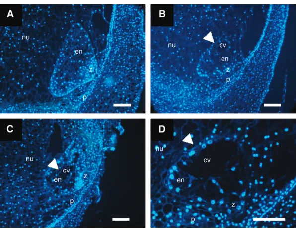
Mutation in bsl1 causes maternal-effect abnormali-ties on seed development: Becausebsl1 causes a ma-ternal effect on seed development, we determined the stage at which developmental abnormalities were first manifest in mutant kernels. For this, we analyzed 1- and 2-dap embryo and syncytial endosperm sections from wild-type plants and plants carrying thebsl1 mutation. However, no abnormalities were detected in embryos or in number of nuclei and their arrangements within syncytial endosperms at these stages (data not shown). In wild-type maize, syncytial endosperms commonly un-dergo coordinated cellularization, initiating from the periphery of the syncytium and progressing centripe-tally until a fully cellular structure is formed (Costaet al.
2003). In this study, fully cellular wild-type endosperms were formed by 3 dap (Figure 3A). By contrast,40% of 3-dap endosperms examined from bsl1/1 plants ex-hibited delayed cellularization, which was indicated by the continued presence of the central vacuole at a time when other sibling endosperms were fully cellular
Figure 3.—Developing 3-dap endosperms segregating from bsl1/1 plants pollinated with wild-type pollen. (A) Wild-type fully cellular endosperm. (B–D) Segregating bsl1mutant en-dosperms showing delayed cellularization—as in-dicated by the prolonged presence of the central vacuole (arrowheads), occurring irregularly, in a noncentripetal fashion. Endosperm sections were stained with DAPI and viewed under fluores-cence microscopy. cv, central vacuole; en, endo-sperm; nu, nucellus; p, pericarp; z, zygote. Bar, 50mm.
TABLE 4
Position of central cell polar nuclei in embryo sacs of wild-type andbsl1/1plants
Plant genotype Central
Apposed to abgerminal wall
Off center, abgerminally positioned
Apposed to adgerminal wall
Wild type 85 0 6 0
(Figure 3, B–D). To determine the ploidy level of these endosperms, we performed a microspectrofluoromet-ric analysis. We found no differences in the nuclear fluorescence intensity (RFU) of these endosperms, indicating that the ploidy levels of sibling wild-type (145.565.3 RFU,n¼98) andbsl1mutant (152.664.5 RFU,n¼127) endosperms were similar.
By 9 dap, bsl1 mutant kernels were macroscopically distinguishable from their wild-type siblings. We there-fore examined the cellular organization of 9-dap sibling mutant and wild-type kernels frombsl1/1plants to de-termine which tissues were most affected by the muta-tion at this stage. Our 9-dap wild-type embryos were at the coleoptile developmental stage (Figure 4A), whereas mutant sibling embryos were retarded and had reached only the transition stage (Figure 4, B and 4C). These embryos typically possessed a morphologi-cally normal suspensor and embryo proper (Figure 4B), although some of the more severe mutants had a smaller than typical embryo proper (Figure 4C).
In mutant endosperms, the most notable and consis-tent defects were observed in the organization of the
BETL. Wild-type endosperms regularly possessed sev-eral layers of elongated and angular BETL cells in the basal region (Figure 4D), whereas we observed patchy distribution of BETL-like cells across fewer cell layers along the basal endosperm of mutant kernels (Figure 4E). Interestingly, bsl1 mutants displayed subtle or no morphological defects in the epidermal aleurone layer or embryo surrounding region (ESR) (data not shown). Abnormalities were, however, noted in the central starchy endosperm (CSE) in more severely per-turbed bsl1/1 kernels, which contained smaller and more irregularly sized cells compared to wild type (Figure 4, F and G).
In summary, our findings show that after fertilization,
bsl1causes retarded growth of embryo and endosperm. Furthermore,bsl1predominantly confers aberrations to the BETL tissue in mutant endosperms.
baseless1alters the spatial distribution of transcripts specific to the basal syncytial and cellular endosperm: To further investigate the effects of thebsl1mutation on early endosperm development, we performed mRNA
expressed in the syncytial endosperm. We examined the localization of two basal-specific transcripts, maizeend1
(seematerials and methods) andMRP1(Gomezet al.
2002), in wild-type syncytia and in syncytia isolated from ears segregating the bsl1 mutant allele. Maize
end1transcript was strongly detected along the entire basal portion of syncytial endosperms from wild-type plants (Figure 5A). By contrast, end1 transcript was localized in the basal region of50% (n¼78) of syn-cytial endosperms examined from plants segregating forbsl1. The remainder exhibited distribution ofend1
transcript either in patches along the basal portion of the syncytium or in the basal medio-lateral margins of the syncytial endosperm (Figure 5, B and C). We found similar results with MRP1, which was weakly expressed in both wild-type and mutant syncytial endosperms exam-ined (data not shown).
In situ hybridization was also performed on 9-dap cellular endosperms to detect a range of transcripts specific to the different domains. Our analysis revealed that several transcripts specific to the aleurone and ESR were correctly localized in wild-type and mutant sibling kernels (data not shown). In stark contrast, we discov-ered that BETL-specific transcripts were not properly localized in mutant endosperms. In wild type,meg1 tran-script was detected throughout the BETL tissue (Figure 5D), whereas inbsl1mutants, this transcript was present only in portions of the abgerminal and adgerminal basal endosperm (Figure 5, E and F). Thus our data indicate that basal endosperm-specific gene expression is se-verely perturbed during free-nuclear and cellular endo-sperm development by mutation inbsl1.
Baseless1 is necessary for the establishment of cor-rect BETL-patterning components: Previous work in maize has shown that interploidy crosses interfere with the normal postfertilization developmental program of the BETL (Charltonet al. 1995; Gutierrez-Marcos
et al.2003). We therefore decided to cross diploid
wild-type and bsl1/1 plants as female by wild-type tetra-ploid plants to investigate whether the maternal defects caused bybsl1could lead to further alterations in BETL development in resulting tetraploid endosperms. By fol-lowing this strategy, we predicted one of three different outcomes: the expected defects in the BETL of tetra-ploid bsl1/bsl1/1/1 endosperms would be similar to those observed in triploidbsl1/bsl1/1 endosperms, be enhanced, or be suppressed. To facilitate this analysis,
bsl1/1plants were initially crossed as females by plants carrying one of two BETL-specific promoter GUS fu-sions, ProMeg1:GUS(Gutierrez-Marcoset al. 2004) or
ProBet1:GUS(Hueroset al.1999a). In wild-type kernels
resulting from crosses between bsl1/1 female hetero-zygotes andProMeg1:GUS-carrying plants, uniform GUS
staining was observed in several layers of the BETL (Figure 6A). As expected from our in situ analysis of basal-specific endosperm transcripts, mutant sibling kernels displayed small patches of GUS precipitate in the lateral margins of the basal endosperm, denoted ‘‘mirror image’’ (Figure 6B). Similar GUS staining pat-terns were observed when using pollen from transge-nic lines carrying the ProBet1:GUS reporter to pollinate
bsl1/1plants (data not shown).bsl1/1,ProMeg1:GUSand 1/1,ProMeg1:GUSdiploid females were then crossed by
tetraploid males and resulting tetraploid endosperms were stained for GUS at different developmental stages. From ears carrying only wild-typeBsl1alleles, all kernels were morphologically similar and GUS staining was often present in discrete clusters along the basal, and in some instances apical, endosperm and denoted ‘‘top– bottom’’ (Figure 6C). By contrast, three distinct classes of seed phenotype were found in ears segregating the
much more unusual and varied GUS staining patterns. We therefore assumed that these kernels were defective for bsl1. The majority of these kernels (25.5 6 4.1%) exhibited small patches of GUS staining in the basal endosperm (mirror image) and, at times, in the apical endosperm (Figure 6E), while others (18.666.5%) dis-played intensely staining puncta scattered throughout the entire endosperm (Figure 6F). Additionally, we noted that the more dramatic GUS staining patterns were observed in the most severely reduced endosperms. Taken together, these findings suggest that not only do the prefertilization defects caused bybsl1greatly perturb BETL development, but also these abnormalities are enhanced in the tetraploid endosperm.
DISCUSSION
bsl1is a novel gametophytic mutant in maize:To gain further understanding of plant reproductive develop-ment, efforts have been made in recent years to identify mutations affecting development and function of the male and female gametophytes (Feldmannet al.1997;
Christensenet al.1998, 2002; Howdenet al.1998; Ebel
et al.2004; Johnsonet al.2004; Pagnussatet al.2005).
For instance, a range of mutations affecting aspects of pollen development, such as the first asymmetric mitotic division, pollen tube growth, or sperm delivery, have been identified thus far (reviewed in McCormick2004).
Our analysis of bsl1 mutants revealed that transmis-sion of the bsl1 mutant allele through the male was reduced to 12%, which would imply that male gameto-phyte development and/or function is perturbed by mu-tation in bsl1. However, no obvious cellular defects in pollen grain morphology or any deficiency in pollen tube growth were detected, suggesting that bsl1 pol-len is not subject to a fundamental developmental or metabolic defect. Instead, we postulate that bsl1 male gametophyte function might be affected either during pollination (e.g., interaction of mutant pollen with silks) or during fertilization (e.g., in delivery and/or function of the sperm cells), where, similarly to maizerop2, mater-nal effect lethal1, andaberrant pollen transmission1mutant pollen (Evansand Kermicle2001; Arthuret al.2003;
Xuand Dooner2006), it is at a competitive
disadvan-tage. By contrast, we noted thatbsl1 caused abnormal development of the female gametophyte. Thus far, an increasing number of female gametophytic mutations affecting most stages of embryo sac development, such as general nuclear division and migration, cellulariza-tion, and/or fusion of the polar nuclei, have been iden-tified (Mooreet al.1997; Christensenet al.1998, 2002;
Figure6.—Effects ofbsl1on BETL-specific transgenic re-porter expression in triploid and tetraploid endosperms. (A and B) 10-dap sibling kernels taken from absl1/1, ProMeg1:
GUS/ProMeg1:GUSplant pollinated by a wild-type diploid plant.
(A) Wild-type triploid endosperm with uniform ProMeg1:GUS
expression (blue) distributed along the basal endosperm. (B) Typical mirror-image GUS expression confined to dis-crete basal areas of a typical bsl1/bsl1/1 endosperm (indi-cated by arrows). (C) 10-dap tetraploid endosperm expressing ProMeg1:GUS in the basal and apical endosperm
(denoted ‘‘top–bottom’’). Tetraploid endosperms were gener-ated from crosses between wild-typeProMeg1:GUS/ProMeg1:GUS
diploid plants and wild-type tetraploid plants. (D–F) Three distinct classes of 10-dap sibling kernels segregating in an ear from absl1/1,ProMeg1:GUS/ProMeg1:GUSplant pollinated
by a wild-type tetraploid plant. (D) Typical kernel of the first class showing top–bottom ProMeg1:GUS expression patterns
similar to those found in tetraploid endosperms shown in C. (E) Abnormal kernel of the second class exhibiting irreg-ular mirror-image GUS staining patterns in the basal endo-sperm. (F) Kernel typical of the third class showing highly scatteredProMeg1:GUSstaining throughout the endosperm
Ebelet al.2004; Pagnussatet al.2005). The majority of
these mutations consequently impair fertilization. Be-cause correct development of the egg apparatus (espe-cially the synergids) is key to successful fertilization (Higashiyama et al.2001; Hucket al. 2003; Rotman
et al.2003; Martonet al.2005), it is likely that the egg
apparatus is unaffected by thebsl1mutation, as fertil-ization ofbsl1embryo sacs is achieved, although subtle defects cannot be ruled out. However, we found notable defects inbsl1central cells, which exhibited abnormal-ities in the position of the polar nuclei, while other cells of the female gametophyte did not show any such phe-notypes. Interestingly, the proportion of fused polar nuclei in embryo sacs from wild-type andbsl1/1plants was similar, as was the fertilization efficiency of wild-type and mutant central cells, together suggesting that most central cell functions are not impaired by thebsl1
mutation. Given that the penetrance of the misplace-ment of polar nuclei in mutant central cells was only 38% (n¼142), it is probable that thebsl1maternal ef-fect on seed development is not a direct consequence of misplacement of the polar nucleiper se. Instead, this result points to underlying developmental abnormali-ties in the bsl1 central cell, which lead to both the maternal effect and misplacement of the polar nuclei. Nonetheless, it is unclear how the sperm are able to locate and fuse with the misplaced polar nuclei inbsl1
embryo sacs as efficiently as in wild type. While a great deal of experimental evidence points to an active role of the female gametophyte in targeting the pollen tube to the synergids for sperm cell release, very little is known about how the sperm nuclei are subsequently targeted to the egg and central cell nuclei to achieve karyogamy after fertilization. One possibility is that this second step is a passive process that depends on the surrounding cytoskeleton to effectively deliver the sperm cells to the appropriate nuclear positions within the embryo sac, a process that in turn is dependent on embryo sac architecture. The displaced cytoplasmic strands inbsl1
central cells support this view, although an alternative explanation of there being an active attraction between the sperm and central cell nuclei cannot be ruled out.
Maternal-effect baseless1 predominantly confers de-velopmental abnormalities to the basal endosperm transfer tissue: The bsl1 mutation caused 50% seed abnormalities when plants carrying the bsl1 defective allele were crossed by wild-type pollen, but not when wild-type plants were crossed bybsl1pollen, indicating that the bsl1 mutation has a maternal effect on seed development rather than a dominant effect. Maternal effects can be caused by mutations in genes expressed either in the surrounding sporophytic tissue (for ex-amples see Felkeret al.1985; Rayet al.1996; Colombo
et al.1997) or in the female gametophyte [examples in-cludecapulet1andcapulet2mutants (Griniet al.2002),
prolifera1(Springeret al.2000; Holdingand Springer
2002), andmaternal effect lethal1(Evansand Kermicle
2001)] and whose gene products are necessary for correct development of embryo and endosperm. The cosegregation of bsl1 alleles with the defective ker-nels demonstrates maternal gametophytic—rather than sporophytic—inheritance.
A distinct class of maternal-effect genes comprises members of thePc-G complex.MEA,FIE, andFIS2are expressed in the central cell before fertilization, where they are required for suppression of autonomous endo-sperm development until fertilization occurs (Ohad
et al.1996; Chaudhuryet al.1997; Grossniklauset al.
1998). During early endosperm development these genes are maternally expressed and paternally silenced. Mutantmea,fie, andfis2endosperms all exhibit retarded growth, while embryos are inviable. Similar tobsl1 mu-tant endosperms, the region thought to be responsible for nutrient transfer is also affected inmea,fie, andfis2
endosperms (Sørensenet al.2001; Guittonet al.2004;
Ingouffet al.2005b). However, the nature of the
aber-rant maternal effects ofbsl1on seed development differs from that reported for defects inPc-G class mutants, in thatbsl1does not induce parthenogenic development of the endosperm. In addition, when bsl1plants were self-fertilized we observed an increase in the proportion of kernels with severe (lethal) developmental abnor-malities, suggesting that paternally contributedBsl1also has a role in kernel development. Because paternally contributed Bsl1 alone was not able to rescue defects caused by maternalBsl1deficiency, it is possible that the paternal contribution either acts to reinforce the mater-nal gametophytic function or performs another role during seed development.
The maternal effect ofbsl1is unique in that the mu-tation specifically causes detrimental effects on BETL development. Because the BETL is the primary site for the transfer of solutes from the mother plant to the seed (Thompsonet al.2001), an impaired BETL would
pro-duce significant changes to nutrient influx, resulting in pleiotropic effects on seed development. This phenom-enon has been observed in other zygotic mutants pos-sessing a defective BETL (Cheng et al. 1996; Maitz
et al.2000; Costaet al.2003) and in maternal-effect
mu-tants with defective connecting maternal sporophytic tissue (Felkeret al.1985). Moreover, an impaired BETL
would certainly account for the slower growth and delayed programmed cell death in bsl1 kernels when compared to wild-type siblings (data not shown). Sim-ilar toreduced grain filling1mutants in maize (Maitzet al.
case for most zygotic defective kernel mutants by using chromosomal translocations to produce nonconcordant seeds with mutant endosperms and wild-type embryos (Neuffer and Sheridan 1980; Chang and Neuffer
1994).
BETL development is under maternal gametophytic control but is also regulated biparentally after fertil-ization: It is currently held that cell fate specification of the maize BETL occurs during early syncytial endo-sperm development (Costa et al.2003). This is most
likely the case in other cereals and thus far a growing number of future transfer cell-specific transcripts that are localized in a polar fashion within barley and wheat syncytial endosperms have been identified (Doanet al.
1996; Dreaet al.2005). Interestingly, we found
irregu-lar distributions of basal-specific endosperm transcripts in syncytia segregating thebsl1mutation, yet we did not detect any morphological abnormalities in these en-dosperms. In addition, irregular localization of BETL-specific transcripts was observed inbsl1mutant cellular endosperms, which also possessed aberrant BETL tis-sue. These findings therefore indicate that BSL1 is neces-sary for correct BETL patterning.
Studies on interploidy crosses in maize have shown that the transfer tissue is particularly sensitive to alter-ations in the 2 m:1 p parental genomic balance in the endosperm (Charltonet al.1995; Hueroset al.1999b;
Gutierrez-Marcos et al. 2003), thus reflecting the
antagonistic influences exerted by maternal and pater-nal genomes following fertilization. When we pollinated
bsl1/1 plants with pollen from tetraploid plants, we found that the introduction of an extra paternal Bsl1
wild-type allele was still not sufficient to compensate for the BETL abnormalities caused by maternal transmis-sion ofbsl1. Although this finding may be indicative of
BSL1 function not being dependent on dosage, the issue remains difficult to resolve since tetraploidyper se
causes patterning defects in the endosperm. Notably, we found more severe defects in the BETL tissue ofbsl1/ bsl1/1/1 tetraploid endosperms than in either bsl1/ bsl1/1 triploid endosperms or 1/1/1/1 tetraploid endosperms. Moreover, these abnormalities ranged in severity, with the frequency of severely defective bsl1/ bsl1/1/1 endosperms (18.6%) roughly correlating with the percentage of severely affected central cells in
bsl1/1plants (i.e., 17.6%, as indicated by displaced polar nuclei). This correlation favors the idea that maternal gametophytic contribution ofBsl1, rather than the num-ber of functionalBsl1alleles in the endosperm, is im-portant for kernel phenotype. Moreover, these data indicate that the prefertilization gametophytic effects of
bsl1lead directly to the formation of a more aberrant BETL. On this basis, it appears that intrinsic informa-tion for BETL patterning is present in the central cell prior to fertilization (see proposed model, Figure 7). Our findings thus contribute significantly to the model currently held for BETL development, which is believed
to occur in an irreversible lineage-dependent manner in response to a combination of developmental cues from the maternal sporophyte and from within the endo-sperm (Costaet al.2003). These data support the
pro-posed model (Birchler1993) that regulation of maize
endosperm development resembles that of the Dro-sophila blastoderm. Early patterning of the maize endo-sperm transfer tissue is under strong maternal control and hence resembles embryonic patterning events in Drosophila, which is achieved before fertilization via the asymmetric localization of maternal determinants within a common cytoplasm ( Johnstone and Lasko
2001; reviewed in Ephrussi and St. Johnston2004).
Certainly, molecular data in Arabidopsis and maize show that there is a significant contribution of mater-nal products to endosperm, as well as embryo (Vielle
-Calzadaet al.2000; Grimanelliet al.2005). In
addi-tion, current genetic data highlight a large number of female gametophytic mutants that exhibit only postfertilization defects (Pagnussat et al. 2005). Our
endosperm (Birchler 1993), in this case to direct
BETL patterning and development.
Analysis ofbsl1central cells and syncytial endosperms further suggests that polarity is perturbed in both struc-tures. We therefore favor a hypothesis in which BSL1 is required for the establishment and/or maintenance of polarity in these reproductive structures. As such, muta-tion inbsl1might affect temporal and/or spatial distribu-tion of BETL factor(s) within the common cytoplasm of the central cell and syncytial endosperm, until endo-sperm cellularization takes place (see proposed model, Figure 7). However, it remains unknown how the orig-inal asymmetry is generated.
In summary, through the study ofbsl1we have found that correct BETL patterning is predetermined in the central cell before fertilization. This information is most likely maintained within the syncytial endosperm, dur-ing which time BETL development becomes finely reg-ulated by both maternally and paternally contributed factors.
We thank Jerry L. Kermicle for advice and support on this project, as well as for seeds of thebsl1mutant. We also thank Hugh G. Dickinson for his support with this work and Richard D. Thompson for seeds carrying theProBet1:GUSreporter construct.
LITERATURE CITED
Arthur, K. M., Z. Vejlupkova, R. B. Meeleyand J. E. Fowler,
2003 Maize ROP2 GTPase provides a competitive advantage to the male gametophyte. Genetics165:2137–2151.
Birchler, J. A., 1993 Dosage analysis of maize endosperm
develop-ment. Annu. Rev. Genet.27:181–204.
Boisnard-Lorig, C., A. Colon-Carmona, M. Bauch, S. Hodge, P.
Doerner et al., 2001 Dynamic analyses of the expression of
the HISTONETYFP fusion protein inArabidopsisshow that syn-cytial endosperm is divided in mitotic domains. Plant Cell13:
495–509.
Brown, R. C., B. E. Lemmonand H. Nguyen, 2003 Events during
the first four rounds of mitosis establish three developmental domains in the syncytial endosperm of Arabidopsis thaliana.
Protoplasma222:167–174.
Chang, M. T., and M. G. Neuffer, 1994 Endosperm-embryo
inter-actions in maize. Maydica39:9–18.
Charlton, W. L., C. L. Keen, C. Merriman, P. Lynch, A. J.
Greenlandet al., 1995 Endosperm development inZea mays:
implication of gametic imprinting and paternal excess in regula-tion of transfer layer development. Development 121: 3089– 3097.
Chaudhury, A. M., L. Ming, C. Miller, S. Craig, E. S. Denniset al.,
1997 Fertilization-independent seed development in Arabidop-sis thaliana.Proc. Natl. Acad. Sci. USA94:4223–4228.
Cheng, W. H., E. W. Taliercioand P. S. Chourey, 1996 The
Min-iature1seed locus of maize encodes a cell wall invertase required for normal development of endosperm and maternal cells in the pedicel. Plant Cell8:971–983.
Christensen, C. A., S. Subramanianand G. N. Drews, 1998
Iden-tification of gametophytic mutations affecting female gameto-phyte development in Arabidopsis. Dev. Biol.202:136–151. Christensen, C. A., S. W. Gorsich, R. H. Brown, L. G. Jones, J.
Brownet al., 2002 Mitochondrial GFA2 is required for synergid
cell death in Arabidopsis. Plant Cell14:2215–2232.
Coleman, A. W., and L. J. Goff, 1985 Mithramycin and 49
,6-diamidino-2-phenylindole (DAPI) as vital stains and for quantitation of nu-clear DNA. Stain Technol.60:145–154.
Colombo, L., J. Franken, A. Van derKrol, P. E. Wittich, H. J. M.
Donset al., 1997 Down-regulation of ovule-specific MADS box
genes from Petunia results in maternally controlled defects in seed development. Plant Cell9:703–715.
Costa, L. M., J. F. Gutierrez-Marcos, T. P. Brutnell, A. J.
Greenlandand H. G. Dickinson, 2003 The globby1–1 (glo1–1)
mutation disrupts nuclear and cell division in the developing maize seed causing alterations in endosperm cell fate and tissue differentiation. Development130:5009–5017.
Costa, L. M., J. F. Gutierrez-Marcos and H. G. Dickinson,
2004 More than a yolk: the short life and complex times of the plant endosperm. Trends Plant Sci.9:507–514.
Doan, D. N., C. Linnestadand O. A. Olsen, 1996 Isolation of
molecular markers from the barley endosperm coenocyte and the surrounding nucellus cell layers. Plant Mol. Biol.31:877– 886.
Drea, S., D. J. Leader, B. C. Arnold, P. Shaw, L. Dolan et al.,
2005 Systematic spatial analysis of gene expression during wheat caryopsis development. Plant Cell17:2172–2185.
Drews, G. N., and R. Yadegari, 2002 Development and function
of the angiosperm female gametophyte. Annu. Rev. Genet.36:
99–124.
Ebel, C., L. Mariconti and W. Gruissem, 2004 Plant
retino-blastoma homologues control nuclear proliferation in the fe-male gametophyte. Nature429:776–780.
Ephrussi, A., and D. St. Johnston, 2004 Seeing is
believ-ing: the bicoid morphogen gradient matures. Cell 116: 143– 152.
Evans, M. M. S., and J. L. Kermicle, 2001 Interaction between
maternal effect and zygotic effect mutations during maize seed development. Genetics159:303–315.
Feldmann, K. A., D. A. Couryand M. L. Christianson, 1997
Ex-ceptional segregation of a selectable marker (KanR) in Arabi-dopsis identifies genes important for gametophytic growth and development. Genetics147:1411–1422.
Felker, F. C., D. M. Petersonand O. M. Nelson, 1985 Anatomy of
immature grains of eight maternal effect shrunken endosperm barley mutants. Am. J. Bot.72:248–256.
Gomez, E., J. Royo, Y. Guo, R. Thompsonand G. Hueros, 2002
Es-tablishment of cereal endosperm expression domains: identifica-tion and properties of a maize transfer cell-specific transcripidentifica-tion factor, ZmMRP-1. Plant Cell14:599–610.
Grimanelli, D., E. Perotti, J. Ramirezand O. Leblanc, 2005
Tim-ing of the maternal-to-zygotic transition durTim-ing early seed devel-opment in maize. Plant Cell17:1061–1072.
Grini, P. E., G. Jurgens and M. Hulskamp, 2002 Embryo and
endosperm development is disrupted in the female gameto-phytic capulet mutants of Arabidopsis. Genetics 162: 1911– 1925.
Grossniklaus, U., J. P. Vielle-Calzada, M. A. Hoeppner and
W. B. Gagliano, 1998 Maternal control of embryogenesis by
MEDEA, a polycomb group gene in Arabidopsis. Science280:
446–450.
Guitton, A. E., D. R. Page, P. Chambrier, C. Lionnet, J. E. Faure
et al., 2004 Identification of new members of Fertilisation Inde-pendent Seed Polycomb Group pathway involved in the control of seed development in Arabidopsis thaliana. Development131:
2971–2981.
Gutierrez-Marcos, J. F., P. D. Pennington, L. M. Costaand H. G.
Dickinson, 2003 Imprinting in the endosperm: a possible role
in preventing wide hybridization. Philos. Trans. R. Soc. Lond. B Biol. Sci.358:1105–1111.
Gutierrez-Marcos, J. F., L. M. Costa, C. Biderre-Petit, B. Khbaya,
D. M. O’Sullivanet al., 2004 Maternally expressed gene1 is a
novel maize endosperm transfer cell-specific gene with a mater-nal parent-of-origin pattern of expression. Plant Cell16:1288– 1301.
Higashiyama, T., S. Yabe, N. Sasaki, Y. Nishimura, S. Miyagishima
et al., 2001 Pollen tube attraction by the synergid cell. Science
293:1480–1483.
Holding, D. R., and P. S. Springer, 2002 TheArabidopsis gene
PROLIFERAis required for proper cytokinesis during seed devel-opment. Planta214:373–382.
Howden, R., S. K. Park, J. M. Moore, J. Orme, U. Grossniklaus
Huck, N., J. M. Moore, M. Federerand U. Grossniklaus, 2003 The
Arabidopsis mutant feronia disrupts the female gametophytic control of pollen tube reception. Development 130: 2149– 2159.
Hueros, G., E. Gomez, N. Cheikh, J. Edwards, M. Weldonet al.,
1999a Identification of a promoter sequence from the BETL1 gene cluster able to confer transfer-cell-specific expression in transgenic maize. Plant Physiol.121:1143–1152.
Hueros, G., J. Royo, M. Maitz, F. Salaminiand R. D. Thompson,
1999b Evidence for factors regulating transfer cell-specific ex-pression in maize endosperm. Plant Mol. Biol.41:403–414. Ingouff, M., J. N. Fitz Gerald, C. Guerin, H. Robert, M. B.
Sorensenet al., 2005a Plant formin AtFH5 is an evolutionarily
conserved actin nucleator involved in cytokinesis. Nat. Cell Biol.
7:374–380.
Ingouff, M., J. Haseloffand F. Berger, 2005b Polycomb group
genes control developmental timing of endosperm. Plant J.42:
663–674.
Johnson, M. A., K.vonBesser, Q. Zhou, E. Smith, G. Auxet al.,
2004 Arabidopsis hapless mutations define essential gameto-phytic functions. Genetics168:971–982.
Johnstone, O., and P. Lasko, 2001 Translational regulation and
RNA localization in Drosophila oocytes and embryos. Annu. Rev. Genet.35:365–406.
Kermicle, J. L., 1978 Imprinting of gene action in maize
endo-sperm, pp. 357–371 inMaize Breeding and Genetics, edited by D. B. Walden. John Wiley & Sons, New York.
Kindiger, B., 1994 A staining procedure for pollen grain
chromo-somes of maize, pp. 476–480 inThe Maize Handbook, edited by M. Freelingand V. Walbot. Springer-Verlag, New York/Berlin/
Heidelberg, Germany.
Maitz, M., G. Santandrea, Z. Zhang, S. Lal, L. C. Hannahet al.,
2000 rgf1, a mutation reducing grain filling in maize through effects on basal endosperm and pedicel development. Plant J.
23:29–42.
Marton, M. L., S. Cordts, J. Broadhvest and T. Dresselhaus,
2005 Micropylar pollen tube guidance by egg apparatus 1 of maize. Science307:573–576.
McCormick, S., 2004 Control of male gametophyte development.
Plant Cell16(Suppl.): S142–S153.
Moore, J. M., J. P. Calzada, W. Gaglianoand U. Grossniklaus,
1997 Genetic characterization of hadad, a mutant disrupting female gametogenesis in Arabidopsis thaliana. Cold Spring Harbor Symp. Quant. Biol.62:35–47.
Neuffer, M. G., and W. F. Sheridan, 1980 Defective kernel mutants
of maize. I. Genetic and lethality studies. Genetics95:929–944. Ohad, N., L. Margossian, Y. C. Hsu, C. Williams, P. Repettiet al.,
1996 A mutation that allows endosperm development without fertilization. Proc. Natl. Acad. Sci. USA93:5319–5324. Olsen, O. A., 2001 Endosperm development: cellularization and
cell fate specification. Annu. Rev. Plant Physiol. Plant Mol. Biol.
52:233–267.
Olsen, O. A., 2004 Nuclear endosperm development in cereals and
Arabidopsis thaliana. Plant Cell16(Suppl.): S214–S227. Pagnussat, G. C., H. J. Yu, Q. A. Ngo, S. Rajani, S. Mayalaguet al.,
2005 Genetic and molecular identification of genes required for female gametophyte development and function in Arabidop-sis. Development132:603–614.
Ray, S., T. Goldenand A. Ray, 1996 Maternal effects of the short
integument mutation on embryo development in Arabidopsis. Dev. Biol.180:365–369.
Rotman, N., F. Rozier, L. Boavida, C. Dumas, F. Berger et al.,
2003 Female control of male gamete delivery during fertiliza-tion in Arabidopsis thaliana. Curr. Biol.13:432–436.
Sørensen, M. B., A. M. Chaudhury, H. Robert, E. Banchareland
F. Berger, 2001 Polycomb group genes control pattern
forma-tion in plant seed. Curr. Biol.11:277–281.
Springer, P. S., D. R. Holding, A. Groover, C. Yordanand R. A.
Martienssen, 2000 The essential Mcm7 protein PROLIFERA
is localized to the nucleus of dividing cells during the G(1) phase and is required maternally for early Arabidopsis development. Development127:1815–1822.
Thompson, R. D., G. Hueros, H. Beckerand M. Maitz, 2001
De-velopment and functions of seed transfer cells. Plant Sci.160:
775–783.
Vielle-Calzada, J. P., R. Baskarand U. Grossniklaus, 2000 Delayed
activation of the paternal genome during seed development. Nature404:91–94.
Walbot, V., and M. M. Evans, 2003 Unique features of the plant life
cycle and their consequences. Nat. Rev. Genet.4:369–379. Xu, Z., and H. K. Dooner, 2006 The maize aberrant pollen
trans-mission 1 gene is a SABRE/KIP homolog required for pollen tube growth. Genetics172:1251–1261.
Yadegari, R., and G. N. Drews, 2004 Female gametophyte
develop-ment. Plant Cell16(Suppl.): S133–S141.