The
bantam
Gene Regulates Drosophila Growth
David R. Hipfner, Katrin Weigmann and Stephen M. Cohen
1European Molecular Biology Laboratory, 69117 Heidelberg, Germany Manuscript received February 12, 2002
Accepted for publication May 9, 2002
ABSTRACT
We report here the consequences of mutations of a novel locus, named bantam, whose product is involved in the regulation of growth in Drosophila.bantam mutant animals are smaller than wild type, due to a reduction in cell number but not cell size, and do not have significant disruptions in patterning. Conversely, overexpression of thebantamproduct using the EP elementEP(3)3622causes overgrowth of wing and eye tissue. Overexpression in clones of cells results in an increased rate of cell proliferation and a matched increase in cellular growth rate, such that the resulting tissue is composed of more cells of a size comparable to wild type. These effects are strikingly similar to those associated with alterations in the activity of the cyclinD-cdk4 complex. However, epistasis and genetic interaction analyses indicate that
bantamand cyclinD-cdk4 operate independently. Thus, thebantamlocus represents a novel regulator of
tissue growth.
M
OST animals grow to a characteristic and repro- andLehner1996). Experimental manipulation of celldivision rates without corresponding changes in cell ducible size. Although the final size of the
com-ponent parts of an animal can be greatly influenced by growth rates can have significant effects on cell size
(Johnstonet al. 1977;Weigmannet al. 1997;Neufeld
environmental factors such as nutrition, organ growth
rates and size are also controlled by mechanisms intrin- et al. 1998). In Drosophila discs, accelerating the cell cycle by genetic means results in normal-sized discs with
sic to the developing organs themselves (Bryant and
more and smaller cells. Conversely, slowing the cell cycle
Simpson1984). The size of a given animal or organ is
produces normal-sized discs with fewer and larger cells determined in large part by the number and size of its
(Weigmannet al. 1997;Neufeldet al. 1998). The fact constituent cells. Consequently, the processes of cell
that final tissue size was unchanged in both cases, within division, cell death, and cell growth must be carefully
limits, indicates that tissue growth is likely not regulated regulated during development to ensure the correct
at the level of cell cycle controlper se.
and proportionate size of the adult animal (Conlon
In contrast, a number of Drosophila genes have been
andRaff 1999).
identified that affect tissue growth directly. These genes The intrinsic and environmental mechanisms
con-all have in common the ability to regulate cellular trolling growth have been the focus of considerable
growth rates. For example, genes encoding components recent attention. Studies of the growth and
develop-of the insulin/phosphatidylinositol-3-kinase (PI3K) sig-ment of Drosophila imaginal discs have begun to
ad-naling pathway have been found to be instrumental in dress the relative importance of cell growth and cell
determining organ and body size. Overactivation of the
division in determining organ and body size (Edgar
pathway in imaginal discs causes tissue overgrowth by
1999;Lehner1999;Oldhamet al. 2000a;Stockerand
increasing the rate of cell growth. In the absence of
Hafen 2000). Imaginal discs are the larval structures
a sufficient corresponding increase in the rate of cell from which all adult epidermal structures of the fly are
division, this causes cells to divide at a larger than nor-derived. These epithelial sacs arise as small clusters of
mal size (Goberdhan et al. 1999; Verdu et al. 1999;
20–50 cells during embryogenesis (Cohen 1993). In
Weinkoveet al. 1999;Gaoet al. 2000). Conversely, de-the span ofⵑ3 days during the larval instars, disc cells
creased pathway activity reduces tissue growth by pro-proliferate rapidly and increase in numberⵑ1000-fold.
ducing smaller cells (Bo¨ hni et al. 1999; Goberdhan
As in higher organisms, imaginal disc cell divisions are
et al. 1999; Montagne et al. 1999; Verdu et al. 1999; regulated at G1-S and G2-M transitions. In addition,
Weinkove et al. 1999; Gao et al. 2000), and in some disc cell divisions are thought to be growth dependent,
cases also by reducing cell number (Bo¨ hniet al. 1999; meaning that the cells normally do not divide until they
Weinkove et al. 1999). The tumor-suppressor genes have grown to a certain critical size or mass (Edgar
TSC1andTSC2restrict tissue growth by regulating cell
growth rates via the insulin/PI3K pathway (Gao and
Pan 2001; Potter et al. 2001; Tapon et al. 2001). In
1Corresponding author:Developmental Biology Program, European
addition to components of the insulin/PI3K pathway, Molecular Biology Laboratory, Meyerhofstrasse 1, 69117 Heidelberg,
Germany. E-mail: cohen@embl-heidelberg.de Drosophila homologs ofras, myc, and TORhave been
(Denefet al. 2000); UAS-GFPNLS (Neufeld et al. 1998);
HS-shown to promote cell growth (Johnston et al. 1999;
FLP1(StruhlandBasler1993);Actin5C⬎CD2⬎GAL4( Pig-Oldhamet al. 2000b;ProberandEdgar2000;Zhang
noni and Zipursky1997); UAS-cycD,UAS-cdk4(Datar et al.
et al. 2000). 2000); andcdk43(Meyeret al. 2000).
In all of the foregoing examples the effects on tissue Mapping and characterization ofP-element insertions in the growth rates were mediated primarily by regulating cell bantamlocus:Insertion sites ofEP(3)3622,EPg(3)30491, and
EPg(3)35007 were determined by plasmid rescue according
growth rates. Consequently, the normal balance
be-to standard procedures. Flanking sequences for banL1170,
tween the rate of cell growth and the rate of cell cycle
EP(3)3208, and EP(3)3219were available from the Berkeley
progression was lost. Cells grew too fast and divided at
Fly Database. ThesePelements are inserted in chromosome
abnormally large sizes. In contrast, the complex com- 3L at cytological position 61C7-8. They are clustered within
posed of Drosophila cyclin D (cycD) and cyclin-depen- 12.3 kb of one another in an interval of 42 kb containing
dent kinase (cdk) 4 controls tissue growth in a manner no known or predicted genes. EP(3)3622 contains two EP
elements inserted in a back-to-back orientation at position
that keeps the rates of cell growth and cell cycle
progres-12,052 of genomic contig AE003469, with one basal promoter
sion in balance (Dataret al. 2000;Meyeret al. 2000).
oriented proximally and one distally. The otherPelements,
Tissue overgrowth due to overactivation of cycD-cdk4
with site of insertion in nucleotides relative toEP(3)3622(⫹,
results from an increase in the number of normal-sized further distal;⫺, further proximal), areEPg(3)35007(⫺874); cells. Mutation of cdk4 reduced tissue size by reducing banL1170(⫺173);EPg(3)30491(⫺12);EP(3)3208(⫹2040); and
EP(3)3219(⫹11,430). ThebanL1170,EP(3)3208, andEP(3)3219
cell number rather than cell size. Thus cycD-cdk4
ap-chromosomes are homozygous lethal, but in each case the
pears to control the rate of growth by coordinated
regu-lethality can be attributed to another locus on the
chromo-lation of cell growth rates and cell cycle progression
some. banL1170, EP(3)3208, and EP(3)3219 are each viable in
rates. With the exception of ras, both these classes of transto theban⌬1deletion. The revertantbanL1170R1
chromo-growth genes (those affecting primarily cell chromo-growthvs. some, generated by excision of thebanL1170Pelement, lost the
those affecting cell growth and division rates) appear banmutant phenotype but remained homozygous lethal.
Generation and molecular characterization of theban⌬1
al-to be primarily involved in growth regulation, as they
lele:To generate mutants for thebanlocus,P-element
exci-have minimal effects upon tissue patterning.
sions of EP(3)3622 were generated. Excisions identified by
To identify additional genes involved in regulating
the loss of the EP-elementmini-w⫹transgene were tested for
imaginal disc growth, we performed a gain-of-function complementation of the deletionDf(3L)Ar11, which removes
genetic screen using the EP method developed by from 61C3-4 to 61E. Excisions failing to complement this
Rorth(1996). When combined with a source of GAL4, deficiency were analyzed by Southern blotting using genomic fragments derived from an EP(3)3622 plasmid rescue
con-the EP element will direct expression of genomic
se-struct. One excision causing early pupal lethality,ban⌬1, was
quences adjacent to its site of insertion. Previous studies
found to delete sequences both proximal and distal to the
have shown that a high proportion of EP elements direct
original EP insertion site. The ends of theban⌬1deletion were
GAL4-dependent overexpression of endogenous genes mapped by genomic PCR on DNA from homozygous mutant
(Rorthet al. 1998). We restricted our analysis to genes third instar larvae with primer pairs spaced at 5- to 10-kb
intervals along the chromosome. Once approximate limits of
involved in growth by screening for EP elements that
the breakpoint were identified, a 2.9-kb PCR product spanning
showed GAL4-dependent effects on tissue size without
the junction was amplified from the same genomic DNA and
disrupting pattern. Here, we report the identification
sequenced.
of a locus that we callbantam (ban), which influences Analysis of adult phenotypes:All crosses for size compari-tissue growth rates. We present evidence thatbantamis son were conducted under identical, uncrowded conditions. involved in coordinately regulating cell growth and cell Crosses withenGAL4for Figure 3, C and D, were carried out at
29⬚. All other crosses were at 25⬚. For theban
complementa-division to regulate the rate of normal tissue growth.
tion analysis, males heterozygous for theP-element insertion being tested and theban⌬1allele were crossed toban⌬1/TM2
females. In this way, each vial contained progeny of the tested MATERIALS AND METHODS
genotype (e.g.,P-element/ban⌬1) andban⌬1/TM2siblings. This
allowed all measurements to be normalized relative toban⌬1/
Fly strains:The EP collection of 2300 lines (Rorthet al.
TM2sibling flies from within the same vial to eliminate the 1998), as well as a new collection of 8500 independent strains
variability in adult body size resulting from differences in carrying insertions of a modified EP element, termed EPg
culture conditions between vials. Relative body mass was
deter-(Mataet al. 2000), were screened. Thesevenless(sev),optomotor
mined by weighing two or three sets of 20 male flies of each
blind, MS1096, and engrailed (en) GAL4 drivers were used
genotype from within a vial and taking the average. Final (Basleret al. 1989;CapdevilaandGuerrero1994;Fietzet
values in Table 1 are based on the average of at least two al. 1995; Lecuit et al. 1996). EP lines were also screened
independent vials. Wing areas were measured using National for modifiers of the effects of overexpression of the tumor
Institutes of Health Image 1.59. To assess female fertility, virgin suppressor gene expanded (genotype: sevGAL4,UAS-expanded,
females (typically 40) of the appropriate genotype were svpAE127,ro1/⫹; ⫹ EP; Boedigheimer and Laughon 1993;
crossed individually to wild-type males, and the number of BlaumuellerandMlodzik2000).banL1170is aP-element
in-viable adult offspring in each vial was countedⵑ20 days later. sertion allele, originally called l(3)L1170, and was obtained
Scanning electron microscopy was performed as described from the Bloomington Drosophila Stock Center. The
armadil-(BlaumuellerandMlodzik2000).
loLacZ, FRT80Bstock was provided by Jessica Treisman. Other
Imaginal disc growth analyses:For all larval analyses, larvae
mutant and transgenic strains are described in the
Embryos were collected for 3 hr. Twenty-four hours later, 65 flies heterozygous for ban⌬1 and the P element were
newly hatched larvae of each genotype were transferred to always normalized to siblings heterozygous forban⌬1and
fresh vials containing yeast paste. Discs from stagedenGAL4
,UAS-theTM2balancer chromosome reared in the same vial.
EGFP/⫹larvae [with or withoutEP(3)3622] were dissected at
EP(3)3622, EP(3)3208, EP(3)3219, EPg(3)30491, and
112⫾1.5 hr after egg laying (AEL) and fixed in 4%
formalde-hyde. Discs were stained with 4⬘,6-diamidino-2-phenylindole EPg(3)35007were each viable in combination withban⌬1.
(DAPI) to visualize the nuclei and analyzed by confocal micros- In addition tol(3)L1170 (which we renamedbanL1170),
copy. The posterior compartment and total disc areas were EP(3)3622, EP(3)3208, EP(3)3219, and EPg(3)30491 measured from the green fluorescent protein (GFP) and blue
caused a reduction in body size whenin transtoban⌬1. In
(DAPI) channels, respectively, using the histogram function
contrast,EPg(3)35007had no effect (Table 1). Although
of Adobe Photoshop.
Flow cytometry:Flip-out clones were induced at 72⫾1.5 smaller, the ban mutant flies were normally
propor-hr AEL in staged larvae of the genotypeHS-FLP1/Act5C ⬎ tioned and the majority did not show significant
pat-CD2 ⬎ GAL4;UAS-GFP [with or without EP(3)3622 or UAS- terning defects, suggesting that the product of theban
Dp110] by heat shock at 38⬚ for 1 hr. A total of 10–15 discs
locus is primarily involved in regulating growth of all
of each genotype were dissected in PBS at 112⫾1.5 hr AEL,
adult structures.
dissociated using trypsin, and stained with Hoechst 33342 as
described (Neufeldet al. 1998). GFP content, cell cycle pro- Another characteristic affected by thebanmutations
files, and forward scatter values were analyzed using a Cytoma- was female fertility. All allelic combinations withban⌬1
tion MoFlo flow cytometer. Experiments were repeated three that decreased adult size also caused a marked decrease times with similar results.
in the average number of viable offspring produced
Measurement of proliferation rates: Flip-out clones were
by mutant females, whereasEPg(3)35007did not (not
induced at 72⫾1.5 hr AEL in staged larvae of the genotype
HS-FLP1/Act5C⬎ CD2⬎GAL4;UAS-GFPNLS [with or without shown). For example, almost allbanL1170/ban⌬1 females
EP(3)3622] by heat shock at 37⬚for 15 min. Discs were dis- were sterile (2.5% fertile), in contrast to the 95% fertility
sected at 112⫾1.5 hr AEL and fixed in 4% formaldehyde. rate of wild-type flies. As with the size reduction pheno-GFP-positive cells within each clone were counted by
epifluor-type associated withbanL1170, this effect on fertility could
escence microscopy. Cell doubling times were calculated using
be completely reverted by precise excision of theP
ele-the formula (log 2/logN)h, whereNis the average number
of cells/clone andh is the hours between heat shock and ment, indicating that both phenotypes are due to the
dissection (Neufeldet al. 1998). The experiment was repeated banL1170insertion.
twice with nearly identical results. We also analyzed flies homozygous for the viableban
allelesEP(3)3622 and EPg(3)30491. In both cases, the homozygous flies showed the same reduced size and
RESULTS
fertility defects as EP/ban⌬1
trans-heterozygotes.
Al-though the growth ofEP(3)3622andEPg(3)30491
homo-bantam mutants produce small flies with fertility
de-fects:In an overexpression screen for genes that affect zygotes was affected to nearly the same extent as in the correspondingEP/ban⌬1flies (Table 1), the fertility of
tissue size, we identified several P-element insertions
mapping within a 12.3-kb interval at cytological location homozygous females of both genotypes was less severely
reduced [50% of females sterile for EPg(3)30491/
61C7-8 (Figure 1).P-element-mediated excision ofEP(3)
3622was used to produce a small deletion (Figure 1). EPg(3)30491 vs. 67.5% sterile for EPg(3)30491/ban⌬1;
35% sterile forEP(3)3622/EP(3)3622 vs. 77.5% sterile The deletion is homozygous lethal at early pupal stages.
Mutant larvae lack detectable imaginal discs. Flies het- for EP(3)3622/ban⌬1; n ⫽ 40 in each case]. Neither
EP(3)3622norEPg(3)30491caused as great a reduction erozygous for the deletion and an independently
iso-latedP-element insertion,l(3)L1170, are viable and nor- in adult size as ban⌬1 when in trans to the other ban
P-element insertions (not shown). We conclude that
mally patterned but are 15% smaller than sibling flies
heterozygous for the deletion alone (Figure 2A, Table both insertions are likely hypomorphic banalleles.
Overexpression ofbantamcauses overgrowth:Several 1). The size reduction phenotype could be completely
reverted by precise excision of thel(3)L1170 Pelement. of thebanEP-element insertions were identified in an
overexpression screen. Four EP insertions produced Flies heterozygous for the deletion and a revertant of
l(3)L1170(calledL1170 R1) are comparable in size to noticeable overexpression phenotypes [EP(3)3622, EP(3) 3208,EPg(3)30491, andEPg(3)35007]. The more distally siblings heterozygous for the deletion alone (Table 1),
indicating that theP-element insertion disrupts a gene insertedEP(3)3219 element did not. When expressed
under the control ofenGAL4,EP(3)3622increased the size
required for normal growth of the fly. We therefore
named the locusbantam, to indicate that the mutants of the posterior compartment in wing imaginal discs
(Figure 3A). Measurement of the relative areas of the are smaller than normal.
We examined in more detail the adult phenotypes posterior and anterior compartments of discs from
en-GAL4/⫹;EP(3)3622/⫹larvae showed that a statistically
sig-resulting from decreasedbanfunction by testing other
Pelements mapping to the deleted region for comple- nificant increase of posterior to anterior area (P:A) ratio
(P ⬍ 0.001; Figure 3B). To examine this phenotype
mentation of theban⌬1excision allele. To eliminate the
variability in adult body size resulting from differences in more detail we measured the effects of EP(3)3622,
Figure 1.—Map of the
bantamlocus (61C7-8). (A)
Map showing the positions of genes surrounding the
EP(3)3622 insertion site.
Orientation and annotation numbers are indicated for several upstream and down-stream genes. The region deleted in the ban⌬1 allele
is indicated below the map. (B) Magnification of the re-gion deleted in the ban⌬1
allele, showing the relative locations ofPelements in-serted in this interval. The orientation of the basal pro-moter is indicated by an arrow for each EP element.
EPg(3)30491andEP(3)3622
each contain twoPelements inserted at the same posi-tion, oriented in opposite directions. EP elements producing a GAL4-dependent overexpression phenotype are indicated in italics.Pelements producing a mutant phenotypein transtoban⌬1are boxed.
the adult wing. Total wing area and the ratio of P:A bantam mutant wings have fewer, but normal-sized,
cells: A number of mutants affecting growth of adult compartment areas were measured. To exclude effects
due to expression ofenGAL4in the region between veins flies have been identified. Viable mutants of Drosophila
mycand certain components of the insulin/PI3K
signal-3 and 4 (Blair 1992), we used the area bounded by
veins 1 and 3 as an estimate of anterior area and the ing pathway have been shown to produce small,
nor-mally patterned adult flies. The decreased size of these area bounded by vein 4 and the posterior margin as an
estimate of posterior area (illustrated in Figure 3D). animals is due in large part to a reduction in the size
of cells in the adult (Chen et al. 1996; Bo¨ hni et al.
enGAL4-driven expression of
EPg(3)35007, EP(3)3208,
EPg(3)30491, and EP(3)3622caused statistically signifi- 1999;Johnstonet al. 1999;Montagneet al. 1999). Flies lacking the product of thecdk4gene are also reduced cant increases in the ratio of P:A areas compared to
enGAL4/⫹ wings (P ⬍0.001; Figure 3C). EP(3)3622had in size. In this case, however, the size deficit is the result
of a decrease in the number of adult cells, rather than the strongest effect and was the only EP line to cause
a statistically significant increase in the overall size of the of effects on cell size (Meyeret al. 2000). We analyzed
wings frombanmutant flies to determine whether the
wing (8%; P ⬍ 0.001; Figure 3C). Overgrowth of the
posterior compartment occurred at the expense of growth deficit in these animals is due to decreased final
adult cell size and/or cell number. Total wing areas the anterior compartment for the other EP lines, since
there was no increase in overall wing size. This was also were determined, and cell size was measured by number
of wing hairs per unit area (each cell in the wing blade the case for EP(3)3622, because the magnitude of the
increase in wing size was less than the relative increase produces a single hair). Measurements for each allele
in transtoban⌬1were normalized toban⌬1/TM2siblings.
in size of the posterior compartment. Only minor
pat-terning abnormalities were observed in these wings (Fig- Wings frombanmutant flies were 9–13% smaller than
ban⌬1/
TM2 siblings (Figure 2B, Table 1). For banL1170,
ure 3D), suggesting that this EP element directs
expres-sion of a factor primarily involved in size regulation. EP(3)3622, andEPg(3)30491overban⌬1, the decrease in
wing size was not due to a decrease in cell size. The The effects ofEP(3)3622overexpression are not
lim-ited to the wing. Expression ofEP(3)3622in cells behind number of hairs per unit area was not significantly differ-ent in these mutantsvs.the corresponding control wings
the morphogenetic furrow using the gmrGAL4 driver
caused bulging of the eye (Figure 3E), suggesting exten- (Table 1). Instead, the decreased wing size was entirely attributable to a reduction in the number of cells in sive overgrowth. The eyes were also externally rough.
EP(3)3622overexpression with appropriate drivers also mutant wings by up to 12%.
The EP(3)3219insertion behaved differently, as the caused duplication or triplication of interommatidial
bristles in the eye and of macrochaete in the notum reduced wing size inEP(3)3219/ban⌬1was due primarily
to a reduction in cell size rather than cell number. Interest-(data not shown). Similar effects on notum
macro-chaete have been described (Abdelilah-Seyfriedet al. ingly,EP(3)3219 had little or no effect on growth when combined with otherP-element alleles [EP(3)3219/banL1170,
2000). These phenotypes are consistent with
expressing clones were consistently slightly smaller than GFP-negative control cells (0.98; Figure 4B), whereas
EP(3)3622-expressing, GFP-positive cells were consis-tently slightly larger (1.01; Figure 4C). ThusEP(3)3622 -expressing cells were 3% larger than control GFP-expressing cells. Although reproducible, it is unclear whether this small difference is meaningful. For com-parison, cells expressing the Dp110 catalytic subunit of PI3K, a known positive regulator of cellular growth rates, were 28% larger than control GFP-expressing cells, con-sistent with previous reports (Weinkoveet al. 1999; Fig-ure 4D). These results indicate thatEP(3)3622 expres-sion has relatively little effect on cell size during the period of wing imaginal disc growth, consistent with the analysis ofbanmutants.
These observations suggest that the mode of action ofbanis distinct from Dp110 and other positive regula-tors of the insulin signaling pathway (Weinkove et al.
1999). Although EP(3)3622 and Dp110 cause tissue
overgrowth, they appear to have different effects at the cellular level. Activation of insulin signaling causes tissue growth by increasing the rate of cell growth more than the rate of cell division so that the overgrown tissue
contains larger cells. EP(3)3622 causes tissue
over-growth, but does not cause a comparable net increase in cell size. This suggested thatEP(3)3622-induced tissue overgrowth is coupled with an increase in the rate of cell division such that the overgrown tissue contains
Figure2.—bantammutations reduce body and wing growth. more cells, as has been demonstrated for the cycD/cdk4
(A) Comparison showing the reduced body size ofbanL1170/
complex (Dataret al. 2000). To test this we measured
ban⌬1male and female flies compared toban⌬1/TM2siblings.
the cell-doubling rate by counting the average number
(B) Comparison of wing sizes frombanL1170/ban⌬1andban⌬1/
of cells in clones allowed to grow for a defined time.
TM2sibling flies. In the overlay thebanL1170/ban⌬1wing is shown
Control GFP-expressing clones orEP(3)3622-expressing
in red and theban⌬1/TM2wing in green.
clones coexpressing GFP were induced in early third instar (72 hr AEL) and allowed to grow for 40 hr. The 187 control clones counted contained an average of 6.2
30491, 102%;EP(3)3219/EP(3)3622, 97%; averages from
two independent vials]. These observations suggest that cells (median⫽ 5), corresponding to a doubling time
of 15.2 hr. In 213 clones expressingEP(3)3622, the
aver-EP(3)3219affects a genetically separable locus and that
it is not an allele ofban. age cell number was 7.0 (median⫽7), corresponding to
a doubling time of 14.2 hr. This difference is statistically
The cellular effects ofbantamoverexpression are
dis-tinct from the insulin signaling pathway: To examine significant (P⬍0.005), and almost identical results were obtained in a second independent experiment. To-how overexpression ofbantamcauses tissue overgrowth,
we examined cell size and cell cycle profile in clones of gether, these results indicate that EP(3)3622 -overex-pressing cells grow and divide more rapidly than control
EP(3)3622-expressing cells.EP(3)3622-expressing clones
marked by coexpression of GFP or control GFP-express- cells. Consistent with this observation,EP(3)3622causes
a large increase in the number of cells in regions of the ing clones were induced at the end of second instar
and allowed to grow until late third instar. Coexpression adult wing in which it has been expressed (not shown). These observations suggest thatbancoordinately
regu-of GFP was used to identify and sort the EP(3)3622
-expressing cells, which were directly compared for cell lates the rates of cell growth and cell division.
bantam does not interact genetically with cyclinD/
cycle phasing and cell sizes with GFP-negative wild-type
control cells from the same disc. There was no apparent cdk4: The growth and fertility phenotypes associated
with gain and loss ofbanfunction are similar to those difference in the distribution of EP(3)3622-expressing
and wild-type cells in the G1, S, and G2 phases of the associated with alterations in the activity of the
cycD-cdk4 complex (Datar et al. 2000; Meyeret al. 2000). cell cycle (Figure 4A). Cell size was compared using
forward scatter values (Neufeld et al. 1998). We ob- Growth-impaired, viable mutants of bothbanandcdk4
are composed of a smaller number of wild-type-sized served a subtle difference between the EP-expressing
TABLE 1 Phenotypic analysis of bantam heteroallelic mutants Viability Body mass Wing-blade area % control C ell area % control Intervein % control Genotype (%) ( n ) a (% control) b (10 6 m 2) c wing area ( m 2) d cell area cell number e cell number ban ⌬
1/⫹
Figure3.—EPs inserted in thebantamlocus cause GAL4-dependent tissue overgrowth in the wing and eye. (A) Imaginal discs from staged larvae expressing EGFP with (right) or with-out (left) coexpression ofEP(3)3622under the control of the enGAL4driver were dissected at 112 hr AEL and fixed. Confocal
images show discs with bright EGFP fluorescence marking the posterior compartment and less intense DAPI staining marking the entire disc (anterior is to the left). (B) Ratios of P:A compartment areas fromenGAL4/⫹control andenGAL4/
⫹;EP(3)3622/⫹discs (n⫽ 26 for each genotype; *,
signifi-Figure 4.—EP(3)3622 expression has little effect on disc cantly different from control;P⬍0.001). (C) Quantitation
cell size. Clones of cells expressing GFP alone or with either of the ratio of P:A compartment sizes and total wing area. EPs
EP(3)3622 or wild-type Dp110 were induced at 72 hr AEL.
were expressed in the posterior compartment of the wing
Discs were dissected at 112 hr AEL and cells were dissociated using theenGAL4driver. Anterior and posterior areas were
mea-and analyzed by flow cytometry. In A–D, data for GFP mea-and EP/ sured from eight female wings of each genotype. Shaded bars:
transgene-expressing clonal cells and nonexpressing control graph of average P:A wing-area ratios for flies expressing
cells from the same discs are in green and red, respectively.
EPg(3)35007,EP(3)3208,EPg(3)30491, orEP(3)3622in the
pos-(A) Distribution of cells in G1 (2C DNA content), S (2C–4C), terior compartment. Solid bars: graph of average total areas
and G2 (4C) phases of the cell cycle forEP(3)3622-expressing of the same set of wings. *, significantly different fromenG4/⫹
and control cells. (B–D) Forward scatter analysis of cells from controls (P ⬍ 0.001). (D) Comparison of enGAL4/⫹ control
discs containing (B) control clones, (C)EP(3)3622-expressing
andenGAL4/⫹;EP(3)3622/⫹wings. Anterior and posterior areas
clones, or (D) Dp110-expressing clones. Numbers represent measured to calculate the P:A area ratios are outlined on the
the ratio of forward scatter values for GFP⫹/GFP⫺cells. Simi-control wing. (E) Scanning electron microscopy images of
lar results were obtained in two additional independent exper-heads from gmrGAL4/⫹ control and gmrGAL4/⫹;EP(3)3622/⫹
iments. flies.EP(3)3622expression in the eye causes roughening and
bulging, suggesting extensive overgrowth.
the cell cycle is apparently uniform, as no alterations in cell cycle phasing are detected. In postmitotic cells The characteristics of tissue growth driven by cycD-cdk4
in the eye, cycD-cdk4 coexpression led to cellular hyper-andbanare indistinguishable. In proliferating epithelial
trophy. The resulting bulging, overgrown eyes are very cells of the wing imaginal disc, both increase rates of
similar in appearance to eyes in whichban overexpres-cell cycle progression and growth in a coordinated
Figure 3E with Figure 3G fromDataret al.2000). These similarities raised the possibility thatban might act to-gether with cycD-cdk4 to promote tissue growth. Conse-quently, we tested for genetic interactions between these growth regulators.
Importantly, both cycD and cdk4 are required to pro-mote tissue growth. Expression of either alone is not sufficient (Dataret al. 2000). Therefore, to determine
whether ban-driven overgrowth was dependent upon
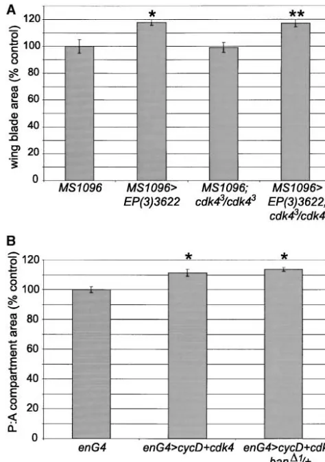
the activity of the cycD-cdk4 complex, we tested whether it could be blocked by removal of one of the components of this complex. We made use of theMS1096GAL4driver,
which directs GAL4 expression in the dorsal compart-ment of the wing disc early in larval developcompart-ment and more broadly throughout the developing wing pouch later (Mila´net al. 1998). Expression ofEP(3)3622with
MS1096GAL4resulted in significant overgrowth of the
en-tire wing. The effect was greater in the dorsal compart-ment, such that the wings curved downwards (not
shown). MS1096GAL4/⫹;EP(3)3622/⫹ wings were 18%
greater in area than controlMS1096GAL4/⫹wings (Figure
5A). Overexpression ofbanin acdk4null mutant back-ground (cdk43/cdk43) had no effect on the overgrowth
phenotype. Wings of MS1096GAL4/⫹;cdk43/cdk43;EP(3)
3622/⫹flies were 18% larger than those ofMS1096GAL4/
⫹;cdk43/cdk43 flies (Figure 5A) and were also curved
downward (not shown). As loss ofcdk4had no apparent effect onban-driven growth, we conclude thatbandoes not promote growth by regulating the activity of the
Figure5.—EP(3)3622and cyclinD-cdk4 act independently
cycD-cdk4 complex.
to drive tissue growth. (A) Wing blade areas were measured
One of the main functions of cycD-cdk4 is believed from control flies and flies expressingEP(3)3622under the to be suppression of the function of “pocket” proteins, control of theMS1096GAL4driver in a wild-type orcdk43mutant
background. Seven female wings of each genotype were
mea-such as pRb. However, genetic analyses in Drosophila
sured. Genotypes: MS1096GAL4/⫹; MS1096GAL4/⫹;EP(3)3622/⫹;
suggested that the effects of cycD-cdk4 in promoting
MS1096GAL4/⫹;cdk43/cdk43; and MS1096GAL4/⫹;cdk43/cdk43;EP(3)
cellular growth are mediated at least in part by unknown
3622/⫹. *, significantly different from MS1096GAL4/⫹ (P ⬍
downstream targets, independent of the fly pRb homo- 0.001). **, significantly different from MS1096GAL4/⫹;cdk43/
log RBF (Datar et al. 2000). We tested whether ban cdk43(P⬍ 0.001). (B) CycD and cdk4 were coexpressed in
the posterior compartment of the wing using theenGAL4driver
might be such a downstream target. Overexpression of
in a wild-type orban⌬1/⫹background. Anterior and posterior
cycD-cdk4 with theenGAL4driver resulted in overgrowth
areas were measured from six female wings of each genotype
of the posterior compartment of the wing, significantly
and expressed as P:A ratios. Genotypes:enGAL4/⫹;enGAL4/⫹
;UAS-increasing the P:A ratio by 11% (Figure 5B). This over- cycD,UAS-cdk4/⫹; andenGAL4/⫹;UAS-cycD,UAS-cdk4/ban⌬1. *,
sig-growth was unaffected by halving the gene dosage of nificantly different fromenGAL4/⫹(P⬍0.001).
ban(Figure 5B).enGAL4
-driven cycD-cdk4 expression was also able to promote posterior compartment overgrowth
to a comparable extent when thebangene dosage was expandedcaused a reduction in eye size relative to wild
type and external roughening and blistering (Figure 6, further reduced in the banL1170
/ban⌬1 allelic
combina-tion, although survival of these flies was poor (not A and B;BlaumuellerandMlodzik2000).
Coexpres-sion of EP(3)3622 almost completely suppressed this
shown). These observations suggest that the
growth-promoting effects of cycD-cdk4 are not dependent upon phenotype, restoring the eye to nearly wild-type size and
appearance (Figure 6C). Reducing ban function had
banlevels.
To further evaluate the relationship between cycD- the opposite effect. Introducing one copy of theban⌬1
allele noticeably reduced the overall eye size and
in-cdk4 and ban, we turned to an independent genetic
assay forbanactivity. Two of thebanEP insertions were creased the blistering in the central and anterior regions of the eye (Figure 6D). In contrast, alterations in cycD-identified initially in a genetic interaction screen as
sup-pressors of the phenotype caused by overexpression of cdk4 activity did not alter the expandedoverexpression
phenotype. Coexpression of cycD-cdk4 with expanded
the expanded tumor suppressor gene. When
Figure6.—bantam, but not cycD-cdk4, inter-acts genetically with the expanded tumor sup-pressor. Scanning electron microscopy images of eyes from female flies raised at 25⬚. Geno-types: (A) sevGAL4/⫹. (B) sevGAL4,UAS-expanded,
svpAE127,ro1/⫹. (C)sevGAL4,UAS-expanded,svpAE127,ro1/
EP(3)3622. (D) sevGAL4,UAS-expanded,svpAE127,ro1/
ban⌬1. (E) sevGAL4,UAS-expanded,svpAE127,ro1
/UAS-cycD, UAScdk4. (F) cdk43/⫹;sevGAL4,UAS-expanded,
svpAE127,ro1/⫹.
previous observations (Dataret al. 2000), but had little that were first identified by in situ hybridization and Northern blot analysis of overexpression RNA with effect on the roughness and blistering (Figure 6E).
Re-moving one copy ofcdk4had no effect (Figure 6F). The flanking genomic sequences as probes, or by RT-PCR
using RNA from overexpressing larvae and an EP-ele-lack of a strong genetic interaction between expanded
and cycD-cdk4 provides additional evidence thatbantam ment specific primer (Rorth1996). However, neither
of these transcripts is able to reproduce the overgrowth is acting independently of this complex to promote
coordinated cell growth and cell cycle progression. phenotype when expressed from a transgene (not
shown). A more complete understanding of the
mecha-Molecular characterization of thebantam locus:The
ban⌬1 deletion removes 21,147 nucleotides, extending nism ofbanaction will await molecular characterization
of the gene product. from 5792 nucleotides proximal to 15,355 nucleotides
distal to theEP(3)3622insertion site, and fails to
com-plement the deficiencyDf(3L)Ar11 that removes from
DISCUSSION
61C3-4 to 61E. Theban⌬1deletion does not extend into
the coding sequences of either of the two identified bantamis required for normal tissue growth:bangene
neighboring genes, CG3200 (Reg-2) or the predicted function appears to be important for regulation of tissue
CG12030 (Figure 1). It is possible that noncoding or growth rates. Several EP elements inserted in this locus,
alternate exons of these genes might be located close most notablyEP(3)3622, are capable of promoting
sub-to the EP elements. However, expression levels of stantial tissue overgrowth in the eye and wing in a
GAL4-CG12030,Reg-2, andCG13893(see Figure 1) were com- dependent manner. Conversely,banmutations decrease parable inban⌬1mutant and wild-type third instar larvae
tissue growth. Mutant phenotypes range from decreased as assessed by Northern blot analysis (not shown). This body size to lethality. The strongest available allele is a
suggests that the mutant phenotypes associated with small deletion that does not remove any known genes.
ban⌬1 are not due to effects on expression of any of This allele is pupal lethal and causes the absence of
these nearby genes. Similarly, we did not detect upregu- detectable imaginal discs. The simplest explanation for
lation ofCG12030, Reg-2, or CG13893 or of the more the reciprocal nature of gain-of-function and
loss-distant genesCG17181,CG12189, orCG12015byin situ of-function phenotypes is thatEP(3)3622is driving
ex-hybridization analysis of wing discs in whichEP(3)3622 pression of the same transcription unit that is affected
was expressed. bybanmutations. This is further supported by the
spe-It is possible that thebanlocus may correspond to a cific and reciprocal nature of the genetic interaction of
gene contained at least in part within theⵑ41-kb inter- gain and loss of banfunction with theexpandedtumor
val betweenCG12030andReg-2, which was not predicted suppressor gene in the eye. However, this remains to be
in the CELERA/BDGP annotation. Although this in- confirmed by molecular characterization of the locus.
tergenic region contains numerous short open reading Growth regulation appears to be a primary function of
frames, we have not found any meaningful homologies ban, asEP(3)3622expression does not cause significant
by BLAST sequence analysis. We have cloned two over- patterning alterations, and ban mutant flies, although
bantamcoordinates cell growth and division rates to partment size, but can increase or decrease the number
regulate tissue growth:Our results suggest thatbanregu- of cells per compartment (Weigmannet al. 1997;
Neu-lates tissue growth by a mechanism that involves coordi- feldet al. 1998). This is consistent with the effects of
nated stimulation of cell growth and cell division.ban Minute mutations that vary the proportion of a
compart-alters tissue growth through effects on cell number ment that can be contributed by the progeny of a single
rather than cell size. Decreased banfunction causes a cell, without affecting compartment size or shape (
Mor-reduction in cell number in the adult wing, but the ataandRipoll1975). However, as first shown by
Leev-surviving cells are of wild-type size, suggesting a coordi- ers et al. (1996), it is possible to alter the size of one
nated decrease in the rate of cell growth and division. compartment relative to another by manipulating
activ-Activation ofEP(3)3622has the opposite effect on cell ity of the insulin/PI3K pathway (Leeverset al. 1996).
number, causing an increase in the rate at which imagi- PI3K-induced overgrowth requires that the pathway be
nal disc cells proliferate. Despite this increased prolifer- activated in all cells of the compartment. Clones of ation rate, cell sizes are little changed. These observa- overgrowing cells do not affect the size of the
compart-tions suggest that the rate of increase in cell division is ment (TelemanandCohen2000). Thus a mechanism
coordinated with the rate of increase of cell mass when must exist that allows a population of cells to measure
banis overexpressed. The effects ofbanon growth and the size of the compartment. Interestingly, it has been
fertility are remarkably similar to those of cycD-cdk4. found that altering the size of the compartment feeds
However, we have found no evidence of a direct connec- back by an unknown mechanism to alter the shape of
tion between cycD-cdk4 andban. It seems unlikely that the Dpp morphogen gradient (Teleman and Cohen
banregulates growth by controlling the activity of cycD- 2000).
cdk4, because ban-driven overgrowth is unaffected in Overexpression of ban with enGAL4 promoted
signifi-the absence of cdk4. Similarly, cycD-cdk4-driven growth cant overgrowth of the posterior compartment. We
is unaffected by reduction ofban, indicating thatbanis noted that posterior compartment overgrowth was
com-unlikely to be a downstream effector. We favor the view pensated for by a nonautonomous reduction in the final
thatbanand cycD-cdk4 act independently. The similar- size of the anterior compartment in most cases. This
ity in their growth phenotypes suggests that they may compensation suggests that total disc size may also be
have some targets in common. However, as attested to regulated to some extent during development. Only in
by the differences in their interactions with expanded, the case of the strongest EP element,EP(3)3622, were
they clearly can act differently as well. total disc and wing size increased.
Growth and pattern formation: The imaginal discs These observations suggest that there may be multiple
are patterned while they grow. The secreted signaling layers of size control operating during imaginal disc
proteins Decapentaplegic (Dpp) and Wingless pattern development. Morphogen gradients influence tissue
the wing and leg discs along their main axes. Dpp and growth. Tissue growth rates influence compartment size
Wingless signaling are also required in some way for and morphogen gradient shape. Finally, size
compensa-disc growth. The parts of the compensa-discs that produce the
tion mechanisms exist to control both compartment appendages are very small in flies lacking either signal
and disc size. At present, little is known about the size-(Spenceret al. 1982;Diaz-Benjumeaet al. 1994;Zecca
sensing mechanisms, except that we can override them
et al. 1995; Neumannand Cohen1996). Cells unable
by stimulating cell and tissue growth rates by various to transduce the Dpp or Wingless signals display
cell-experimental means. Identifying how size is measured autonomous defects in proliferation and are lost from
during tissue growth poses a significant challenge. the disc (Peifer et al. 1991;BurkeandBasler1996).
We thank Ann Atzberger for flow cytometric analyses, Ann Mari To date it has not been reported whether loss of cells
Voie for preparing transgenic fly strains, and Christine Blaumueller under these conditions is due to reduced proliferation
for fly stocks and advice; Enrique Martin-Blanco for sharing informa-or to reduced survival. However, recent studies suggest tion on theEP(3)3622insertions prior to publication; and Pernille that Dpp signaling may directly influence cell prolifera- Rørth and members of the lab for helpful discussions. K.W. and D.R.H. tion in the wing disc (Martin-CastellanosandEdgar were fellows of the European Molecular Biology Organization. D.R.H. was a fellow of the Human Frontiers Science Program Organization. 2002). Wingless signaling has been shown in one
situa-tion to repress growth at late stages of wing develop-ment, in part by negative regulation ofdmycexpression
(JohnstonandEdgar1998;Johnstonet al. 1999). If LITERATURE CITED
Dpp and Wingless act directly to regulate tissue growth,
Abdelilah-Seyfried, S., Y. M. Chan, C. Zeng, N. J. Justice, S. we would expect them to coordinately regulate cell Younger-Shepherdet al., 2000 A gain-of-function screen for growth and cell division rates. It will be of interest to genes that affect the development of the Drosophila adult
exter-nal sensory organ. Genetics155:733–752.
learn whether ban and/or cycD-cdk4 mediate the
Basler, K., P. SiegristandE. Hafen, 1989 The spatial and temporal growth effects of these signaling molecules.
expression pattern ofsevenlessis exclusively controlled by
gene-Compartments and imaginal discs as units of size internal elements. EMBO J.8:2381–2386.
Blair, S., 1992 engrailedexpression in the anterior lineage
com-ment of the developing wing blade of Drosophila. Developcom-ment coordinates mitosis and morphogenesis in Drosophila by
regulat-115:21–33. ing string/CDC25 proteolysis. Cell101:511–522.
Blaumueller, C. M., andM. Mlodzik, 2000 The Drosophila tumor Meyer, C. A., H. W. Jacobs, S. A. Datar, W. Du, B. A. Edgaret al., suppressorexpandedregulates growth, apoptosis, and patterning 2000 Drosophila cdk4 is required for normal growth and is during development. Mech. Dev.92:251–262. dispensable for cell cycle progression. EMBO J.19:4533–4542. Boedigheimer, M., andA. Laughon, 1993 Expanded: a gene in- Mila´n, M., F. Diaz-BenjumeaandS. M. Cohen, 1998 Beadexencodes volved in the control of cell proliferation in imaginal discs. Devel- an LMO protein that regulates Apterous LIM-homeodomain
ac-opment118:1291–1301. tivity in Drosophila wing development: a model for LMO
onco-Bo¨ hni, R., J. Riesgo-Escovar, S. Oldham, W. Brogiolo, H. Stocker gene function. Genes Dev.12:2912–2920.
et al., 1999 Autonomous control of cell and organ size by Montagne, J., M. J. Stewart, H. Stocker, E. Hafen, S. C. Kozma
CHICO, a Drosophila homolog of vertebrate IRS1-4. Cell 97: et al., 1999 Drosophila S6 kinase: a regulator of cell size. Science
865–875. 285:2126–2129.
Bryant, P. J., andP. Simpson, 1984 Intrinsic and extrinsic control Morata, G., andP. Ripoll, 1975 Minutes: mutants of Drosophila of growth in developing organs. Q. Rev. Biol.59:387–415. autonomously affecting cell division rate. Dev. Biol.42:211–221. Burke, R., andK. Basler, 1996 Dpp receptors are autonomously Neufeld, T. P., A. F. de la Cruz, L. A. JohnstonandB. A. Edgar, required for cell proliferation in the entire developing Drosoph- 1998 Coordination of growth and cell division in the Drosophila
ila wing. Development122:2261–2269. wing. Cell93:1183–1193.
Capdevila, J., andI. Guerrero, 1994 Targeted expression of the Neumann, C. J., andS. M. Cohen, 1996 Distinct mitogenic and cell signalling molecule decapentaplegic induces pattern duplica- fate specification functions ofwinglessin different regions of the tions and growth alterations in Drosophila wings. EMBO J.13: wing. Development122:1781–1789.
4459–4468. Oldham, S., R. Bohni, H. Stocker, W. BrogioloandE. Hafen,
Chen, C., J. JackandR. S. Garofalo, 1996 The Drosophila insulin 2000a Genetic control of size in Drosophila. Philos. Trans. R. receptor is required for normal growth. Endocrinology137:846– Soc. Lond. B Biol. Sci.355:945–952.
856. Oldham, S., J. Montagne, T. Radimerski, G. ThomasandE. Hafen,
Cohen, S. M., 1993 Imaginal disc development, pp. 747–841 in 2000b Genetic and biochemical characterization of dTOR, the
Drosophila Development, edited by A.Martinez-Ariasand M.Bate. Drosophila homolog of the target of rapamycin. Genes Dev.14:
Cold Spring Harbor Laboratory Press, Cold Spring Harbor, NY. 2689–2694.
Conlon, I., andM. Raff, 1999 Size control in animal development. Peifer, M., C. Rauskolb, M. Williams, B. Riggleman and E.
Cell96:235–244. Wieschaus, 1991 The segment polarity genearmadillointeracts
Datar, S. A., H. W. Jacobs, A. F. de La Cruz, C. F. LehnerandB. A. with thewinglesssignaling pathway in both embryonic and adult Edgar, 2000 The Drosophila cyclin D-cdk4 complex promotes pattern formation. Development111:1029–1043.
cellular growth. EMBO J.19:4543–4554. Pignoni, F., andS. L. Zipursky, 1997 Induction of Drosophila eye Denef, N., D. Neubueser, L. PerezandS. M. Cohen, 2000 Hedge- development bydecapentaplegic.Development124:271–278.
hog induces opposite changes in turnover and subcellular local- Potter, C. J., H. HuangandT. Xu, 2001 Drosophila Tsc1 functions ization of Patched and Smoothened. Cell102:521–531. with Tsc2 to antagonize insulin signaling in regulating cell Diaz-Benjumea, F. J., B. CohenandS. M. Cohen, 1994 Cell interac- growth, cell proliferation, and organ size. Cell105:357–368.
tions between compartments establishes the proximal-distal axis Prober, D. A., and B.A. Edgar, 2000 Ras1 promotes cellular growth of Drosophila limbs. Nature372:175–179. in the Drosophila wing. Cell100:435–446.
Edgar, B. A., 1999 From small flies come big discoveries about size Rorth, P., 1996 A modular misexpression screen in Drosophila control. Nat. Cell Biol.1:E191–193.
detecting tissue specific phenotypes. Proc. Natl. Acad. Sci. USA Edgar, B. A., andC. F. Lehner, 1996 Developmental control of 93:12418–12422.
cell cycle regulators: a fly’s perspective. Science274:1646–1652.
Rorth, P., K. Szabo, A. Bailey, T. Laverty, J. Rehmet al., 1998 Fietz, M. J., A. Jacinto, A. M. Taylor, C. AlexandreandP. W.
Systematic gain-of-function genetics in Drosophila. Development Ingham, 1995 Secretion of the amino-terminal fragment of
125:1049–1057. Hedgehog protein is necessary and sufficient forhedgehog
signal-Spencer, F., M. Hoffman and W. M. Gelbart, 1982 Decapen-ling in Drosophila. Curr. Biol.5:643–650.
taplegic: a gene complex affecting morphogenesis inDrosophila
Gao, X., andD. Pan, 2001 TSC1andTSC2tumor suppressors
antago-melanogaster.Cell28:451–461.
nize insulin signaling in cell growth. Genes Dev.15:1383–1392.
Stocker, H., andE. Hafen, 2000 Genetic control of cell size. Curr. Gao, X., T. P. NeufeldandD. Pan, 2000 DrosophilaPTENregulates
Opin. Genet. Dev.10:529–535. cell growth and proliferation through PI3K-dependent and
-inde-Struhl, G., and K. Basler, 1993 Organizing activity ofwingless
pendent pathways. Dev. Biol.221:404–418.
protein in Drosophila. Cell72:527–540. Goberdhan, D. C., N. Paricio, E. C. Goodman, M. MlodzikandC.
Tapon, N., N. Ito, B. J. Dickson, J. E. TreismanandI. K. Hariharan, Wilson, 1999 Drosophila tumor suppressorPTENcontrols cell
2001 The Drosophila tuberous sclerosis complex gene homo-size and number by antagonizing the Chico/PI3-kinase signaling
logs restrict cell growth and cell proliferation. Cell105:345–355. pathway. Genes Dev.13:3244–3258.
Teleman, A. A., andS. M. Cohen, 2000 Dpp gradient formation in Johnston, G. C., J. R. PringleandL. H. Hartwell, 1977
Coordina-the Drosophila wing imaginal disc. Cell103:971–980. tion of growth with cell division in the yeastSaccharomyces cerevisiae.
Verdu, J., M. A. Buratovich, E. L. WilderandM. J. Birnbaum, Exp. Cell Res.105:79–98.
1999 Cell-autonomous regulation of cell and organ growth in Johnston, L. A., andB. A. Edgar, 1998 Wingless and Notch regulate
Drosophila by Akt/PKB. Nat. Cell Biol.1:500–506. cell-cycle arrest in the developing Drosophila wing. Nature394:
Weigmann, K., S. M. CohenandC. Lehner, 1997 Cell cycle progres-82–84.
sion, growth and patterning in imaginal discs despite inhibition Johnston, L. A., D. A. Prober, B. A. Edgar, R. N. EisenmanandP.
of cell division after inactivation of Drosophila Cdc2 kinase. Devel-Gallant, 1999 Drosophila myc regulates cellular growth
dur-opment124:3555–3563. ing development. Cell98:779–790.
Lecuit, T., W. J. Brook, M. Ng, M. Calleja, H. Sunet al., 1996 Two Weinkove, D., T. P. Neufeld, T. Twardzik, M. D. Waterfieldand distinct mechanisms for long-range patterning by Decapen- S. J. Leevers, 1999 Regulation of imaginal disc cell size, cell taplegic in the Drosophila wing. Nature381:387–393. number and organ size by Drosophila class I(A) phosphoinositide Leevers, S. J., D. Weinkove, L. K. MacDougall, E. HafenandM. D. 3-kinase and its adaptor. Curr. Biol.9:1019–1029.
Waterfield, 1996 The Drosophila phosphoinositide 3-kinase Zecca, M., K. BaslerandG. Struhl, 1995 Sequential organizing Dp110 promotes cell growth. EMBO J.15:6584–6594. activities ofengrailed, hedgehoganddecapentaplegicin the Drosoph-Lehner, C. F., 1999 The beauty of small flies. Nat. Cell Biol.1: ila wing. Development121:2265–2278.
E129–130. Zhang, H., J. P. Stallock, J. C. Ng, C. ReinhardandT. P. Neufeld,
Martin-Castellanos, C., andB. A. Edgar, 2002 A characterization 2000 Regulation of cellular growth by the Drosophila target of of the effects of Dpp signaling on cell growth and proliferation rapamycin dTOR. Genes Dev.14:2712–2724.
in the Drosophila wing. Development129:1003–1013.