BOOKCHAPTER
Title Deficits in Voice-Identity Processing: Acquired and Developmental
Phonagnosia
Author names Claudia Roswandowitza,b* Corrina Maguinnessa*
Katharina von Kriegsteina,c,d
Author affiliations a Max Planck Institute for Human Cognitive and Brain Sciences, Stephanstraße 1a, 04103 Leipzig, Germany bInternational Max Planck Research School on
Neuroscience of Communication, Stephanstraße 1a, 04103 Leipzig, Germany
c
Humboldt University zu Berlin, Rudower Chaussee 18, 12489 Berlin, Germany
d
Technische Universität Dresden, Faculty of Psychology, Bamberger Str. 7, 01187 Dresden
*both authors contributed equally to this work
Correspondence should be addressed to Claudia Roswandowitz ([email protected]) and Katharina von Kriegstein ([email protected])
This is a draft of a chapter that has been accepted for publication by Oxford University Press in the forthcoming book The Oxford Handbook of Voice Perception edited by Sascha Frühholz and Pascal Belin due for publication in November 2018.
1
Abstract
The voice contains elementary social communication cues, conveying speech, as well as paralinguistic information pertaining to the emotional state and the identity of the speaker. In contrast to vocal-speech and vocal-emotion processing, voice-identity processing has been less explored. This seems surprising, given the day-to-day significance of person recognition by voice. A valuable approach to unravel how voice-identity processing is accomplished is to investigate people who have a selective deficit in recognising voices. Such a deficit has been termed phonagnosia. In the present chapter, we provide a systematic overview of studies on phonagnosia and how they relate to current neurocognitive models of person recognition. We review studies that have characterised people who suffer from phonagnosia following brain damage (i.e. acquired phonagnosia) and also studies, which have examined phonagnosia cases without apparent brain lesion (i.e. developmental phonagnosia). Based on the reviewed literature, we emphasise the need for a careful behavioural characterisation of phonagnosia cases by taking into consideration the multistage nature of voice-identity processing and the resulting behavioural phonagnosia subtypes.
Keywords: phonagnosia, acquired, developmental, apperceptive, associative, voice-identity processing, speaker recognition, core-voice system, extended system
Introduction
Recognising a person by voice is a skill, which humans master with ease. However, for some people this skill can be impaired. This deficit is termed ‘phonagnosia’ (Van Lancker and Canter, 1982); originating from the Greek words ‘φώνημα’ or ‘phone’ meaning voice or sound and the term agnosia (αγνώσις). Agnosia is commonly used for conditions in which the recognition of stimuli is disturbed (Lissauer, 1890; Freud, 1891). In phonagnosia, the ability to process other vocal information e.g., gender, age, and emotion as well as speech, music, and facial information is largely preserved (Neuner and Schweinberger, 2000; Garrido et al., 2009; Roswandowitz et al., 2014). Phonagnosia can occur after brain damage (i.e. acquired phonagnosia) (Assal
et al., 1976; Van Lancker and Canter, 1982; Neuner and Schweinberger, 2000) or in the absence of brain insult (i.e. developmental phonagnosia) (Garrido et al., 2009; Roswandowitz et al., 2014). The disorder has currently two major sub-classifications: apperceptive and associative phonagnosia. In apperceptive phonagnosia, the deficit lies in the perceptual analysis of voice features, whereas the association of semantic information to a voice is intact (Hailstone et al., 2011; Roswandowitz et al., 2014; Xu et al., 2015). While associative phonagnosia is understood as a failure to recognise a voice as familiar (familiarity decision) and to associate semantic information to a voice (semantic processing), though the perception of the voice is unaffected (Hailstone et al., 2010; Hailstone et al., 2011; Roswandowitz et al., 2014).
Though phonagnosia may offer a unique instance to study auditory person recognition, the number of scientific investigations so far has been limited. This might be caused by the following factors: (i) Phonagnosia has been under scientific investigation for a rather short time. The first study on acquired phonagnosia was published in 1976 (Assal et al., 1976) and on developmental phonagnosia in 2009 (Garrido et al., 2009). (ii) Testing of voice-recognition deficits is relatively difficult, as standard tests are not readily available and are often language- dependent (but see Aglieri et al., 2017). (iii) Cases of phonagnosia are rare, although this perceived rarity may be more related to a low self-awareness, rather than a low prevalence rate, of voice-identity processing disorders (Roswandowitz et al., 2014).
In the following chapter, we provide a systematic overview of investigations on phonagnosia and how they relate to current models of voice-identity processing. We begin by introducing a neurocognitive model of voice-identity processing and provide an overview of the
behavioural tests, which are used to assess cases and subtypes of phonagnosia. We then review clinical studies, which documented cases of acquired phonagnosia, before turning to focus on recently reported cases of developmental phonagnosia. We discuss the reviewed findings within the context of current voice-identity processing models and conclude with proposing future research directions.
Model of voice-identity processing
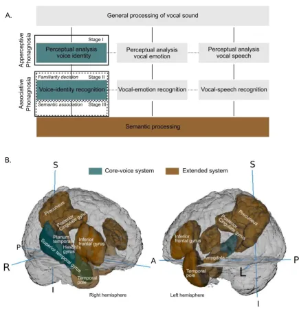
Recognising voices at the individual level is a challenge for the perceptual and cognitive system. Each voice that we hear shares the same basic perceptual features across individuals (acoustic parameters such as pitch and timbre (Lavner et al., 2001; López et al., 2013)); and thus the brain is tasked with representing a unique voice in memory, by perceiving and representing often subtle differences in these features across individuals (Belin et al., 2011). Furthermore, it is not sufficient that we simply recognise a voice as familiar. Rather, successful voice recognition also involves linking the familiar voice to stored knowledge, or semantics, including where we know the voice from, what the person looks like, are they a friend or a foe? Thus, voice recognition can be conceived as a multistage process, which begins with the encoding of the incoming vocal signal and ends in successful identification of the voice at the level of a specific individual identity. In Figure 1A we present a cognitive model of voice-identity processing and highlight candidate brain regions in Figure 1B, which may support this multistage process. We also outline how subtypes of phonagnosia, apperceptive and associative, may arise due to dysfunction at different
stages of voice-identity processing.
Figure 1. Neurocognitive model of voice processing. A. A model (adapted from Ellis et al. (1997), Belin et al. (2004), Blank et al. (2014), and Neuner and Schweinberger (2000), based on a seminal model of face processing outlined by Bruce & Young, (1986)) which describes the cognitive processes involved in voice-identity processing. B. Overview of potential brain structures supporting voice-voice-identity processing, as evidenced in neuroimaging studies with neurotypical participants. R= Right; L = Left; S = Superior; I = Inferior; A = Anterior; P = Posterior.
According to the model (Figure 1A), the vocal sound undergoes an initial general processing phase. This processing may be partly shared and partly independent from the processing of other sound sources, including object sounds or music. After this initial phase, voice-identity processing begins. Stage I: Here, the perceptual system analyses complex spectrotemporal acoustical properties of the incoming vocal sound, which support identity processing. This stage encompasses ‘structural encoding’ (see e.g. Neuner and Schweinberger, 2000), where invariant properties of the voice (vocal properties which remain constant across different speech utterances or changes in prosody) are extracted. These properties are merged to create a coherent voice percept. The merged voice properties may be contrasted against a ‘prototype’ voice (Lavner et al., 2001; Andics et al., 2010; see prototype encoding of voices; Latinus et al., 2013; for review see Maguinness et al., 2018). The prototype voice may represent an average approximation of the voices the listener has encountered or it may reflect a “very common voice” (Lavner et al., 2001). The computed acoustical differences between the voice percept and the prototype voice can be passed on for analysis to support identity- recognition at later stages of processing. Other features of the vocal sound, which support vocal emotion and speech processing are also analysed at this stage but are argued to be processed in partly independent but interacting systems (von Kriegstein et al., 2010; Kreitewolf et al., 2014). The stage I of processing is suggested to be supported by brain regions of a core-voice system. Potential candidate brain regions are the posterior and mid regions of superior temporal gyrus/sulcus (STG/S) (e.g., Belin et al., 2000; von Kriegstein and Giraud, 2004; Warren et al., 2006; Pernet et al., 2015; Roswandowitz et al., 2018) and auditory regions such as the planum temporale (von Kriegstein et al., 2006b; Warren et al., 2006) and Heschl’s gyrus (Formisano et al., 2008; Bonte et al., 2014), predominantly in the right hemisphere (Figure 1B). Apperceptive phonagnosia may emerge due to dysfunction at this early stage of processing (Figure 1A, Stage I). Poor perceptual analysis of the voice may result in a weak representation of the voice- individuating features, which may impact negatively on later stages of processing (voice-identity recognition).
Stage II: At the stage of voice-identity recognition, a sense of familiarity is generated if the computed voice percept closely resembles a stored voice representation. These voice representations may be stored as relatively unique ‘reference patterns’ for each known voice- identity (see Lavner at al. 2001). This process is likely supported by anterior and mid regions of
STG/S including parts of the anterior temporal lobe (most likely superior lateral part, see e.g. Belin and Zatorre, 2003; von Kriegstein et al., 2003) in the core-voice system, while more posterior regions are concerned with perceptual voice analysis (Belin and Zatorre, 2003; von Kriegstein et al., 2003; Andics et al., 2013; Latinus et al., 2013; Schall et al., 2015). A deficit at this stage would give rise to deficient familiarity decisions despite a successfully analysed vocal percept. We will call this familiarity-associative phonagnosia (Figure 1A, Stage II). Disrupted access to the stored voice-identity representations constrains the ability to judge whether the voice has been encountered before.
Stage III: After the voice has been recognised as familiar it is linked to stored multi- modal semantic information characterising the person identity. This multi-modal information is processed in an extended system (semantic processing), which is proposed to share connections with the core-voice system. Regions concerned with vocal emotion and speech recognition may also share connections with this extended system. Potential brain candidates for the extended system include supra-modal regions encompassing discrete regions of the temporal pole, precuneus/posterior cingulate, amygdala, and inferior frontal gyrus (Shah et al., 2001; von Kriegstein and Giraud, 2006; Andics et al., 2010; Latinus et al., 2011; for review see Blank et al., 2014). Dysfunction at this stage of processing, i.e. poor connectivity between the core-voice and extended system (Figure 1A, Stage III), may underpin cases of semantic-associative phonagnosia which are characterised by a deficit in associating semantic information to a voice, which has been successfully perceived and categorised as familiar1. Note that we focus here on
the auditory modality, for reviews on how voice information is linked to face representations at several stages of processing see (von Kriegstein, 2011; Blank et al., 2014; Maguinness and von Kriegstein, 2017).
1
The classification of subtypes of phonagnosia is informed by the visual agnosia literature (Lissauer, 1890; De Renzi et al., 1991). There, an ‘apperceptive’ agnosia is consistently categorised as a perceptual processing deficit (i.e. Figure 1A, Stage I) (Warrington, 1975; De Renzi, 1986; De Renzi et al., 1991). However, there is much discrepancy regarding the definition of ‘associative’ agnosia, specifically within the realm of prosopagnosia (‘face-blindness’), a visual parallel disorder to phonagnosia. Classically, associative (prosop)agnosia has been defined as a failure to link an analysed percept to stored multi- modal semantic information (i.e. Figure 1A, Stage III) (Warrington, 1975; Warrington and Shallice, 1984). However, others have stated that this poor semantic association should be labelled ‘amnestic’ prosopagnosia and that ‘associative’ prosopagnosia rather reflects a failure to link the analysed percept to a stored facial representation i.e. impaired familiarity decisions (i.e. Figure 1A, Stage II) (Fox et al., 2008;
Stollhoff et al., 2011; Avidan and Behrmann, 2014). Here, we propose to resolve the discrepancy by adopting the general label of ‘associative phonagnosia’ as a deficit, which encompasses a failure to attribute meaning to the successfully analysed vocal percept. This may arise due to 1) impaired familiarity decisions or 2) impaired semantic association to the vocal identity. To avoid confusion, we will call the first familiarity-associative phonagnosia and the second semantic-associative phonagnosia.
Tests of voice-identity processing
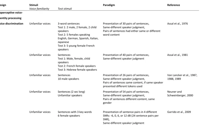
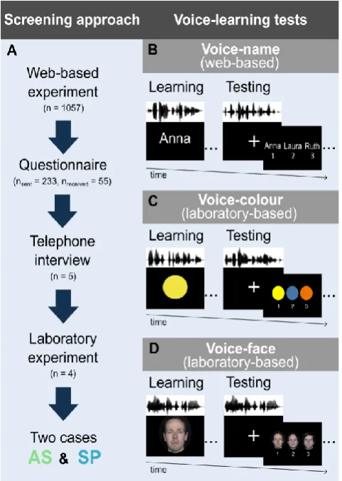
Given the theoretical framework proposed in Figure 1A, tests for phonagnosia need to be designed to address the multistage nature of voice-identity processing. Currently, employed voice-processing tests (summarised in Table 1) include measures which can evaluate: 1) the perceptual analysis of the vocal signal, achieved through means of unfamiliar voice discrimination and unfamiliar speaker change detection tests. Such tests can reveal apperceptive impairments; 2) a sense of familiarity with the encoded familiar vocal percept (i.e. familiarity decision); 3) the ability to link the encoded familiar vocal percept to identity-specific person knowledge (i.e. semantic association). Familiar voice-recognition tests are commonly used to examine both familiarity decisions and semantic association. These tests often assess both associative abilities, e.g. first listeners indicate a sense of familiarity towards a voice and then associate semantic knowledge to the familiar voice. Voices presented in those tests may involve famous, personally familiar or newly learned speakers’ voices.
Table 1. Overview of the tasks and stimuli used for assessing apperceptive (upper section) and associative (lower section) voice-identity processing.
Design Stimuli Paradigm Reference
Voice familiarity Test stimuli Apperceptive
voice-identity processing
Voice discrimination Unfamiliar voices 3-word sentences
Test 1: 2 male, 2 female, 2 child speakers
Test 2: 5 females speaking English, German, Spanish, Italian, Japanese
Test 3: 5 young female French speakers
Presentation of 30 pairs of sentences, Same-different speaker judgment,
Pairs of sentences had either same or different word content
Assal et al., 1976
Unfamiliar voices Sentences
Test 1: Male, female, child speakers
Test 2: French female speakers Test 3: Hebrew female speakers
Presentation of 40 pairs of sentences, Same-different speaker judgment
Assal et al., 1981
Unfamiliar voices Sentences
10 male speakers
Presentation of 26 pairs of sentences, Same-different speaker judgment,
Pairs of sentences same content, if same speaker presented different tokens used
Van Lancker et al., 1987, 1988, 1989
Unfamiliar voices Sentences (2 sec long)
Unfamiliar speakers
Presentation of 54 pairs of sentences, Same-different speaker judgment, Pairs of sentences different content, same gender
Neuner and
Schweinberger, 2000
Unfamiliar voices Sentences with 3 key words
6 female speakers
Presentation of sentence pairs in 4 different SNRs: -6, 0, 6, or 12 dB (24 sentence pairs per SNR),
Same-different speaker judgment
Unfamiliar voices Sentences
21 female speakers
Presentation of NV sentence pairs with 6, 16, or 48 frequency channels (24 sentence pairs per frequency channel level)
Same-different speaker judgment
Garrido et al., 2009
Unfamiliar voices 2-word sentences
3 male speakers
Brief familiarisation with the voice identities (passive listening), followed by:
Presentation of 54 pairs of sentences, Same-different speaker judgment
Roswandowitz et al., 2014
Unfamiliar voices Sentences
5 female speakers
Presentation of target voice, followed by: (i) 5, 10 or 20 second interval, and (ii)
presentation of 2 test voices (40 trials per interval duration)
2AFC speaker matching task
Xu et al., 2015
Speaker change detection
Unfamiliar voices 4 min long text Text included 24 speaker changes,
Speaker change detection
Assal et al., 1981
Unfamiliar voices High frequent words (names of
weekdays and months) Female speakers
Sequences of words including speaker changes - 24 trials of weekdays
- 24 trials of months, Speaker change detection
Hailstone et al., 2010
Unfamiliar voices High frequent words (names of
weekdays) Female speakers
Test 1: Naturalistic stimuli Test 2: Fixed f0 (220Hz)
Sequences of words including speaker changes Test 1: 28 trials
Test 2: 12 trials,
Speaker change detection
Hailstone et al., 2011
Associative
voice-identity processing
Familiar voice recognition
Famous voices 7 celebrity voices,
Male speaker
For each voice, cross-modal matching on 4-choice response array (voice – face/name) (semantic association)
Famous voices 25 celebrity voices, Male speaker, 4 sec long samples
For each voice, cross-modal matching on 4-choice response array (voice – face/name) (semantic association),
Debriefing of subjective familiarity with celebrities (van Lancker et al., 1989)
Van Lancker et al., 1987, 1988, 1989
Famous and unfamiliar voices
32 celebrity voices, 32 unfamiliar voices, Female and male speaker, 2 sec long samples
After voice presentation,
(i) familiarity judgment (familiarity decision), (ii) if familiar, voice naming (semantic association) Neuner and Schweinberger 2000 Personally familiar and unfamiliar voices Per participant:
1 familiar voice, 5 unfamiliar voices,
Voice samples consisted of vowels, CVC syllables, words, and sentences
After voice presentation, familiarity judgment (familiarity decision)
Lang et al., 2009
Famous and unfamiliar voices
48 celebrity voices 48 unfamiliar voices 7 sec long samples
After voice presentation,
(i) familiarity judgment (familiarity decision), (ii) if familiar, voice identification (provide name or other biographical detail) (semantic
association)
Garrido et al., 2009
Famous and unfamiliar voices
24 celebrity voices, Female and male voices
After voice presentation, familiarity judgment (familiarity decision)
Hailstone et al., 2010, 2011
Famous and unfamiliar voices
24 celebrity voices (same as above)
After voice presentation,
(i) voice identification (provide name or other biographical detail)
(ii) cross-modal matching on a response array (voice – face/name)
(semantic association)
Hailstone et al., 2010, 2011
Famous and unfamiliar voices
42 celebrity voices, 20 unfamiliar voices, Female and male voices, 5 sec long samples
After voice presentation
(i) familiarity judgment (familiarity decision) (ii) if familiar,
voice identification (provide name or other biographical detail; Roswandowitz et al., 2014)
cross-modal matching on a response array (voice
– face/name; Roswandowitz et al. 2018)
(semantic association),
Debriefing of subjective voice familiarity of celebrities
Famous and unfamiliar voices
100 celebrity voices 100 unfamiliar voices 6-8 sec long samples
Celebrity face-name composite displayed (1,2 or 4 identity composites), followed by 2 voice samples, then:
(i) indicate which of the voices is a celebrity (familiarity decision)
(ii) cross-modal matching of famous voice to face/name composite (1,2 or 4 options) (semantic association)
Xu et al., 2014, see also Herald et al., 2014
Newly-learned voice recognition
Newly-learned voices
6 unfamiliar female speakers Sentences with 3 key words
Cross-modal learning: name and voice, followed by:
(i) Voice recognition task (is the voice the same as the target speaker?)
(ii) Voice recognition task (what name matches the voice identity? 6 options)
(iii) Old/new task (is the voice new or has it been heard before i.e. old?)
Garrido et al., 2009
Newly-learned voices
3 male and 3 female unfamiliar speakers per test (voice-name/ voice-face test)
Voice-name test:
Cross-modal learning: simultaneous voice-name presentation,
Testing: voice - name matching Voice-face test:
same paradigm, just with voice-face associations
Roswandowitz et al., 2014, 2018
Acquired phonagnosia
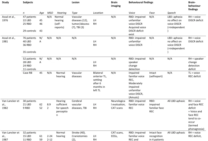
Lesion studies on phonagnosia allow a strong interpretation about brain regions required to identify voices. In the following, we review brain lesion studies which aimed to characterise the cognitive and neural mechanisms supporting voice-identity processing. The term phonagnosia implies a modality-specific deficit requiring many different control tests. However, the number of control tests or self-reports assessing other person-recognition or speech- processing abilities varies across clinical studies. Thus, whether the reported cases of acquired phonagnosia are also associated with other impairments often remains unclear in particular in those studies that do not include a systematic investigation of control abilities. For an overview of the reviewed lesion studies on voice-identity processing see Table 2.
Table 2. Overview about lesion studies on voice-identity processing.
Study Subjects Lesion Brain
imaging
Behavioural findings
Brain-behaviour findings
n Age MSO Hearing Type Location Voice Face Speech
Assal et al., 1976 47 patients 22 LBD 25 RBD 29 controls 45 46 43
N/A Normal hearing (self-reports) Vascular diseases (12), tumor/abscess (7), TBI (3)
LH RH
N/A RBD: impaired
unfamiliar voice DISCR Acquired voice DISCR deficit exist
N/A LBD: aphasia
no affect on voice DISCR (=independent )
RH = voice DISCR deficit
Assal et al., 1981 76 patients 40 LBD 36 RBD 35 controls N/ A
N/A N/A N/A
LH RH
N/A RBD: impaired
unfamiliar voice DISCR
N/A LBD: aphasia
no affect on voice DISCR (=independent )
RH = voice DISCR deficit 52 patients 28 LBD 24 RBD 11 controls N/ A
N/A N/A N/A
LH RH
N/A RBD: impaired
speaker change detection
N/A N/A RH = speaker
change detection deficit
Case RB 45 N/A Normal
hearing Vascular diseases Bilateral anterior TL, settling after 6 months in left TL
N/A Impaired
familiar voice REC, Moderately impaired unfamiliar voice DISCR, (Amusic) Intact (selfreport)
N/A TL = voice
REC deficit
Van Lancker et al., 1982 30 patients 21 LBD 9 RBD 62 52 8.9 2 Hearing sufficient for speech perceptio n Cerebral vascular diseases, TBI LH RH Neurologica l evaluation, CAT scans RBD: impaired familiar voice REC RBD: impaired familiar face REC
All LBD aphasic RH = voice and face REC deficit = Voice and face REC tend to co-occur (termed phonagnosia) Van Lancker et
al., 1987 32 patients 15 LBD 11 RBD 61 59 2-24 2-12 Normal hearing Stroke (40), Craniotomies (2), LH RH CAT scans, EEGs, RBD: impaired familiar voice REC and Intact face recognition in 4 patients
6 BBD
48 controls 69
64
1-24 haemorrhage
(1),
meningioma (1), tumor (1)
Both Hs neurological
evaluations unfamiliar voice DISCR, LBD: impaired unfamiliar voice DISCR, intact familiar voice REC in 14 patients dissociation between familiar voice REC and unfamiliar voice DISCR
LH + RH = voice DISCR deficit
Van Lancker et al., 1988 6 case reports 2 LBD 1 RBD 3 BBD 30 controls 65 (52 – 82) 50-85
0.5 –
year s after TSO Normal hearing reported for 2 cases
Stroke (5) Haemorrhage (1) Mainly temporal, parietal, frontal lobe
CTs 4 BBD:
impaired unfamiliar voice DISCR, 3 RBD: impaired familiar voice REC, 1 RBD: impaired in both tasks In 5 patients dissociation between familiar voice REC and unfamiliar voice DISCR
N/A 4 LBD with
aphasia and voice DISCR deficit
Right PL = voice REC deficit, Bilateral TL = voice DISCR deficit
Van Lancker et al., 1989 56 patients 25 LBD 25 RBD 6 BBD 48 controls 61 63 71 64
N/A. N/A Cerebral
infarction Lesions classified in parietal, temporal, and temporo-parietal lesions
CTs BBD: impaired
unfamiliar voice DISCR, RBD: impaired familiar voice REC
N/A All LBD aphasic Quantitative
Neuner and Schweinberger , 2000 36 patients 16 LBD 13 RBD 7 BBD 20 controls 48 44
8.2 N/A anemic infarct
(10),
haemorrhage (10),
subarachnoid haemorrhage (5), TBI (5), hypoxia (2), TBI with hypoxia (1), encephalitis (2), tumor (1)
LH RH Both Hs
Surgery reports, CTs, or MRI scans 4 patients: selective voice REC deficit (intact face, name, and sound REC), 2 RBD, 1 LBD: impaired in familiar voice REC, 1 RBD: impaired in familiar voice REC and unfamiliar voice DISCR
In 4 patients intact face-identity processing
N/A RH = voice
REC deficit
Lang et al., 2009 20 patients 11 LBD 9 RBD 17 controls 66 64 64 3.1 1.6
N/A Ischemic
infarcts LH: MCA (6),
PCA (2), LSA (3)
RH: MCA (9), PCA (1)
N/A RBD: impaired
familiar voice REC
LBD: intact familiar voice REC
N/A All LH aphasic
and intact voice REC
RH = voice REC deficit
Hailstone et al., 2010
Case QR 61 - Normal
hearing Behavioural variant frontotemporal dementia Right anterior TL extending to TL (STG)
MRI scans Impaired
familiarity and REC of familiar voices, Intact unfamiliar voice DISCR (impaired music instrument processing) Impaired familiarity, moderately impaired REC of familiar faces, Intact unfamiliar face DISCR
N/A Right
anterior TL and STG = voice REC deficit (associative phonagnosia) Case KL 24 controls
72 - Normal
hearing Frontotempora l lobar degeneration Bilateral anterior TL atrophy extending to inferior temporal cortices (incl. FFA)
MRI scans Impaired
familiarity and REC of familiar voices, Intact unfamiliar voice DISCR Impaired familiarity and REC of familiar faces, Intact unfamiliar face DISCR
N/A Bilateral
Hailstone et al., 2011 36 patients 14 FTLD 22 Alzheimer’ s 35 controls 64 67 64 - - Normal
hearing Frontotempora
l lobar degeneration Alzheimer’s disease Bilateral anterior TL atrophy (14) hippocampa l atrophy (16), generalized cerebral atrophy (4) MRI scans (11 FTLD, 18
Alzheimer’s)
FTLD: impaired familiarity, REC of familiar voices, intact unfamiliar voice DISCR Alzheimer’s: impaired familiarity, REC of familiar voices, impaired unfamiliar voice DISCR FTLD + Alzheimer’s: impaired familiar face familiarity, REC, and apperceptiv e face processing
N/A VBM analysis
Right anterior TL = voice, name and face REC, Right inferior PL (angular gyrus) = unfamiliar voice DISCR Roswandowitz et al., 2018
58 patients focal brain lesions
31 RBD 27LBD
48 46 Normal
hearing (covariate in VLSM analysis) ischemic stroke (34), intracerebral haemorrhage (6), subarachnoid haemorrhage (6), TBI (7), tumor (4) Bilateral TL and right inferior PL well covered by lesions MRI scans (56), CT scans (2) Worse performance in voice-name test in patients compared to controls (Roswandowit z et al., 2014) RBD worse in voice REC of recently familiarised voices than LBD
9% of patients report poor voice REC after lesion onset 5% of patients report poor face REC after lesion onset No severe aphasia VLSM analysis Right mid/posterio r TL = selective voice REC deficit Right inferior PL = impaired voice-face integration
All brain-behaviour findings rely on descriptive brain-behaviour associations if not stated otherwise.
MSO= Months since onset, LBD = Left-brain damaged patients, RBD = Right-brain damaged patients, BBD = Bilateral-brain damaged patients, N/A = not
Apperceptive voice-identity processing in acquired phonagnosia
Although the term phonagnosia was first mentioned in 1982 by van Lancker and Canter, examinations on voice-identity processes had begun almost a decade previously. These first studies addressed mainly perceptual aspects of voice-identity processing (e.g. unfamiliar voice discrimination). In 1976 the Swiss neurosurgeon Par G. Assal and his colleagues (Assal et al., 1976) published the first study on acquired phonagnosia. They investigated 47 patients with unilateral brain lesions, including 25 patients with lesions in the right hemisphere (right brain damaged or RBD) and 22 patients with lesions in the left hemisphere (left brain damaged or LBD) as well as 29 healthy age- and handedness-matched controls. This study was centred on three main questions: (i) Does a deficit in voice discrimination after brain damage exist? (ii) Is the voice-discrimination deficit associated with right hemispheric lesions? (iii) Are voice-identity and language processes dissociable mechanisms? The authors showed that patients with brain lesions performed significantly worse than healthy controls on discrimination tasks with unfamiliar voices. Participants were tested on discrimination between unfamiliar adult male, female, and children’s voices as well as on discrimination among only unfamiliar female voices either speaking different languages or the same language (i.e. French) (Table 1). RBD patients performed significantly below controls on all three tests (i.e. based on Tuckey-Hayes statistics), whereas LBD patients only performed worse than controls when discriminating female voices speaking different languages. A direct statistical patient group comparison however was not conducted. This was the first indication that impaired apperceptive voice-identity processing exists after brain damage and that it might be predominantly a function of the right hemisphere. Although no information about precise lesion locations was available the authors attempted to localise right hemispheric lesions relevant for voice discrimination with a dichotic listening test. RBD patients performed worse in voice discrimination if voices were presented to the left in comparison to the right ear. The authors speculated that voice discrimination may be assigned to the right temporal lobe.
Further, addressing the relation between voice-identity and language processes, the authors directly compared voice-discrimination abilities between LBD patients with and without aphasia (i.e. speech and language disorder caused by brain damage predominantly to the language-dominant left hemisphere). Performance in the voice-discrimination tests was similar for aphasic and non-aphasic LBD patients. This was a first indication of the separability of voice-
identity processing from language abilities. In RBD patients language abilities were not considered, probably because it is unlikely that aphasia occurs in RBD patients. But in RBD patients visual abilities were tested. This was done with a visual figure/ground discrimination task (Poppelreuter test) and a visual-spatial memory task. Results showed that unfamiliar voice- discrimination performance was significantly worse in RBD patients with impaired visual processing than in RBD patients with intact visual processing. Whether the RBD patients with intact visual processing had nevertheless voice-discrimination difficulties in contrast to healthy controls was not tested.
Five years later, Assal and colleagues (1981) elaborated on their pioneering study by assessing apperceptive voice mechanisms by testing voice-discrimination abilities alongside the ability to detect a change in speaker identity (Table 1). This time, Assal et al. investigated unfamiliar voice discrimination in a sample of 76 patients (40 LBD, 36 RBD) and 35 healthy controls and unfamiliar speaker-change detection in 52 patients (28 LBD, 24 RBD) and 11 healthy controls. The authors replicated their previous findings: (i) They found a right hemispheric dominance for apperceptive voice-identity processing. This time, the authors showed that RBD patients were impaired on both apperceptive voice tasks, i.e. unfamiliar voice discrimination and unfamiliar speaker change detection. Importantly, in contrast to the previous study, this time a direct statistical group comparison between RBD and LBD patients on voice discrimination yielded a significant group difference (ANOVA at α = 0.05): RBD patients performed worse than LBD patients. (ii) Based on the dichotic listening results, the authors again suggested an important role of the right temporal lobe (this time more specifically of the temporo-parietal region) during voice discrimination. (iii) Again they noted that dissociation between speech and voice-identity processing was evident in this cohort; voice-discrimination performance was not different between aphasic and non-aphasic LBD patients.
The first case report of acquired phonagnosia: The case RB
Assal et al. (1981) also reported the first case study of acquired phonagnosia; case RB. He was a 45-year-old male, managing director, had musical training, and normal hearing abilities. After brain injury resulting from vascular disease, RB reported difficulties in music and irony perception, voice recognition, as well as speech and sound perception. While RB recovered from the latter two difficulties one month after lesion onset, he continued to evidence a strong
deficit in recognising familiar voices and a moderate deficit in discriminating voices compared to controls which was based on numerical group difference inspection. Unfortunately, details on the test designs were not reported. Interestingly, face recognition was tested and intact. A brain scan (not specified by the authors, but likely a CT scan) originally revealed bilateral cortico- subcortical lesions in the anterior temporal lobe, initially more pronounced in the right hemisphere that after six weeks resolved into a lesion predominantly in the left temporal lobe.
This first case report on acquired phonagnosia implicated a role for the temporal lobe in voice-identity processing and showed that voice-identity processing can be impaired while leaving face-identity processing intact. Further, the case report gave a first indication that voice recognition (associative voice-identity processing) and voice discrimination (apperceptive voice- identity processing) might be dissociable mechanisms.
In our view, it is remarkable that in these first studies Assal and colleagues asked questions that have traced all future studies on voice-identity processing. However, to date, these studies are relatively unknown in the field, probably because they are reported in French only. Apperceptive and associative voice-identity processing in acquired
phonagnosia
Van Lancker and colleagues took research on phonagnosia a decisive step further. Van Lancker and Canter (1982) investigated associative voice-identity processing in 30 patients with focal brain lesions (21 LBD/ 9 RBD) with a familiar voice-recognition test (Table 1). All LBD patients had aphasia. One aim of the study was to assess whether familiar voice recognition is primarily assigned to the right hemisphere, as found in the prosopagnosia (i.e. face-identity processing deficit) literature (De Renzi, 1986; Damasio et al., 1990; De Renzi et al., 1991). Further, van Lancker and Canter were interested in the relation between voice- and face-identity processing. Therefore, patients were tested on their voice- and face-recognition abilities. In both tasks, patients were asked to match a celebrity voice/face to a written name (Table 1). A deficit in the voice- and face-recognition task was more prevalent in RBD than in LBD patients. 4/9 RBD patients were impaired on familiar voice recognition. Only in 1 RBD patient this deficit was selective to voice recognition; the remaining three RBD patients also had a deficit in face recognition. In contrast, only 1/21 LBD patients had impaired familiar voice recognition and another one had impaired familiar face recognition. The authors concluded that associative
voice-identity processing can be assigned to the right hemisphere. Further, the authors suggested that voice- and face-recognition deficits tend to co-occur and that both may rely on neuronal mechanisms within the right hemisphere. Further, the authors conclude that voice recognition might be dissociable from left-hemisphere language functions as 20 of 21 aphasic LBD patients had intact voice recognition. Interestingly, there were two cases in which voice-recognition impairments seemed to be selectively impaired, i.e. with intact face recognition (1 RBD, 1 LBD). The behavioural profile of these two patients might be indicative of specific neural mechanisms for familiar voice recognition that can be dissociated from those supporting language and face- recognition abilities.
Next, Van Lancker and Kreiman (1987) directly compared the relation between apperceptive and associative voice-identity processing by testing unfamiliar voice discrimination and familiar voice recognition in the same patients. Although both abilities were located in the right hemisphere in previous studies (Assal et al., 1976; Assal et al., 1981; Van Lancker and Canter, 1982), the case RB had indicated a potential dissociation between both mechanisms (Assal et al., 1981). Van Lancker and colleagues tested 32 patients (15 LBD, 11 RBD, 6 bilateral brain damaged (BBD)) and healthy age- and education-matched controls (n = 48) on both unfamiliar voice discrimination and familiar voice recognition. All LBD patients had aphasia. In contrast to previous findings, patients with lesions in the left or right hemisphere were similarly impaired (relative to the control group; 2-way repeated measure ANOVA at α = 0.01) in the unfamiliar voice-discrimination task. In contrast, only RBD patients showed impaired familiar voice recognition, as compared to controls (Van Lancker and Kreiman, 1987). LBD patients’ familiar voice-recognition performance was similar to controls. Looking at the cases individually, 14 of the 32 patients showed dissociable behavioural performances in the unfamiliar voice-discrimination and familiar voice-recognition task (1 RBD, 6 LBD, and 3 BBD; no lesion lateralisation on the remaining 4 patients was reported). They had impaired voice discrimination and intact voice recognition or vice versa. Of the 10 patients for whom they reported individual results, worse voice discrimination was associated with LBD and worse voice recognition with RBD. BBD patients had both worse voice discrimination and recognition. Moreover, there was no correlation between the discrimination and recognition performance in patients. Taken together, these results suggested that both apperceptive and associative voice- identity processing might be underpinned by dissociable cognitive and neuroanatomical mechanisms.
To assess the selectivity of a given voice-identity processing deficit, 4 patients with a severe deficit in either voice discrimination or recognition were also tested on their face recognition and -discrimination abilities as well as environmental sound processing. A voice- specific deficit pattern emerged; face and sound processing was intact in those patients suggesting a fairly selective phonagnosia.
To reveal which anatomical regions within the respective hemisphere sub-serve apperceptive and associative voice-identity processing, van Lancker and colleagues (1988) studied 6 brain-lesioned cases for which CT scans were available. Patients were tested on both unfamiliar voice discrimination (apperceptive voice-identity processing) and familiar voice recognition (associative voice-identity processing) (Table 1). Patients’ performance was compared to 30 healthy age-matched control participants. 5 of the 6 patients showed a clear discrepancy between the ability to discriminate unfamiliar voices and to recognise familiar voices (i.e. more than 2 SDs away from the controls’ mean difference in test scores). Van Lanker et al. noted that the 3 patients who were exclusively impaired on unfamiliar voice discrimination had a lesion overlap in the temporal lobe of either the left or the right hemisphere (i.e. including anterior, mid and posterior regions) and were aphasic.
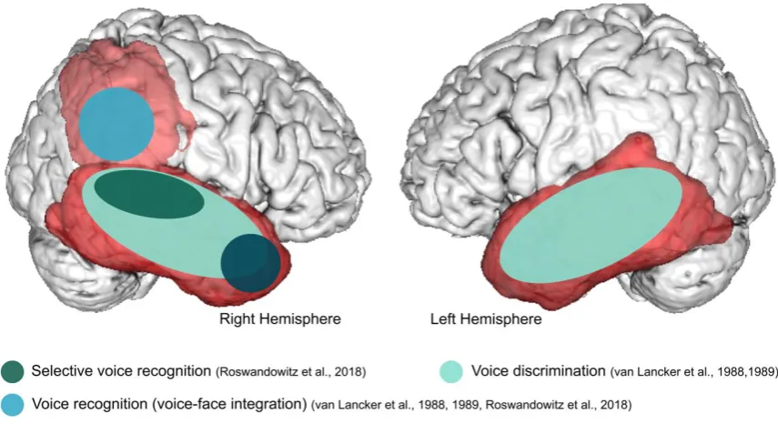
In contrast, the 2 patients with selectively impaired familiar voice recognition had in common lesions, which were located exclusively in the right hemisphere, including the posterior part of the temporal and parietal lobe structures such as the superior portion of the angular gyrus and the posterior supramarginal gyrus. The one patient who did not show dissociation between voice discrimination and recognition, being impaired on both tasks, had a lesion in the right mid/ posterior temporal lobe and the right parietal lobe including the superior angular gyrus and the supramarginal gyrus. The authors (Van Lancker et al., 1988) discuss a relevant role of the bilateral temporal lobe for unfamiliar voice discrimination (apperceptive voice-identity processing) and of the lateral parietal lobe in the right hemisphere for familiar voice recognition (associative voice-identity processing) (Figure 2). This study provided supporting evidence for distinct mechanisms underlying apperceptive and associative voice-identity processing.
In a follow up study, Van Lancker et al. (1989) aimed to quantitatively confirm the descriptive behavioural and neuroanatomical dissociation between apperceptive and associative voice-identity processing. To allow a quantitative brain-behaviour analysis, they tested a large
sample of 56 brain-damaged patients. 44 patients (23 LBD, 15 RBD, 6 BBD) were tested on both an unfamiliar voice-discrimination and familiar voice-recognition task (Table 1). 12 patients (2 LBD, 10 RBD) were tested only on familiar voice recognition. All LBD patients were aphasic. Results were compared between lesion groups and healthy age- and education-matched control participants. Behavioural results showed that both LBD and RBD patients performed worse on the unfamiliar voice-discrimination task compared to controls (2-way repeated measure ANOVA at α = 0.05). On the familiar voice-recognition task, only RBD patients were impaired, relative to controls. In line with previous findings (Van Lancker and Kreiman, 1987; Van Lancker et al., 1988), unfamiliar voice discrimination (apperceptive voice-identity processing) was assigned to lesions in both the left or right hemispheres and familiar voice recognition (associative voice- identity processing) only to lesions in the right hemisphere. Next, they investigated the neuroanatomic substrates underlying this behavioural pattern. Based on 43 available CT scans, lesions were classified according to the lobe with the largest extend of the lesion. According to their hypothesis, a lesion in the right parietal lobe was significantly associated with a deficit in associative voice-identity processing (familiar voice-recognition task). All 9 patients with a right parietal lobe lesion showed impaired familiar voice recognition; as did 7 of 43 patients having the lesion elsewhere. Unfortunately, the authors did not report the lesion location of those 7 patients. It would have been interesting to observe whether lesions in these additional 7 cases were located adjacent to the right parietal lobe or in other regions such as the temporal lobe as suggested by Assal
et al. (1981) and by neuroimaging findings (Figure 1 B).
The analysis of apperceptive voice-identity processing was based on 25 CT scans. Confirming the authors’ hypothesis, 13 patients with a lesion in either the left or right temporal lobe performed worse in discriminating unfamiliar voices compared to controls. There were also 4 patients with temporal lobe lesion and preserved task performance. These patients had lesions exclusively in the left hemisphere indicating a higher relevance of the right hemisphere during unfamiliar voice discrimination. Of the patients having their lesion outside the temporal lobe, 9 had high and 4 had low scores on the discrimination task. Of these 4 patients with impaired voice discrimination, lesions were adjacent to the temporal lobe. In summary, van Lancker and colleagues provided quantitative evidence that lesions in the right parietal lobe were associated with associative voice-identity processing and lesions in either the left or right temporal lobe with apperceptive voice-identity processing (Figure 2).
Group evidence for selective voice-identity processing impairments
Previous studies were not conclusive as to whether phonagnosia may reflect a modality specific disorder. For example, while the case of RB (Assal et al., 1981) and 6 patients in van Lancker et al. (Van Lancker and Canter, 1982; Van Lancker and Kreiman, 1987) suggested dissociation between voice- and face-identity processing, the patient group reported by van Lancker and Canter (1982) showed that voice- and face-identity deficits can co-occur. The same diversity emerged when considering the relation between voice-identity processing and other auditory processing abilities, such as speech, sound, emotion, and music processing (e.g., case RB, Assal et al., 1981; and the 4 cases in Van Lancker and Kreiman, 1987).
To systematically assess the relation between voice-identity and identity processing of other sensory modalities as well as other auditory processes, Neuner and Schweinberger (2000) developed a comprehensive behavioural test battery. They studied 36 brain-lesioned patients (16 LBD, 13 RBD, and 7 BBD) for which brain surgery reports, CT or MRI scans were available and 20 healthy controls (matched in age, gender, and education). The test battery assessed apperceptive (discrimination tasks) and associative (familiarity decision and semantic association tasks) abilities of persons’ voices, faces, and names (Table 1). In addition, the test battery included control tests on word, picture, and sound recognition to investigate the specificity of a given person-recognition deficit. In 13/36 patients, familiar voice recognition assessed by a familiarity decision task was significantly worse compared to controls’ performance (cut-off for impairment: patient scores below the control mean at α = 0.05 and 0.01). However, only 4 of the 13 patients showed a selective form of phonagnosia, with impaired familiar voice recognition, but intact sound, face, and name recognition. Unfortunately, for these cases, semantic association scores were not reported. 1 of these 4 patients also showed an overlapping impairment in voice discrimination; the lesion was located in the right hemisphere (Table 1). 2 of the 4 patients with selective familiar voice recognition deficits had a lesion located in the right hemisphere and one in the left hemisphere. Neuner and Schweinberger (2000) made large strides in investigating the specificity of phonagnosia. Their systematic investigation attested that phonagnosia can be witnessed as a specific deficit independent of nonverbal sound, face, and name recognition.
A study by Lang and colleagues (2009) specifically examined the relation between voice- identity and speech processing. In this study, familiar voice recognition was assessed (Table 1).
The study included 20 brain-damaged patients (11 LBD, 9 RBD) and 17 healthy age-matched controls. The two groups were matched for lesion location and extent. Left-brain damaged patients were tested for aphasia (Aachen Aphasia Test). The results yielded a familiar voice- recognition deficit in RBD relative to performance in LBD patients and controls (one-factorial ANOVA at α = 0.05). In contrast, LBD patients and healthy controls performed equally well on familiar voice recognition. The authors concluded that in LBD patients aphasia (5 amnestic, 5 Wernicke, 1 Broca aphasia) was not associated with familiar voice-recognition deficits. However, whether there is a double dissociation between voice-identity and speech processing remains open as language abilities were not assessed in RBD patients. Lesions in the right hemisphere were mostly confined to the supply areas of the middle cerebral artery and similar lesions in the left hemisphere did not affect familiar voice recognition. Unfortunately, more exact lesion location was not reported.
Case report evidence for selective voice-identity processing impairments
Hailstone et al. (2010) comprehensively evaluated voice-identity processing and several control tasks in 2 patients with neurodegenerative diseases (frontotemporal dementia) and 24 healthy age-matched controls. The authors assessed apperceptive (unfamiliar speaker-change detection) and associative (familiarity decision and semantic association) voice-identity processing as well as face, name, music, and sound processing (Table 1). Patient QR, 61-years old, had bilateral fronto-temporal atrophy, accentuated in the right anterior temporal lobe but extending posteriorly within the temporal lobe. Patient KL, 72-years old, had bilateral, predominantly anterior temporal lobe atrophy, which was more marked on the right hemisphere and in the inferior temporal cortices including the fusiform gyrus. In both patients, processing of familiar voices (familiarity decision, semantic association) was severely impaired in contrast to controls (modified t-test for single case studies at α = 0.05; Crawford and Howell, 1998). In addition, both patients as compared to controls were impaired in familiar face and name processing. However, QR’s face and name abilities were superior to KL’s. This indicates a more selective phonagnosia in QR and a rather multi-modal identity processing deficit in KL. The person-identity processing deficits observed in QR and KL seemed to be restricted to associative processes. Apperceptive processing of voices (including perceptual processing of vocal-identity, vocal-gender, and speaker-size information) and faces were preserved in both. Hence, the authors classify the patients’ deficits as associative agnosias. Both patients also
showed intact vocal-emotion recognition abilities. Processing of musical instruments in an auditory and visual task design however was affected in QR and KL. The authors suggested that the bilateral anterior temporal lobe is involved in supporting multiple aspects of person knowledge including voices, faces, and names with a right hemispheric dominance for aspects of nonverbal person knowledge.
Statistical brain lesion-behaviour relation: multimodal person recognition deficit
In the past decade sophisticated statistical approaches have been developed for high- resolution structural MRI group studies to afford more robust and objective associations between brain structure and behavioural performance (VBM: Ashburner and Friston, 2000; VLSM: Bates et al., 2003). The first study assessing a statistical voxel-wise association between brain structure and voice-identity processing was published in 2011 by Hailstone and colleagues (Hailstone et al., 2011). 36 patients with neurodegenerative diseases (14 Frontotemporal lobar degeneration (FTLD), 22 Alzheimer’s disease) and 35 healthy controls (matched in age, gender, handedness, and education) were tested on a comprehensive behavioural test battery. For all 16 FTLD and 20 Alzheimer’s disease patients, a high-resolution structural MRI scan was available. FTLD patients had atrophy in the anterior temporal lobes of both hemispheres. Of the Alzheimer’s diseases patients, 16 had hippocampal atrophy and 4 patients had generalised cerebral atrophy. Participants were tested on apperceptive (unfamiliar speaker-change detection) and associative (familiarity decision and semantic association) voice-identity processing (see Table 1). To assess the selectivity of a given voice-identity processing deficit, within and across modalities, the test battery included tests on other measures of vocal processing (including speaker-size and vocal- gender information) as well as tests on face and name processing. In the associative voice tasks, both disease groups performed significantly worse compared to controls (z-tests and 95% Wald- type confidence intervals at α = 0.05 and 0.001). However, the deficits were more profound in the FTLD than the Alzheimer’s patients. A more heterogeneous pattern emerged for the apperceptive tests. During speaker-change detection and vocal-gender perception, only Alzheimer’s patients were impaired. However, apperceptive face processing was impaired in both disease groups.
By applying voxel-based morphometry, the authors presented neuroanatomical evidence that the anterior temporal lobe (predominantly of the right hemisphere), as well as the right fusiform gyrus, plays an important role in associative person recognition across different modalities, including voices, faces, and names (Figure 2). This is consistent with previous reports of associative person-recognition deficits with anterior temporal lobe lesions in neurodegenerative disease (Gainotti et al., 2003; Gainotti et al., 2008; Hailstone et al., 2010). For apperceptive voice-identity processing (speaker-change detection), the right inferior parietal lobe (i.e. angular gyrus) was found to be relevant (Figure 2). In light of the previous findings (Van Lancker et al., 1988; Van Lancker et al., 1989), association of the parietal lobe with apperceptive voice processing is unexpected. However, based on patients’ atrophy descriptions, lesions in the Hailstone et al. (2011) study covered mostly the anterior temporal lobes and thus results on parietal lobes might have to be interpreted with caution.
Statistical brain lesion-behaviour relation: selective voice-identity recognition deficit
In a recent study Roswandowitz et al. (2018) aimed to identify which lesion locations may cause a selective deficit in person-identity processing, which is confined to the auditory domain i.e. to voice-identity recognition. The authors were in particular interested in examining the contribution of the right inferior parietal lobe and the temporal lobe to voice-identity recognition (Fig. 1 B, Stage II). Based on the acquired phonagnosia cases described above (see section ‘Apperceptive and associative voice-identity processing in acquired phonagnosia’), the right inferior parietal lobe is crucial for voice-identity recognition. Conversely, neuroimaging studies on neurotypicals have consistently identified recruitment of the temporal lobe during voice-identity recognition tasks (see section ‘Model of voice-identity processing’). To resolve this discrepancy of regions critical for voice-identity recognition, Roswandowitz et al. conducted a voxel-based lesion-behaviour mapping study in a cohort of 58 patients with unselected unilateral focal brain lesions (31 RBD, 27 LBD patients) and high-resolution structural brain images. The study included a comprehensive behavioural test battery including recognition tasks of recently-familiarised, i.e. newly-learned (voice-name, voice-face association learning) and familiar voices (famous voice recognition) as well as visual (face-identity recognition) and acoustic control tests (vocal-pitch and vocal-timbre discrimination). Voxel-based lesion- symptom mapping (VLSM) analyses revealed a strong association between lesions in the right
mid/posterior temporal and right inferior parietal lobe and the recognition of both recently- familiarised and familiar voices. However, a selective voice-recognition deficit, that was independent of face-identity processing and acoustical analyses of voice-identity features such as pitch and timbre, was associated only with lesions in the right mid/posterior temporal lobe. This finding implicated an obligatory function for the temporal lobe to voice-identity processing, making it the most likely key structure of the core-voice system. In contrast, lesions in the right inferior parietal lobe were associated with reduced voice-identity recognition when voices were associated with a face. This finding is similar to the earlier van Lancker studies where lesions in the right inferior parietal lobe were associated with reduced performances in tasks where patients had to match a famous voice to a display of faces (and their names) (Van Lancker et al., 1988; Van Lancker et al., 1989). Thus, the right inferior parietal lobe might have a facultative role during voice-identity processing only when additional face information is available. The study by Roswandowitz et al. is the first to provide group evidence for an association between spatially well-defined brain lesions and selective voice-identity processing impairments (Fig. 2).
Figure 2. Schematic overview of studies reporting lesion locations associated with the respective voice- identity processing and multi-modal person recognition deficit. The temporal lobe is indicated by the dark red map and the right inferior parietal lobe by the light red map.
Developmental phonagnosia
Developmental phonagnosia has been discovered only recently (Garrido et al., 2009; Herald et al., 2014; Roswandowitz et al., 2014; Xu et al., 2015). Current prevalence estimates suggest that anything within the range of 0.2 % (Roswandowitz et al., 2014), 1 % (Xu et al., 2015) to 3.2 % (Shilowich and Biederman, 2016) of the population may have this deficit. While the precise aetiology of the deficit is unknown, it is possible that phonagnosia may have a heritable component, as has been observed in developmental prosopagnosia (Duchaine et al., 2007; Grueter et al., 2007; Schmalzl et al., 2008; Lee et al., 2010). In the following pages, we review the first documented cases of developmental phonagnosia, which have allowed for an examination of the nature and specificity of this developmental deficit.
The case of KH
Garrido and colleagues reported the first case of developmental phonagnosia, case KH (Garrido et al., 2009). KH was a 60-year-old female who worked as a successful manager. She presented with a life-long impairment in voice recognition and reported that she failed to even recognise her daughter’s voice on the phone. To confirm and assess the specificity of her self- report deficit, KH and a group of age-matched controls (n = 8) undertook a detailed behavioural battery of vocal-, visual-, and auditory-processing tests. As suspected, compared to controls, KH was significantly impaired in familiar voice-identity recognition. Specifically, her ability to judge whether a voice was famous or not was close to chance. This indicated weak feelings of familiarity towards known voice identities (i.e. impaired familiarity-association). In addition, her retrieval of names for the famous voices was negligible; KH could only accurately recall the name of one of the 48 presented famous identities, indicating impaired semantic-association. Her poor performance could not simply be explained by a lack of exposure to the vocal identities in every-day life2. When exposure to voices was explicitly controlled in a task, which required the
learning of new unfamiliar speakers’ voices with their corresponding name, KH's performance remained significantly poorer than age-matched controls (n = 8) for both naming and judging the familiarity (old/new judgment) of the speakers. Interestingly, KH's ability to discriminate between unfamiliar voices, that is to say whether two voice samples were articulated by the same
2In a post-test KH was asked to indicate if she had significant exposure in daily life to the voices, which
she failed to name during testing. Taking this assessment into account, KH named only 3.85 % (i.e. 1 of 26) of the identities, which she stated she had significant exposure to.
or a different speaker, was similar to controls under optimal listening conditions (task as described in Neuner and Schweinberger (2000), see Table 1). However, she was impaired in discriminating between identities when the task was made more difficult through the inclusion of auditory noise. When examining KH performance across tasks, the authors found no statistical evidence for dissociation between familiar voice recognition (associative voice-identity processing) and unfamiliar voice discrimination (apperceptive voice-identity processing).
Garrido and colleagues also examined whether KH’s voice-identity processing impairment could be mediated by a higher-order multimodal person-recognition deficit, and, or a general deficit in vocal or auditory processing. Interestingly, KH’s memory for faces was either superior to, or within the normal range of, controls. Her recognition and processing of general auditory information, including environmental sounds and musical excerpts, was normal; as was her ability to extrapolate vocal cues to support gender and emotion categorisation. In terms of speech processing, KH’s performance was within the control range on a number of tasks, including vowel identification and the matching of verbal content to a visual target image. However, her performance under more challenging listening conditions was less clear. KH was impaired, relative to controls, in perceiving speech, which was embedded in auditory noise, although this impairment was not consistent across all levels of auditory noise. For example, KH’s speech perception was impaired relative to controls for intermediate noise levels (SNR -3, SNR 3 dB), while her performance at the highest (SNR -6 dB) and lowest (SNR 6 dB) levels of auditory noise appeared normal. The authors attributed this poor performance to possible testing fatigue.
The case of KH suggested that developmental phonagnosia could represent a deficit in the processing of vocal identity, which was not mediated by a general deficit in the processing of auditory information, nor by a higher-level multimodal deficit affecting identity recognition across the visual and auditory domain. However, evidence for a possible dissociation between voice and speech processing, as well as voice recognition and voice discrimination, would become clearer in the following years, as more cases of developmental phonagnosia came to the attention of researchers (Roswandowitz et al., 2014; Roswandowitz et al., 2017).
The case of AN
AN was a 20-year-old female university student who presented with a deficit in familiar voice recognition (Herald et al., 2014; Xu et al., 2015). Intriguingly, AN stated that she was not particularly aware of her deficit growing up as she had not thought that people could recognise an individual without seeing their face. Indeed, AN’s face recognition was normal, as she obtained high scores on tests of familiar face recognition and naming (Xu et al., 2015). Xu et al.
(2015) and Herald et al. (2014) formally tested AN’s familiar voice-recognition performance through a web-based experiment. In each trial, participants listened to samples of two voices; one celebrity and one non-celebrity voice. In parallel, 1, 2, or 4 celebrity face-name composites were presented. Participants first (i) decided, which of the two voices the celebrity voice was (i.e. familiarity decision) and then (ii) they indicated, which celebrity face-name composite matched the familiar rated voice. Relative to controls (n = 21, age range = 19-73 years), AN was markedly impaired in her ability to match the voices, which she classified as familiar with the correct celebrity face and name. Unfortunately, it was not explicitly reported whether her familiarity judgements towards the famous voices were also impaired. Conversely, AN's accuracy was similar to age-matched controls (n = 9) when the task was to choose, which of two unfamiliar voice samples matched a target voice. The target and test samples contained different verbal content. Given the dissociation between deficient familiar voice recognition and intact unfamiliar voice matching her behavioural profile is most likely indicative of an associative voice-identity processing impairment. Unfortunately, AN’s abilities in other auditory tasks such as speech, emotion, and music processing were not formally assessed leaving open the possibility of additional impairments in other aspects of auditory processing.
The authors also examined the neuronal mechanisms underlying AN’s voice-recognition deficit using two functional imaging experiments (Xu et al., 2015). They employed (i) a standard functional localiser known to elicit voice-sensitive responses in the temporal voice areas (TVAs) of the STS/G (Belin et al., 2000; see also Belin, Chapter 3, this edition) and (ii) a voice-imagery task. The study included AN and 9 controls (22-31 years). Functional imaging during the first experiment of passive listening to vocal, as compared to non-vocal, sounds (Belin et al., 2000) demonstrated typical responses in AN in the TVAs, located bilaterally along the temporal lobes (Xu
et al., 2015). The second fMRI experiment assessed functional responses during voice imagery. Here, participants were presented with pictures of familiar persons’ faces and names