Application of acute stroke imaging
Selecting patients for revascularization therapy
Tiesong Shang, MD, PhD
Dileep R. Yavagal, MD
ABSTRACT
Due to the dynamic and versatile characteristics of ischemic penumbra, selecting the right acute ischemic stroke (AIS) patients for revascularization therapy (RT) based on initial available imaging can be challenging. The main patient selection criterion for RT is the size of the mismatch between the potentially salvageable tissue (penumbra) and the irreversibly damaged tissue (core). The goal of revascularization RT is to “freeze” the core and prevent it from extending to the penumbral tissue. Penumbral imaging selection of AIS patients for RT, using magnetic resonance or CT-based studies, may provide more clinical benefit to the appropriate patients, although direct evi-dence is pending. Not all penumbra-core mismatches beyond 3 hours are equal and need treatment, and defining which mismatches to target for RT is the current goal of ongoing clinical trials. In addition to “penumbral”-based imaging, large vessel occlusion and clot length estimation based on CT angiography and noncontrasted ultrathin CT scan has been used to identify patients who are refractory to systemic thrombolysis and may be eligible for endovascular therapy. The application of various imaging modalities in selecting and triaging AIS patients for RT is dis-cussed in this review. Larger prospective randomized trials are needed to better understand the role of various imaging modalities in selecting AIS patients for RT and to understand its influence on clinical outcome.Neurology®2012;79 (Suppl 1):S86–S94
GLOSSARY
AIS⫽acute ischemic stroke;BBB⫽blood–brain barrier;CBF⫽cerebral blood flow;CBV⫽cerebral blood volume;CTA⫽CT angiography;CTP⫽CT perfusion;DWI⫽diffusion-weighted imaging;ET⫽endovascular therapy;ICM⫽imaging-clinical mismatch;MCA⫽middle cerebral artery;MERCI⫽Mechanical Embolus Removal in Cerebral Ischemia;MRA⫽magnetic resonance angiography;MRP⫽magnetic resonance perfusion;mRS⫽modified Rankin Scale;MTT⫽mean transit time; NCCT⫽noncontrasted CT;PWI⫽perfusion-weighted imaging;rtPA⫽recombinant tissue plasminogen activator;sICH⫽
symptomatic intracranial hemorrhage;SVIN⫽Society of Vascular and Interventional Neurology;THERAPY Trial⫽Assess the Penumbra System in the Treatment of Acute Stroke trial;T-max⫽time to maximum.
Although acute ischemic stroke (AIS) is the leading cause of disability worldwide, treatment
options for AIS remain limited to systemic and local revascularization therapies. Currently, the
only US Food and Drug Administration–approved treatment for AIS is IV recombinant tissue
plasminogen activator (rtPA), which must be administered within 3 hours of symptom onset.
1Results from the ECASS III trial have since expanded treatment to 4.5 hours; however, the
majority of AIS patients still do not receive IV rtPA because of this narrow time window.
2Endovascular therapy (ET) may be considered for AIS patients for whom IV rtPA fails, who do
not qualify for IV rtPA treatment, or who present beyond the IV rtPA time window. Despite
the relatively high reported recanalization rate of ET (48% to 87%), good clinical outcome is
limited to 25% to 41%.
3–9One possible explanation of the discrepancy between recanalization
and clinical outcome rates is the presence of variable sizes of salvageable brain tissue
(penum-bra) in the ischemic territory. Penumbra-based imaging, large-vessel occlusion, and clot length
estimation based on CT angiography (CTA) and ultrathin noncontrasted CT (NCCT) head
scanning have been used to identify patients who are refractory to systemic thrombolysis and
may be appropriate for ET, thus potentially improving clinical outcome. Selecting patients on
the basis of vessel occlusion and clot length is the main target for an ongoing clinical trial,
From the Departments of Neurology (T.S., D.R.Y.) and Neurosurgery (D.R.Y.), University of Miami, Miami, FL.
Go to Neurology.org for full disclosures. Disclosures deemed relevant by the authors, if any, are provided at the end of this article. Correspondence & reprint
Assess the Penumbra System in the
Treat-ment of Acute Stroke (THERAPY trial).
10The THERAPY trial is based on an AIS
study that found limited response to systemic
thrombolysis in patients with a clot length of
ⱖ
8 mm identified on an ultrathin NCCT
head scan.
11Establishing the best AIS imaging protocol,
whether CT- or MRI-based, to identify
sal-vageable tissue, vessel occlusion, and clot
length prior to interventional treatment may
be useful in avoiding futile therapy and
im-proving clinical outcome. However, AIS
pre-treatment imaging needs to fulfill certain
prerequisites, including providing reliable and
consistent results, ultrafast acquisition and
postprocessing, and 24/7 availability, prior to
its wide application in future trials and
rou-tine adaptation in daily clinical practice.
In this article, which is based on a
presenta-tion during the Society of Vascular and
Inter-ventional Neurology (SVIN) roundtable
meeting and is presented here as a
comple-mentary part of the imaging section of this
supplement, we present an overview of the
ap-plication of various imaging modalities in
se-lecting and triaging AIS patients for ET.
Additional discussion is found in the editorial
and accompanying articles in this imaging
section.
THE CONCEPT OF PENUMBRA The concept of penumbra was first described in the late 1970s to early 1980s.12,13The penumbra is an area of vulnera-ble brain tissue that surrounds the central area of an infarct core. In the central core of infarction, irrevers-ible tissue damage occurs when cerebral blood vol-ume (CBV) decreases dramatically and cells die rapidly. However, in the penumbra, the injury is thought to be reversible as cerebral blood flow (CBF) is reduced to a critical point where the cells are dys-functional and possibly “stunned.” This area has some energy to maintain membrane potential but not enough to perform physiologic function. With-out effective treatment, cell death occurs and the penumbra will be recruited into the infarct core. Pen-umbra is estimated to be present in up to 80% of AIS patients.12,13 The duration of penumbra is variable, with studies reporting time ranges of 6 to 24 hours after symptom onset.12,13“Core freeze” and “penum-bra reperfusion” are the main targets of AIS thera-peutic intervention. Identifying the extent of the
penumbra and tailoring therapeutic options accord-ingly might aid in avoiding unnecessary therapy and complications and may potentially improve clinical outcome.
Imaging identification of penumbra. CT without contrast is used in AIS patients to rule out hemor-rhage, large ischemic infarct, and other underlying pathology that contraindicates IV rtPA treatment; however, it may provide only limited information regarding the penumbra. Advanced neuroimaging could provide a more comprehensive survey of real-time pathophysiology of AIS, with accurate identifi-cation of the site of arterial occlusion, evaluation of hemodynamic and pathophysiologic impact on the brain parenchyma, and identification of the penumbra. These imaging techniques include magnetic resonance angiogram (MRA), magnetic resonance perfusion (MRP), CTA, and CT perfu-sion (CTP).14 –20The penumbra is defined for clin-ical and practclin-ical purposes as the mismatch between the infarct core and hypoperfused region, which can be ap-preciated on diffusion-weighted imaging (DWI) and perfusion-weighted (PWI) MRI, respectively.14 –18On CTP, the infarct core correlates with CBV and CBF, and the hypoperfused area (penumbra) correlates with CBF, time to maximum (T-max), and mean transit time (MTT) values.18 –20The penumbra is the region of difference between these presumed areas of core infarct and hypo/slow perfusion. MRP and CTP have rela-tively good correlation in identifying the penumbra.20 Using either imaging modality (CTP or MRP) to triage AIS patients for ET beyond the traditional time win-dow appears to be a safe and feasible approach.21In a multicenter registry that applied either magnetic reso-nance or CT penumbral imaging triage in 237 AIS pa-tients treated with ET who presented beyond 8 hours of symptoms onset, 73.84% had successful revasculariza-tion, with an 8.86% symptomatic intracranial hemor-rhage (sICH) rate. The 3-month mortality rate was 21.5%, and good clinical outcomes (modified Rankin Scale [mRS] score of 2 or less) were seen in 45% of patients.21
In the subsequent sections, we summarize a pro-posed CT- and MRI-based AIS imaging triage proto-col for revascularization therapy.
concentration and resolution from the tissue allows the CT software to create time-vs-contrast concen-tration curves. This is determined by inputting man-ually or automatically standard arterial and venous regions of interest, such as the anterior cerebral artery and the torcular herophili.23
MTT is defined as the time it takes the contrast to flow through arterial to venous regions in seconds, whereas CBV is the total volume of blood in a region of brain (mL of blood per 100 g of brain tissue). CBF is defined as the volume of blood that passes through a volume of brain tissue over time and is a calculated number of CBV over time (MTT).23The mismatch be-tween CBV, CBF, and MTT may be used to estimate the core and penumbra. The penumbra may be identified as MTT⫺CBV or (1⫺CBV/MTT).18–20,22,23
CTP has value in identifying significant penumbra-core mismatch and in predicting clinical outcome on the basis of the core infarct volume. In 1 study, none of the patients presenting with AIS with a CTP in-farct core volume of⬎100 mL had a good clinical
outcome (as defined by an mRS scoreⱕ2) despite ET to the occluded middle cerebral artery (MCA).24 In addition to a large-volume infarct seen on CTP predicting the lack of clinical response to ET, CTP seems to predict the prognosis of ischemia seen on the admission CTP scan when compared to the in-farct volume on DWI. Among 22 AIS patients treated with ET, for patients whose recanalization failed, the final infarct volume on DWI correlated with the total volume on the initial CTP scan CBV map, whereas those whose recanalization was suc-cessful had a smaller final infarct volume on DWI than the initial CBV map.25
In a study of 99 consecutive patients with anterior circulation AIS who had undergone ET with prein-tervention CTP, the rates of recanalization and clini-cal outcomes were compared with the Mechaniclini-cal Embolus Removal in Cerebral Ischemia (MERCI), Multi-MERCI, and Penumbra trials.26Successfully recanalized patients had a significantly higher rate of good outcome: 67% vs 46% in MERCI, 49% in
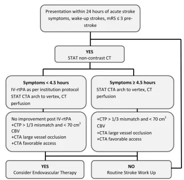
Figure 1 Suggested CT-based imaging triage protocol for endovascular acute ischemic stroke therapy
This is a proposed algorithm; hospitals may implement all or parts of this protocol as it applies to their practice pattern and available resources. Abbreviations: CBV⫽cerebral blood volume; CTA⫽CT angiography; CTP⫽CT perfusion; mRS⫽
Multi-MERCI, and 29% in Penumbra. The rate of fu-tile recanalization was 33%, compared with 54% for MERCI, 51% for Multi-MERCI, and 71% for Pen-umbra. A small CBV abnormality (p⬍0.0001) and large MTT-CBV mismatch (p⬍0.0001) were strong predictors of a good clinical outcome (mRS scoreⱕ2).26
Although retrospective studies and case series may support the use of CTP, there are limited prospective data on the use of CTP in identifying significant mis-match to triage AIS patients prior to ET in random-ized clinical trials. Other limitations of CTP include the coverage area and tissue sampling by CTP, which
is usually not sufficient to cover all ischemic regions; the lack of standardized optimal processing; and vari-ations in software, which may produce different CBV, CBF, and MTT maps.27
Despite these limitations, the advantages of CTP-based AIS triage are its wide availability (24/7), ultrafast acquisition and processing, and rapid in-terpretation. The CTP protocol appears to be safe, and there are no differences in the risk of acute ne-phropathy in hospitalized patients who did or did not receive AIS CT protocol, including those who are followed by conventional angiography.28CT
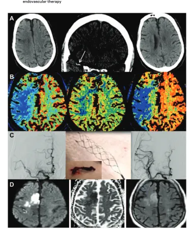
ra-Figure 2 An example of applying the CT protocol to triage patients with acute ischemic stroke for endovascular therapy
diation dose and safety concerns are balanced, given the benefit of potentially better guidance for ET.29
The data obtained from CT-based AIS triage pro-tocol (structural, vascular, and physiologic) imaging are particularly valuable when there is an unquestion-ably large area of mismatch or large area of infarct volume.
The CT-based AIS triage protocol algorithm, proposed in figure 1, is a suggested outline that can be modified by different institutions according to their available resources. AIS patient CT scan triage by care providers should take the utmost precedence, given the futility of delayed intervention and narrow therapeutic margin when performed for small areas of mismatch or large infarct volume. The CT and MRI radiology technologists are an instrumental part of the stroke team. Hospitals should have technol-ogists trained and geared toward the concept of
high-urgency AIS imaging triage for emergency performance and processing of CT imaging. In this proposed protocol (figure 1), patients who present within 24 hours of stroke symptoms onset are evalu-ated with NCCT, CTA from the arch to the vertex, and CTP. Rapid CT imaging, postprocessing with maximum-intensity projection, multiplanar reforma-tions, and CTP maps need to be in place as part of the routine AIS protocol. If the patient has significant mis-match on CTP and large-vessel occlusion on CTA without clinical improvement or large infarct volume, then ET may be considered (figure 1).
In figure 2, we present an example of a patient who was triaged with a CTP-based protocol. The patient presented with left hemiplegia, drowsiness, and gaze deviation to the left at 5 hours after symp-tom onset. A CT scan showed a small cortical stroke with a 9-mm right MCA clot. The CTP shows a
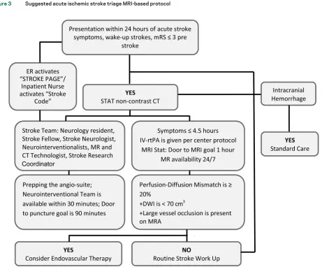
Figure 3 Suggested acute ischemic stroke triage MRI-based protocol
significant area of mismatch between the CBV and MTT or CBF. The angiogram demonstrated right M1 occlusion with complete recanalization by means of a Solitaire stent-retriever device (Covidien, CA) after 1 pass. The final infarct volume on MRI scan corresponded to the initial CT scan and CBV.
In summary, the AIS CTP-based protocol appears to be easily applied, safe, and feasible. The limitations to widely adapting CTP are the lack of optimal standard-ization of the processing, formatting, and variable threshold parameters to different CTP maps.
MRI PERFUSION PROTOCOL The approach of using MRI as the basis for selecting AIS patients for reperfusion treatment has been evaluated in several clinical trials.14 –22,30 –32 Diffusion-weighted Imaging Evaluation for Understanding Stroke Evolution (DEFUSE), Echoplanar Imaging Thrombolysis Evaluation Trial (EPITHET), and Desmoteplase in Acute Ischemic Stroke 2 (DIAS-2) are prospective trials that implemented penumbral imaging in select-ing patients for revascularization therapy.30 –32
The treatable penumbra was identified as a hypoper-fused region of at least 20% larger volume than the in-farct core; this threshold is considered to have sufficient volume of brain tissue that is at risk and is the target of reperfusion therapy. In the DEFUSE and EPITHET trials, patients were treated with IV rtPA within 6 hours after onset of symptoms. In DIAS-2, patients were
ran-domized by MRP and CTP mismatch criteria and treated with desmoteplase within 9 hours after symp-tom onset. A meta-analysis of these trials showed im-proved recanalization/reperfusion but demonstrated no significant improvement in clinical outcome with mismatch-based selection.33The sICH rate significantly increased after IV rtPA in this meta-analysis.33There were several potential explanations for these results. In the DEFUSE and EPITHET trials, the PWI volume was calculated with use of a T-max threshold⬎2 sec-onds delay, which may overestimate the penumbra size and include areas of oligemia. It was proposed later that a T-max⬎6 seconds delay might be more reliable and appropriate in evaluating penumbra.34The rate of large-vessel occlusion was 30% in the DIAS-2 trial,32which could explain the good clinical outcome in the control group. The hypothesis is that the penumbra without vessel occlusion rarely evolves into an infarction.
Another MRI-based selection strategy is to select AIS patients who have a moderate to severe deficit with limited abnormality on DWI for ET, and imaging-clinical mismatch (ICM). In a small study of 11 patients presenting beyond 8 hours of symp-tom onset who had ICM and underwent ET, a de-crease in the NIH Stroke Scale score⬎4 points was achieved in 72% of the subjects without sICH.35
The added value in MRI triage of AIS is the po-tential of excluding patients who have stroke mimics
Figure 4 Patient presenting with acute onset of global aphasia and right hemiparesis
and do not need ET. Stroke mimics were reported to be 3% to 11%.36 MRI could also exclude patients who are likely to be harmed by revascularization therapy. It is expected that about 10% to 18% of patients with mismatch will be excluded from ET due to the presence of a large DWI lesion on MRI.37 In a retrospective study of AIS, the diagnosis was changed after MRI in 20 (21%) of 97 patients, and the treatment plan was changed in 25 (26%) of the 97 patients.16The MRI data in this study were ac-quired in less than 15 minutes, a time that is hard to achieve in everyday practice.16
In addition to selecting patients who may benefit from ET, MRI-based triage may aid in excluding pa-tients who are at risk for sICH. The rate of sICH in ET trials was 8% to 11%,3–9and it was 3.5% to 9.5% in mismatch-guided IV thrombolysis trials.30 –32The sICH was related to many factors, including age, baseline stroke severity, hyperglycemia, and onset-to-treatment time. As shown in EPITHET and DEFUSE, DWI vol-ume is a very important predictor for sICH. A large infarct volume size, as defined by a DWI⬎100 mL or PWI ⬎100 mL with a T-max ⬎8 seconds, was considered to be a malignant mismatch.38This is ap-proximately greater than one-third the volume of the MCA territory and is found to have a high risk of ICH. Recent studies showed that a DWI lesion vol-ume ⬎70 cm3 has a high specificity for poor
out-come, with or without revascularization therapy.39 Furthermore, early blood– brain barrier (BBB) dis-ruption in AIS has a propensity for hemorrhage after reperfusion therapy. Detecting BBB permeability by MRP may identify patients who are at risk of hemor-rhage.39, 40One study using CTP demonstrated that BBB disruption has a sensitivity of 100% and a spec-ificity of 79% for sICH.41
The disadvantages of MRI-based protocol include the lack of 24/7 availability, lack of standardization of the software and threshold for postprocessing, and lack of feasibility in patients with metallic implant or claustrophobia. In addition, MRI-based AIS triaging protocol may delay revascularization therapy. In 1 single-center study of 87 consecutive AIS patients un-dergoing MRI-based protocol for ET triage, the time from MRI order to last MRI was approximately 100 minutes.42 This time remained unchanged in each consecutive 6-month period following implementa-tion of the MRI selecimplementa-tion strategy, indicating that the process to obtain the emergency MRI in AIS patients does not accelerate with increased experience over time.42It remains unclear if this added time for MRI is counterbalanced by significant benefit from MRI selection.
However, the sensitivity and specificity of MRI stroke imaging are high, and the information provided
on the underlying hemodynamics, pathophysiology, and vasculature are invaluable and can be incorporated into AIS triage protocols in centers where expedited and 24/7 MRI resources are available.
A proposed MRI-based triage protocol for ET in AIS is summarized in figure 3; the suggested protocol involves emergent MRI with DWI, PWI, fluid-attenuated inversion recovery, gradient echo, and MRA of the Circle of Willis in AIS patients who present within 24 hours from symptom onset. The presence of penumbra is determined by PWI-DWI mismatch or ICM. Patients receive IV rtPA prior to ET if qualified. Patients are selected for ET if the mis-match isⱖ20% by visual measurement, infarct volume is⬍70 cm3, or clinical symptoms are disproportionate
to the DWI volume. An example of a case treated with use of this MRI protocol is presented in figure 4.
DISCUSSION AIS patient selection for revascular-ization therapy using CT-based or MRI-based pen-umbral and vessel imaging protocols may be useful in avoiding futile therapy and improving clinical out-come. Imaging-based ET clinical trials and standard-ization of AIS acute imaging/triaging protocols across endovascular centers will further our current understanding of imaging-based patient selection for ET.
AUTHOR CONTRIBUTIONS
Both authors participated in the design, writing, and editing of the final manuscript.
ACKNOWLEDGMENT
The authors thank Alicia C. Castonguay, PhD, for her help with English language and grammar editing, and Osama O. Zaidat, MD, MS, for editorial input and providing figure 2.
DISCLOSURE
Dr. Shang reports no disclosures. Dr. Yavagal received honorarium from Penumbra Inc. for consultation and speaking; serves as an Associate Edi-tor forFrontiers in Endovascular Neurology; and serves as a consultant to Penumbra Inc., Codman Neurovascular, Micrus Inc., Genentech, and Boston Scientific.Go to Neurology.org for full disclosures.
Received June 28, 2011. Accepted in final form July 2, 2012.
REFERENCES
1. NINDS Study Group. Tissue plasminogen activator for acute ischemic stroke. N Engl J Med 1995;333:1581– 1587.
2. Hacke W, Kaste M, Bluhmki E, et al.; ECASS Investiga-tors. Thrombolysis with alteplase 3 to 4.5 hours after acute ischemic stroke. N Engl J Med 2008;359:1317–1329. 3. Furlan A, Higashida R, Wechsler L, et al. Intra-arterial
prourokinase for acute ischemic stroke: the PROACT II study: a randomized controlled trial: Prolyse in Acute Ce-rebral Thromboembolism. JAMA 1999;282:2003–2011. 4. Ogawa A, Mori E, Minematsu K, et al.; MELT Japan
the Middle Cerebral Artery Embolism Local Fibrinolytic Intervention Trial (MELT) Japan. Stroke 2007;38:2633– 2639.
5. IMS Trial Investigators. Combined intravenous and intra-arterial recanalization for acute ischemic stroke: the inter-ventional management of stroke study. Stroke 2004;35: 904 –911.
6. IMS II Trial Investigators. The International Management of Stroke (IMS) II Study. Stroke 2007;38:2127–2135. 7. Smith WS, Sung G, Starkman S, et al.; for the MERCI
Trial Investigators. Safety and efficacy of mechanical em-bolectomy in acute ischemic stroke: results of the MERCI Trial. Stroke 2005;36:1432–1438.
8. Smith WS, Sung G, Saver J, et al., for the Multi MERCI investigators. Mechanical thrombectomy for acute isch-emic stroke: final results of the Multi MERCI Trial. Stroke 2008;39:1205–1212.
9. The Penumbra Pivotal Stroke Trial Investigators. The Penumbra Pivotal Stroke Trial: safety and effectiveness of a new generation of mechanical devices for clot removal in intracranial large vessel occlusive disease. Stroke 2009;40: 2761–2768.
10. Assess the Penumbra System in the Treatment of Acute Stroke (THERAPY). Available at: clinicaltrials.gov/ct2/show/ NCT01429350?term⫽Therapy⫹trial&recr⫽Open&rslt⫽ Without&type⫽Intr&cond⫽Stroke&intr⫽Penumbra& rank⫽1.
11. Riedel CH, Zimmermann P, Jensen-Kondering U, Stin-gele R, Deuschl G, Jansen O. The importance of size: suc-cessful recanalization by intravenous thrombolysis in acute anterior stroke depends on thrombus length. Stroke 2011; 42:1775–1777.
12. Astrup J, Siesjo¨ BK, Symon L. Thresholds in cerebral isch-emia: the ischemic penumbra. Stroke 1981;12:723–725. 13. Astrup J, Symon L, Branston NM, Lassen NA. Cortical
evoked potential and extracellular K⫹and H⫹at critical levels of brain ischemia. Stroke 1977;8:51–57.
14. Chemmanam T, Campbell BC, Christensen S, et al; EPITHET Investigators. Ischemic diffusion lesion re-versal is uncommon and rarely alters perfusion-diffusion mismatch. Neurology 2010;75:1040 –1047. 15. Jovin TG, Yonas H, Gebel JM, et al. The cortical ischemic
core and not the consistently present penumbra is a deter-minant of clinical outcome in acute middle cerebral artery occlusion. Stroke 2003;34:2426 –2433.
16. Heidenreich JO, Hsu D, Wang G, et al. Magnetic reso-nance imaging results can affect therapy decisions in hy-peracute stroke care. Acta Radiol 2008;49:550 –557. 17. Wechsler LR. Imaging evaluation of acute ischemic stroke.
Stroke 2011;42:S12–S15.
18. Bandera E, Botteri M, Minelli C, Sutton A, Abrams KR, Latronico N. Cerebral blood flow threshold of ischemic penumbra and infarct core in acute ischemic stroke: a sys-tematic review. Stroke 2006;37:1334 –1339.
19. Murphy BD, Fox AJ, Lee DH, et al. Identification of pen-umbra and infarct in acute ischemic stroke using com-puted tomography perfusion-derived blood flow and blood volume measurements. Stroke 2006;37:1771–1777. 20. Schaefer PW, Barak ER, Kamalian S, et al. Quantitative assessment of core/penumbra mismatch in acute stroke: CT and MR perfusion imaging are strongly correlated when sufficient brain volume is imaged. Stroke 2008;39: 2986 –2992.
21. Jovin TG, Liebeskind DS, Gupta R, et al. Imaging-based endovascular therapy for acute ischemic stroke due to proximal intracranial anterior circulation occlusion treated beyond 8 hours from time last seen well: retrospective mul-ticenter analysis of 237 consecutive patients. Stroke 2011; 42:2206 –2211.
22. Axel L. Cerebral blood flow determination by rapid-sequence computed tomography: theoretical analysis. Ra-diology 1980;137:679 – 686.
23. Konstas AA, Goldmakher GV, Lee TY, Lev MH. Theo-retic basis and technical implementations of CT perfusion in acute ischemic stroke, part 1: theoretic basis. AJNR Am J Neuroradiol 2009;30:662– 668.
24. Lev MH, Segal AZ, Farkas J, et al. Utility of perfusion-weighted CT imaging in acute middle cerebral artery stroke treated with intra-arterial thrombolysis: prediction of final infarct volume and clinical outcome. Stroke 2001; 32:2021–2028.
25. Wintermark M, Reichhart M, Thiran JP, et al. Prognostic accuracy of cerebral blood flow measurement by perfusion computed tomography, at the time of emergency room admission, in acute stroke patients. Ann Neurol 2002;51: 417– 432.
26. Rai AT, Raghuram K, Domico J, Hobbs G, Carpenter J. Pre-intervention triage incorporating perfusion imaging improves outcomes in patients undergoing endovascular stroke therapy: a comparison with the device trials. J Neu-rointerv Surg Epub 2012 Feb 18.
27. Fahmi F, Marquering HA, Streekstra GJ, et al. Differences in CT perfusion summary maps for patients with acute ischemic stroke generated by 2 software packages. AJNR Am J Neuroradiol Epub 2012 May 3.
28. Lima FO, Lev MH, Levy RA, et al. Functional contrast-enhanced CT for evaluation of acute ischemic stroke does not increase the risk of contrast-induced nephropathy. AJNR Am J Neuroradiol 2010;31:817– 821.
29. Mnyusiwalla A, Aviv RI, Symons SP. Radiation dose from multidetector row CT imaging for acute stroke. Neurora-diol 2009;51:635– 640.
30. Albers GW, Thijs VN, Wechsler L, et al. Magnetic reso-nance imaging profiles predict clinical response to early reperfusion: the Diffusion And Perfusion Imaging Evalua-tion For Understanding Stroke EvoluEvalua-tion (DEFUSE) Study. Ann Neurol 2006;60:508 –517.
31. Davis SM, Donnan GA, Parsons MW, et al. Effects of alteplase beyond 3h after stroke onset in the Echoplanar Imaging Thrombolytic Evaluation Trial (EPITHET): a placebo-controlled randomized trial. Lancet Neurology 2008;7:299 –399.
32. Hacke W, Furlan AJ, Al-Rawi Y, et al. Intravenous des-moteplase in patients with acute ischaemic stroke selected by MRI perfusion-diffusion weighted imaging or perfu-sion CT (DIAS-2): a prospective, randomised, double-blind, placebo-controlled study. Lancet Neurol 2009;8: 141–150.
33. Mishra NK, Albers GW, Davis SM, et al. Mismatch-based delayed thrombolysis: a meta-analysis. Stroke 2010;41: e25– e33.
34. Olivot JM, Mlynash M, Thijs VN, et al. Optimal Tmax threshold for predicting penumbral tissue in acute stroke. Stroke 2009;40:469 – 475.
stroke based on clinical-diffusion mismatch. AJNR Am J Neuroradiol 2009;30:1024 –1027.
36. Chernyshev OY, Martin-Schild S, Albright KC, et al. Safety of tPA in stroke mimics and neuroimaging-negative cerebral ischemia. Neurology 2010;74:1340 –1345. 37. Yoo AJ, Verduzco LA, Schaefer PW, Hirsch JA, Rabinov
JD, Gonza´lez RG. MRI-based selection for intra-arterial stroke therapy: value of pretreatment diffusion-weighted imaging lesion volume in selecting patients with acute stroke who will benefit from early recanalization. Stroke 2009;40:2046 –2054.
38. Mlynash M, Lansberg MG, De Silva DA, et al.; DEFUSE-EPITHET Investigators. Refining the definition of the malignant profile: insights from the DEFUSE-EPITHET pooled data set. Stroke 2011;42:1270 –1275.
39. Tanne D. Imaging blood-brain barrier disruption: an evolv-ing tool for assessevolv-ing the risk of hemorrhage after thromboly-sis. Nat Clin Pract Neurol 2008;4:644 – 645.
40. Bang OY, Buck BH, Saver JL, et al. Prediction of hemor-rhagic transformation after recanalization therapy using T2*-permeability magnetic resonance imaging. Ann Neu-rol 2007;62:170 –176.
41. Hom J, Dankbaar JW, Soares BP, et al. Blood-brain barrier permeability assessed by perfusion CT predicts symptomatic hemorrhagic transformation and malignant edema in acute ischemic stroke. AJNR Am J Neuroradiol 2011;32:41– 48. 42. Pham T, Shang T, Milan D, et al. MRI turn-around time
DOI 10.1212/WNL.0b013e3182695800
2012;79;S86-S94
Neurology
Tiesong Shang and Dileep R. Yavagal
Application of acute stroke imaging: Selecting patients for revascularization therapy
This information is current as of September 24, 2012
Services
Updated Information &
http://n.neurology.org/content/79/13_Supplement_1/S86.full including high resolution figures, can be found at:
References
http://n.neurology.org/content/79/13_Supplement_1/S86.full#ref-list-1 This article cites 38 articles, 25 of which you can access for free at:
Subspecialty Collections
roke
http://n.neurology.org/cgi/collection/other_cerebrovascular_disease__st Other cerebrovascular disease/ Stroke
http://n.neurology.org/cgi/collection/mri MRI
http://n.neurology.org/cgi/collection/dwi DWI
e
http://n.neurology.org/cgi/collection/all_cerebrovascular_disease_strok All Cerebrovascular disease/Stroke
following collection(s):
This article, along with others on similar topics, appears in the
Permissions & Licensing
http://www.neurology.org/about/about_the_journal#permissions its entirety can be found online at:
Information about reproducing this article in parts (figures,tables) or in
Reprints
http://n.neurology.org/subscribers/advertise
Information about ordering reprints can be found online:
rights reserved. Print ISSN: 0028-3878. Online ISSN: 1526-632X.
1951, it is now a weekly with 48 issues per year. Copyright Copyright © 2012 by AAN Enterprises, Inc.. All ® is the official journal of the American Academy of Neurology. Published continuously since