Implications of the spatial and temporal regulation
of Hox genes on development and evolution
JAMES CASTELLI-GAIR*
Department of Zoology, University of Cambridge, Cambridge, United Kingdom
ABSTRACT Nearly 20 years have passed since Ed Lewis revealed the importance of Hox genes in the specification of different segments in the anterior-posterior axis of the fly. Pioneering studies by several authors, among others García-Bellido and his student Ginés Morata, helped to elaborate a theory of segmental specification that was strengthened with the arrival of molecular techniques to the field of Developmental Biology. The conservation of Hox genes in metazoans at the level of sequence, function and complex organization has resulted in the export of this Drosophila theory as a paradigm to interpret the development of axial specification in organisms less amenable to experimental study. There are two main ways to interpret how Hox genes work in Drosophila. One considering Hox genes as "segment identity" factors giving global properties to the segments in which they are active. Another considering Hox genes as encoding spatially restricted transcription factors required for a number decisions taken at the cellular level. Here I use published and unpublished experimental data to illustrate that early activation of the Hox genes does not establish a gene code that leads to "segment identity". I will stress the point that Hox expression patterns develop with the embryo, that there are many genes involved in this modulation, and that the changing pattern of expression is important to achieve the final shape of the animal. I will show that, by interpreting Hox gene function in this way, some apparently paradoxical results in the Hox field can be answered. Finally, I discuss the implications of dynamic Hox gene expression on the evolution of segment morphology.
KEY WORDS:
gene regulation, hox development, evolution
0214-6282/98/$10.00 © UBC Press
Printed in Spain
*Address for reprints: Department of Zoology, University of Cambridge, Cambridge CB2 3EJ, United Kingdom. FAX: 44-1223-336676. e-mail: [email protected]
Introduction
A wild type fly has a well defined head, thorax and abdomen. Genetic analysis showed that flies lacking the three Hox genes of the bithorax complex: Ultrabithorax (Ubx), abdominal-A (abd-A) and Abdominal-B (Abd-B), are only composed of a head and an extended thorax as all the abdominal segments resemble thoracic segments (Lewis, 1978; Sánchez-Herrero et al., 1985). Lewis’ work led García-Bellido to propose that Hox genes regulate body morphology acting as selectors of alternative developmental path-ways (García-Bellido, 1975). What they would do is to regulate other genes under their command so that in a certain segment a particular Hox gene would modulate downstream genes involved in making the abdomen, while in a more anterior segment another Hox gene will modulate genes that generate a thorax. These genetic propositions implied that, in Drosophila, the Hox genes encode regulatory proteins, and that they are expressed in different patterns along the anterior-posterior axis. Both proposals were confirmed by molecular biology studies: the Hox genes encode
transcription factors active in different segments where they con-trol the activation of target genes required for the development of the structures typical of that segment.
An important experiment that influenced our current views on segment specification was the observation that, in the epidermis, Hox gene function is cell autonomous. Morata and García-Bellido showed that if a small group of cells stop expressing the Ubx gene in T3 they would form T2 structures (Morata and García-Bellido, 1976). Comparing the number of cells of the wing and the haltere at different stages in development, they found that the differences between these homologous thoracic appendages are established very early in development. However, they also showed that Ubx function is required continuously to maintain cell affinities and patterns of cell division during development. Consistent with these observations, molecular studies proved that the Hox genes are expressed from early embryogenesis until the end of development (reviewed in Akam, 1987).
controlled by two classes of genes. One class, required only transiently to activate hox expression, is integrated by the genes involved in subdividing the embryo into segments (Nüsslein-Volhard et al., 1984; Ingham and Martínez-Arias, 1986; White and Lehmann, 1986; Harding and Levine, 1988; Irish et al., 1989; Jack and McGinnis, 1990; Casares and Sánchez-Herrero, 1995). The other class, required for the maintenance of Hox gene expression, is integrated by genes of the Polycomb and Trithorax groups which are dispensable for the early activation (Struhl and Akam, 1985; Wedeen et al., 1986; Breen and Harte, 1993).
These results put together have given rise to the Hox code theory (reviewed in Lawrence and Morata, 1994): The early activa-tion of a Hox gene by the segmentaactiva-tion genes in a metamere provides the cells expressing it with a "segmental identity". This identity is maintained by the Polycomb and trithorax group genes by keeping the early state of Hox expression during development. A cell therefore has the same identity all along development and accordingly gives rise to the structures specific of that segment. As each segment expresses different combinations of Hox genes, each has a different code resulting in the anterior posterior diver-sification of the segments of the animal.
The Hox code theory initially developed in Drosophila is now used as a paradigm to explain how Hox genes work in other organisms. It is therefore important to highlight the points that the Hox code hypothesis cannot explain but are never discussed in reviews about Drosophila development. While in earlier work Hox genes were seen as affecting the development of particular struc-tures (Ventral pits, Keilin organs... Lewis, 1978) or the cellular properties of cells (cell affinities, mitotic rates... García-Bellido, 1975; Morata and García-Bellido, 1976), the focus has later shifted to metameric units studied as a whole. We do not talk about cell properties or particular structures and organs but of "segment identity". While this view is easier to use when talking in abstract terms it does not always work when applied to the development of particular organs. In what follows I will discuss why we should study Hox genes not as regulators of segment identity but as spatially restricted transcription factors involved in the control of cell behaviors and the specification of segment structures.
Relativity in the development of segment identity: the
function of HOX transcription factors changes during
development
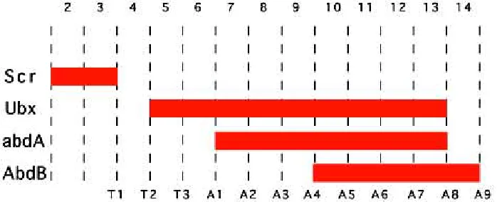
Figure 1 represents the common summary about Hox gene expression in Drosophila presented in reviews (Akam, 1987;
Lawrence and Morata, 1994; Carroll, 1995). Ubx is required for the development of T3 and A1, abd-A for A2-A7 and Abd-B for A5-A9. This summary compiles data that include experimental results obtained in the embryo and in the larva and its weakness is, paradoxically, its simplicity and elegance. The model in Figure 1 shows a static view of Hox expression in which Hox genes are continuously expressed within the same parasegmental bounda-ries. This situation is not real, as all Hox genes have a temporal and spatial expression that does not fit with that idealization.
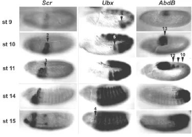
To illustrate some exceptions I will concentrate on the expres-sion of the Scr, Ubx and Abd-B genes (Fig. 2). First, in all cases the Hox genes are expressed in fewer segments at early than at late stages of development. Second, at a single time point, a segment is a mosaic of cells expressing and not expressing a given Hox gene. Third, although the expression of Hox genes is frequently parasegmental, there are many cases in which this is not true (Fig. 3 and Martínez-Arias et al., 1987; DeLorenzi and Bienz, 1990; Irvine et al., 1991).
Such exceptions complicate the understanding of segment specification. For example, what is the segment identity of a cell that has had two different Hox codes during development? What happens to cells of the same segment which express different Hox codes? These can be viewed as minor exceptions when looking at the global Hox expression, but should not be ignored as they can result in dramatic morphological changes when considering the development of a particular organ (see below).
As the protein patterns are changing with the developmental stage of the embryo, unless we define experimentally when HOX protein function is required, we are not able to decide what aspects of these expression patterns are significant for development. Because during embryogenesis the embryo is forming a collection of different cells (muscle, epidermis, nervous system) the require-ment of HOX function has to be answered independently for each different cell type. As doing this complicates matters we should first find out if such complex approach is necessary.
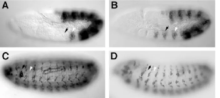
We can test if the conventional model of Hox gene function presented in Figure 1 can predict the outcome of a simple experi-ment: If we prevent the seventh abdominal segment (A7) from expressing Abd-B, would this segment transform to a more anterior segment? The answer is yes and no. In Figure 4 we see the result of preventing Abd-B expression in A7 in the embryo and in the adult. Such a mutation strongly affects the A7 segment in the adult while many A7 structures in the embryo remain normal (PNS, CNS, oenocytes) (Heuer and Kaufman, 1992). The usual response to this observation has been to suggest that the homeotic change in
these cells is too subtle to be detected. A simpler answer is that we do not see changes because there are none. In the wild type fly Abd-B is only required in A7 for the formation of some structures. These include the larval denticle belts and the cells that will form the adult epidermis. The normal development of A7 structures like the PNS or the oenocites does not require Abd-B; in fact unrestricted expression of ABD-B protein disrupts the normal development of A7 (Lamka et al., 1992; Castelli-Gair et al., 1994).
This observation explains the morphology of the A7 segment in the wild type embryo. The PNS in the abdomen is identical from A2 to A7 (Ghysen et al., 1986) despite the fact that A2-A4 do not express Abd-B and A5-A8 do (Figs. 1 and 2). This does not mean that Abd-B is dispensable for PNS development as Abd-B mutants affect the PNS of A8 (Fig. 4). Why do A7 and A8 respond in such a different way to Abd-B expression? One possible answer could be that there is a difference in the temporal expression of Abd-B between A7 and A8 (Fig. 2). Expression is detectable previous to 6 h in A8 and it is only later seen in A7 and more anterior segments in a restricted spatial expression.
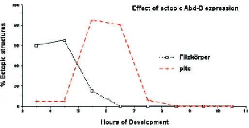
To test if Abd-B expression at different stages of development has different effects on development we can control when we express Abd-B using a heat shock promoter. The results of expressing ectopically Abd-B for one hour at different stages of development are shown in Figure 5. Unrestricted expression of ABD-B at 3-5 h of development results in the appearance in anterior segments of the posterior spiracles normally restricted to A8. Unrestricted expression at 5-7 h does not result in the appear-ance of ectopic posterior spiracles but affects sensory organ development. In the abdomen of wild type flies basiconical sensilla are only present in segments A8 and A9, while in embryos heat shocked from 5-7 h they also appear from A1-A7. Unrestricted expression at 8 h results in none of the previous transformations but still affects the development of denticle belts.
These experiments should not be over-interpreted as the transient Abd-B expression they generate is an artificial situation never present in normal embryos. However, these results high-light the fact that cells respond differently to Hox expression at different stages of development. Therefore we cannot talk about a "Hox code" because in a particular group of cells the outcome of Hox expression changes with time.
In the above example the PNS morphology can be affected by Hox function at a stage when Abd-B is not expressed in A7. At a later stage when ABD-B is expressed in A7 it will not be capable of affecting PNS morphology, but will be able to affect the elements that give rise to larval denticle belts and imaginal abdominal cuticle.
Fig 2. Patterns of expression of Scr, Ubx and Abd-B protein during early embryogenesis.Each column shows the expression of a different Hox gene at progressively later stages of develop-ment. The expression is very dynamic and comes up in different segments at different stages. Scr is first expressed in PS2, later it expands to PS3. At stage 9 Ubx expression is confined to the abdo-men (PS6-13), at stage 10 it expands to the thorax up to PS5. At stage 15 on-wards Ubx is expressed in PS4, a region where no Ubx function has been found. Abd-B expression at early stages is con-fined to the last abdominal segments (PS 13-14). At later stages Abd-B is ex-pressed also in PS10-12. In many (para)segments there are cells express-ing and not expressexpress-ing a particular Hox gene. This mosaic expression is particu-larly clear for Scr in PS3; for Ubx in PS5 and for Abd-B in PS10-12.
Other Hox genes also have different developmental effects when expressed at different times. Ubx has three different effects on leg development. In A1 Ubx prevents leg formation; in T3 Ubx is required continuously for the normal leg morphology while in T2 Ubx has to be active before 6 h of development and then turned off (Morata and Kerridge, 1981).
The regulation of the Distalless (Dll) gene, a gene required for leg development that is a direct target of Ubx, illustrates at the molecular level how dynamic Hox expression influences the morphology of the animal (Castelli-Gair and Akam, 1995). UBX represses Dll expression in A1 but not in T3 despite the fact that UBX is expressed in both segments in the relevant cells. The reason for this difference resides in the temporal expression of Ubx in T3 and A1 and in the regulation of Dll itself. The expression of Dll in the leg primordia is controlled by two enhancers (Vachon et al., 1992). One enhancer is sufficient for the early activation of Dll in both the thoracic and abdominal segments, but the binding of UBX protein to this enhancer blocks Dll expression in the abdominal segments (Vachon et al., 1992). Blocking of the early Dll enhancer does not occur in T3 as no Ubx is expressed in T3 at this stage of development. The early enhancer is not sufficient to maintain Dll expression in the leg primordia for which a second enhancer is required. The second enhancer is not repressed by UBX and depends on Dll autoregulation (Castelli-Gair and Akam, 1995). The use of the two enhancers and the temporal expression of Ubx in T3 and A1 explains the different effects of UBX protein on the regulation of Dll. If Dll gets activated in the leg primordia before UBX is expressed, the DLL protein autoregulatory loop will maintain Dll expression independently of UBX presence at later stages (the situation found in T3). On the other hand if UBX gets activated earlier, the initial expression of Dll will be blocked and the feedback loop will not occur, preventing the formation of a leg (the situation found in A1). In this way differences in timing of expression of about one hour can have drastic consequences on the morphology of the organism.
Not only leg development depends on the correct temporal modulation of Ubx expression. In a single PNS organ, the dorsal Kölbchen, ABD-A or UBX expression has different temporal effects (Castelli-Gair et al., 1994). Early expression affects the dorsal-ventral migration of the sensory organ but not the specifi-cation of the organ as abdominal or thoracic; while later expres-sion will select the abdominal differentiation of the sensory organ. This case is different from that described for the leg, in the sense that the location and differentiation of the sense organ seem to be independent decisions, while the presence or absence of a leg seems to involve a program that is turned on and maintained: a developmental subroutine. However, in both cases minor changes in Hox expression can have important effects on the final morphol-ogy of a segment, as this will be the result of a complex outcome that involves the cell’s competence to take certain decisions.
Recent results studying the regulation of the mouse Hoxd-11 gene prove that tight temporal regulation of Hox gene expression is also required for the normal segment morphology in verte-brates. The deletion of a conserved enhancer results in the local delay of Hox expression. Although those cells do express normal levels of Hox genes at later stages, this delay in Hox expression results in the transformation of sacral vertebrae into lumbar morphology (Zákány et al., 1997).
The results summarized above do not imply that every minor detail of the Hox expression pattern has an influence on segment morphology. Ectopic induction of Hox genes does not have an effect at all stages of development. Also, in the wild type fly some Hox genes are expressed in regions were they do not affect the segment morphology. For example, Ubx is expressed in T1 at late stages of development without influencing its development. The most extreme case of this is that of the proboscipedia gene. While proboscipedia is expressed in the head of both embryo and larva, the expression in the embryo is not required for the development of the larval head, while larval expression is required for the adult head.
In summary, Hox genes are not controlling segment identity, but cellular behavior that will result in a certain segment morphol-ogy. This is what Hox genes do in unsegmented organisms like C. elegans (Salser and Kenyon, 1996) and is probably what they did in the common ancestor of all metazoans. Segment identity is a subjective concept that originates from the observation that in a particular species, a number of cell characteristics are always associated in a given segment.
Modulation of Hox expression
The above results show that the spatial and temporal regula-tion of Hox gene expression has to be finely tuned during normal development . In Drosophila, it is thought that we have already got a good understanding of how Hox gene expression is controlled. In the blastoderm, the genes that are involved in subdividing the embryo into segments also activate Hox expression in restricted anterior-posterior domains. This is achieved by the combination of activator genes (fushi-tarazu, evenskipped, tailless, etc) in conjunction with repressors (hunchback) that establish the do-mains of Hox expression . Both activators and repressors are only transiently expressed. As, in general, Hox genes do not autoregulate, the patterns that segmentation genes establish have to be maintained later in development by other genes. These are the genes of the Polycomb and trithorax groups.
The above elements suggest an early crystallized pattern of Hox expression that is propagated during development, but there must be other genes responsible for the subtle Hox gene modu-lation. Which are these genes? It has been proposed that the segmentation genes of the segment polarity class are involved in the local modulation of Hox patterns (Martínez-Arias and White, 1988). In contrast to other segmentation genes, segment polarity genes are not expressed transiently, but they are maintained during development. The best documented case is the down regulation of the expression of Ubx in the posterior compartment by the segment polarity gene engrailed (en). As we have seen this down regulation is important for the formation of the legs, due to the modulation of the levels of UBX protein (Mann, 1994) or timing of Ubx expression (Castelli-Gair and Akam, 1995). In en mutants the expression of Ubx in A1 increases (Martínez-Arias and White, 1988; Mann, 1994). Conversely ectopic expression of en using a heat shock promoter results in the down regulation of Ubx in all segments as a result of which the leg marker Dll is expressed in all the abdominal segments (Mann, 1994).
We know very few genes modulating Hox expression. The complex modulation of Hox expression suggests that they must form a large and diverse class of genes responsible for the temporal as well as dorsal-ventral and even cell specific regula-tion. The reason for the scarce knowledge of Hox modulators probably resides in the subtlety of their effects on development. Good candidates to be Hox modulators are the zinc-finger pro-teins encoded by the spalt (sal) and sal-related (sal-r) genes (Kühnlein et al., 1994; de Celis et al., 1996). In sal mutants (or both) Ubx becomes expressed in more anterior segments (Fig. 6). sal has been classified as a Pc group gene (Casanova, 1989). Several reasons suggest that sal is not a Pc group gene but forms part of this less well characterized class of Hox modulators. First, genes of the Pc-G are required to repress all Hox genes in regions where they were not initially activated; in contrast, sal mutants
only affect Ubx expression while Scr, abd-A, and Abd-B expres-sion is not affected (data not shown). Casanova suggested that Scr is expressed ectopically in double mutants for sal and the bithorax complex (Casanova, 1989) but ectopic Scr expression can be detected in bithorax complex mutants alone (Pelaz et al., 1993). Second, in sal mutations the spatial expression of Ubx in PS3 and PS4 mimics the spatial expression of Ubx in PS5, suggesting that sal interacts with the subset of enhancers respon-sible for that pattern. Third, the result of this anterior Ubx expres-sion is not a complete transformation of one segment into another. While the PNS and the denticle belt patterns in the thoracic cuticle in sal mutants are normal the formation of the anterior spiracle is repressed (Fig. 6) resulting in the formation of a segment with mosaic characteristics not to be found in the wild type fly.
The existence of a large number of modulator genes with small effects on the Hox regulatory regions provides a plasticity to the evolution of different segment morphologies that is unlikely to have been missed in evolution. Minor changes in the cis regulatory elements of the target genes to which these modulator proteins bind could result in the modification of segment morphology without creating animals of improbable viability.
Hox gene regulation and the evolution of body shape
From early studies it was realized that the Hox genes could be paramount in the evolution of body shapes. Ed Lewis proposed that the evolution of the insects body plan from a precursor Arthropod had depended on the appearance of new Hox genes by duplication of ancestral ones. These new genes would be ex-pressed in more posterior segments and their evolution would result in the appearance of new segmental structures. Initially, with only one Hox gene all the trunk segments would look alike. After duplication, the gene would acquire new functions leading to the appearance of a distinct thorax and abdomen, further duplica-tion and divergence events leading to the transformaduplica-tion of four winged insects into two winged flies. Mutations in Hox genes would reverse evolutionary processes resulting in atavistic
formations (García-Bellido, 1977). That is, these mutations in Hox genes would take the organism "back" in evolution.
The finding that Hox genes are present in vertebrates and in non segmented animals (McGinnis et al., 1984; Costa et al., 1988) seemed inconsistent with the view that Arthropod body shape and Hox genes evolved together: if the ancestors of insects already had a complete set of Hox genes, their invention could not be responsible for the evolution of segment diversity in arthropods. Despite of this, Hox genes have a great potential to change body shapes as shown by the effect of mutations and it is unlikely that they have not participated directly in some aspects of evolution. Hox genes are unlikely to have participated in the sudden invention of a new type of segment but, as we have seen, by modulating its patterns of expression in time or space they can generate changes in either a specific cell type or a structure.
Modulation of Hox expression can be achieved by minor changes in an enhancer. The cis regulatory elements in Drosophila have enhancers with major effects in body shape which are in general those involved in gene maintenance. There is also evidence of a different class of enhancer that is acting in a redundant or collabo-rative way. For example, a construct containing 8.8 Kb of Ubx cis regulatory sequences (the BRE element) contains enhancers capable of driving expression in A1 (PS6) in the embryo; a mutation (the bx34e-prv allele) that removes these sequences develops a
normal A1 segment showing that other enhancers can compen-sate for the absence of the BRE element (Peifer and Bender, 1986; Qian et al., 1991). Experiments studying the adult structures of A1 also suggest the presence of redundant Ubx cis regulatory ele-ments. There are two Ubx cis regulatory regions that when re-moved independently do not affect the development of A1, how-ever, when removed simultaneously the A1 segment is completely missing (Castelli-Gair et al., 1992). Many other examples of enhancers acting collaboratively have been described. It is at this level that subtle evolution could act.
There are several cases in which changes in Hox spatial expression have been correlated with the evolution of segment morphology. A very nice case has been shown in Crustaceans (Averof and Patel, 1997). Genes homologous to Drosophila’s Ubx and abd-A are expressed in the thorax in a region that normally forms swimming appendages. However, in some species one or
more thoracic appendages are transformed into feeding append-ages (maxillipeds). The number of maxillipeds varies among species. Studying thirteen different species of Crustaceans Averof and Patel show that UBX /ABD-A protein expression correlates with thoracic segments bearing swimming appendages, while thoracic segments that do not express these proteins are trans-formed into maxillipeds.
Another case has been shown in Lepidoptera (Warren et al., 1994). Butterfly caterpillars have abdominal prolegs. Prolegs differ from thoracic legs in morphology but both express the homolog of Dll. As we have previously seen Dll is repressed in the abdomen of Drosophila by UBX and ABD-A. Why is Dll expressed in the abdomen of butterflies? Warren and collaborators show that Dll activation in the abdomen of the butterfly is delayed with respect to the thorax. At later stages however, ABD-A protein is down regulated in the abdomen allowing activation of Dll.
These two examples suggest that changes of segment pat-terns during evolution could have been due to the modulation of Hox expression. However, a note of caution should be said in the interpretation of expression patterns in the absence of experi-mental data. Not all expression cases will turn out to be so clearly cut. For example, most insects have wings in T2 and T3. In flies the T3 pair of wings has evolved into specialized balancing structures, the halteres. Because when flies do not express Ubx in T3 the halteres are transformed into wings, one could predict that butterflies do not express the homolog of Ubx in T3. This is not the case, butterflies do express Ubx in the T3 wings (Warren et al., 1994), a fact that is not surprising if we think on the possible existence of developmental subroutines as that described for Dll in the leg. It is under this light that we can interpret this result. Ubx in butterflies would not prevent the development of wings in T3 if Ubx is expressed at a moment in which wing development has been irreversibly triggered.
The application of molecular techniques to the study of com-parative development is going to provide many interesting data about how developmental mechanisms evolved. Unfortunately, the interpretation of these data will depend very much on the views of the researcher. To take full advantage of these advances we should try to use model organisms where hypothesis can be tested or to develop experimental approaches in organisms where such
approaches are not available. This will be the only way to get a clear understanding of the evolution of developmental mechanisms.
Materials and Methods
The Abd-BM5 and Abd-BD14 double heterozygous flies were used for
Figure 4c and Abd-BM1 null allele for Figure 4d (Casanova et al., 1986;
Zavortink and Sakonju, 1989). Two sal mutant alleles were used salIIB57 and
Df(2L)5 a deletion removing both sal and sal-r (Nüsslein-Volhard et al., 1984; de Celis et al., 1996). The following antibodies were used: a polyclonal anti-CUT (Blochinger et al., 1990), and the monoclonals FP3.38 anti-UBX (White and Wilcox, 1984), 6H4 anti-SCR (Glicksman and Brower, 1988) and 1A2E9 anti-ABD-B (Celniker et al., 1989).
For heat shock experiments eggs from HS-ABD-Bm F4 flies (Lamka et al., 1992) were collected every hour and aged for different periods at 25°C. A single 1 h heat shock was induced by immersion in a 37°C water bath. Heat shocked eggs were transferred to agar plates and allowed to develop prior to cuticle preparation. Cuticle preparations and antibody stainings were done as described in Castelli-Gair and Akam (1995).
Acknowledgments
I thank Hilary Reed and Alfonso Martínez Arias for discussing the manuscript; Ana Busturia and José Félix de Celis and the Tübingen stock centre for stocks; Rob White, D. Brower, Mar Ruíz Gómez for antibodies and Michael Akam in whose laboratory some of these experiments were done. This work was supported by The Royal Society.
References
AKAM, M. (1987). The molecular basis for metameric pattern in the Drosophila embryo. Development 101: 1-22.
AVEROF, M. and PATEL, N.H. (1997). Crustacean appendage evolution associated with changes in Hox gene expression. Nature 388: 682-686.
BLOCHINGER, K., BODMER, R., JAN, L.Y. and JAN, Y.N. (1990). Patterns of expression of Cut, a protein required for external sensory organ development in wild-type and cut mutant Drosophila embryos. Genes Dev. 4: 1322-1331.
BREEN, T.R. and HARTE, P.J. (1993). trithorax regulates multiple homeotic genes in the bithorax and Antennapedia complexes and exerts different tissue-specific, parasegment-specific and promoter-specific effects on each. Development 117: 119-134.
CARROLL, S.B. (1995). Homeotic genes and the evolution of arthropods and chordates. Nature 376: 479-485.
CASANOVA, J. (1989). Mutations in the spalt gene of Drosophila cause ectopic expression of Ultrabithorax and Sex combs reduced. Roux Arch. Dev. Biol. 198: 137-140.
CASANOVA, J., SÁNCHEZ-HERRERO, E. and MORATA, G. (1986). Identification and Characterisation of a Parasegment Specific Regulatory Element of the Abdominal-B Gene of Drosophila. Cell 47: 627-636.
CASARES, F. and SÁNCHEZ-HERRERO, E. (1995). Regulation of the infra-abdomi-nal regions of the bithorax complex of Drosophila. Development 121: 1855-1866.
CASTELLI-GAIR, J. and AKAM, M. (1995). How the Hox gene Ultrabithorax specifies two different segments: The significance of spatial and temporal regulation within metameres. Development 121: 2973-2982.
CASTELLI-GAIR, J., CAPDEVILA, M.P., MICOL, J.L. and GARCÍA-BELLIDO, A. (1992). Positive and negative cis-regulatory elements in the bithoraxoid region of the Drosophila Ultrabithorax gene. Mol. Gen. Genet. 234: 177-184.
CASTELLI-GAIR, J., GREIG, S., MICKLEM, G. and AKAM, M. (1994). Dissecting the temporal requirements for homeotic gene function. Development 120: 1983-1995.
CELNIKER, S.E., KEELAN, D.J. and LEWIS, E.B. (1989). The molecular genetics of the bithorax complex of Drosophila: characterization of the products of the Abdominal-B domain. Genes Dev. 3: 1424-1436.
COSTA, M., WEIR, M., COULSON, A., SULSTON, J. and KENYON, C. (1988). Posterior pattern formation in C. elegans involves position-specific expression of a gene containing a homeobox. Cell 55: 747-756.
DE CELIS, J.F., BARRIO, R. and KAFATOS, F.C. (1996). A gene complex acting downstream of dpp in Drosophila wing morphogenesis. Nature 381: 421-424.
DELORENZI, M. and BIENZ, M. (1990). Expression of Abdominal-B homeoproteins in Drosophila embryos. Development 108: 323-329.
GARCÍA-BELLIDO, A. (1975). Genetic control of wing disc development in Dro-sophila. In Cell Patterning (Elsevier, Amsterdam) Ciba Found. Symp. 29: 161-178.
GARCÍA-BELLIDO, A. (1977). Homoeotic and atavic mutations in insects. Am. Zool. 17: 613-629.
GHYSEN, A., DAMBLY-CHAUDIERE, C., ACEVES, E., JAN, L.Y. and JAN, Y.N. (1986). Sensory neurons and peripheral pathways in the Drosophila embryo. Roux Arch. Dev. Biol. 195: 281-289.
GLICKSMAN, M.A. and BROWER, D.L. (1988). Expression of the Sex combs reduced protein in Drosophila larvae. Dev. Biol. 127: 113-118.
HARDING, K. and LEVINE, M. (1988). Gap genes define the limits of Antennapedia and Bithorax gene expression during early development in Drosophila. EMBO J. 7: 205-214.
HEUER, J.G. and KAUFMAN, T.C. (1992). Homeotic genes have specific functional roles in the establishment of the Drosophila embryonic peripheral nervous system. Development 115: 35-47.
INGHAM, P.W. and MARTÍNEZ-ARIAS, A. (1986). The correct activation of Antennapedia and bithorax complex genes requires the fushi tarazu gene. Nature 324: 592-597.
IRISH, V.F., MARTÍNEZ-ARIAS, A. and AKAM, M. (1989). Spatial regulation of the Antennapedia and Ultrabithorax homeotic genes during Drosophila early develop-ment. EMBO J. 8: 1527-1539.
IRVINE, K.D., HELFAND, S.L. and HOGNESS, D.S. (1991). The large upstream control region of the Drosophila homeotic gene Ultrabithorax. Development 111: 407-424.
JACK, T. and MCGINNIS, W. (1990). Establishment of the Deformed expression stripe requires the combinatorial action of coordinate, gap and pair-rule proteins. EMBO J. 9: 1187-1198.
KÜHNLEIN, R.P., FROMMER, G., FRIEDRICH, M., GONZÁLEZ-GAITÁN, M., WEBER, A., WAGNER-BERNHOLZ, J.F., GEHRING, W.J., JÄCKLE, H. and SCHUH, R. (1994). spalt encodes an evolutionary conserved zinc finger protein of novel structure which provides homeotic gene function in the head and tail region of the Drosophila embryo. EMBO J. 13: 168-179.
LAMKA, M.L., BOULET, A.M. and SAKONJU, S. (1992). Ectopic expression of UBX and ABD-B proteins during Drosophila embryogenesis: competition, not a func-tional hierarchy, explains phenotypic suppression. Development 116: 841-854.
LAWRENCE, P.A. and MORATA, G. (1994). Homeobox genes: their function in Drosophila segmentation and pattern formation. Cell 78: 181-189.
LEWIS, E.B. (1978). A gene complex controlling segmentation in Drosophila. Nature 276: 565-570.
MANN, R.S. (1994). engrailed-mediated repression of Ultrabithorax is necessary for the parasegment 6 identity in Drosophila. Development 120: 3205-3212.
MARTÍNEZ-ARIAS, A. and WHITE, R.A.H. (1988). Ultrabithorax and engrailed expression in Drosophila embryos mutant for segmentation genes of the pair-rule class. Development 102: 325-338.
MARTÍNEZ-ARIAS, A., INGHAM, P.W., SCOTT, M.P. and AKAM, M.E. (1987). The spatial and temporal deployment of Dfd and Scr transcripts throughout develop-ment of Drosophila. Developdevelop-ment 100: 673-683.
MCGINNIS, W., LEVINE, M.S., HAFEN, E., KUROIWA, A. and GEHRING, W.J. (1984). A conserved DNA sequence in homoeotic genes of the Drosophila Antennapedia and bithorax complexes. Nature 308: 428-433.
MORATA, G. and GARCÍA-BELLIDO, A. (1976). Developmental analysis of some mutants of the bithorax system of Drosophila. Roux Arch. Dev. Biol. 179: 125-143.
MORATA, G. and KERRIDGE, S. (1981). Sequential functions of the bithorax complex in Drosophila. Nature 290: 778-781.
NÜSSLEIN-VOLHARD, C., WIESCHAUS, E. and KLUDING, H. (1984). Mutations affecting the pattern of larval cuticle in Drosophila melanogaster. I. Zygotic loci on the second chromosome. Roux Arch. Dev. Biol. 193: 267-282.
PEIFER, M. and BENDER, W. (1986). The anterobithorax and bithorax mutations of the bithorax complex. EMBO J. 5: 2293-2303.
hunchback and other segmentation genes. EMBO J. 10: 1415-1425.
SALSER, S.J. and KENYON, C. (1996). A C. elegans Hox gene switches on, off, on, and off again to regulate proliferation, differentiation and morphogenesis. Devel-opment 122: 1651-1661.
SÁNCHEZ-HERRERO, E., VERNÓS, I., MARCO, R. and MORATA, G. (1985). Genetic organisation of the Drosophila bithorax complex. Nature 313: 108-113.
STRUHL, G. and AKAM, M. (1985). Altered distributions of Ultrabithorax transcripts in extra sex combs mutant embryos of Drosophila. EMBO J. 4: 3259-3264.
VACHON, G., COHEN, B., PFEIFLE, C., MCGUFFIN, M.E., BOTAS, J. and COHEN, S.M. (1992). Homeotic genes of the bithorax complex repress limb development in the abdomen of the Drosophila embryo through target gene Distal-less. Cell 71: 437-450.
WARREN, R.W., NAGY, L., SELEGUE, J., GATES, J. and CARROLL, S. (1994).
WEDEEN, C., HARDING, K. and LEVINE, M. (1986). Spatial Regulation of Antennapedia and Bithorax Gene Expression by the Polycomb Locus in Dro-sophila. Cell 44: 739-748.
WHITE, R.A. and LEHMANN, R. (1986). A gap gene, hunchback, regulates the spatial expression of Ultrabithorax. Cell 47: 311-321.
WHITE, R.A.H. and WILCOX, M. (1984). Protein products of the bithorax complex in Drosophila. Cell 39: 163-171.
ZÁKÁNY, J., GÉRARD, M., FAVIER, B. and DUBOULE, D. (1997). Deletion of a HoxD enhancer induces transcriptional heterochrony leading to transposition of the sacrum. EMBO J. 16: 4393-4402.