Technical Article
A simple and efficient cryopreservation method for primate
embryonic stem cells
TSUYOSHI FUJIOKA
1,2, KENTARO YASUCHIKA
1, YUKIO NAKAMURA
2, NORIO NAKATSUJI
3and HIROFUMI SUEMORI*
,11Laboratory of Embryonic Stem Cell Research, Stem Cell Research Center, Institute for Frontier Medical Sciences, Kyoto University, Japan, 2Cell Engineering Division, BioResource Center, RIKEN, Japan and 3Division of Differentiation and Development, Institute for Frontier
Medical Sciences, Kyoto University, Japan.
ABSTRACT Human embryonic stem (ES) cells have the potential to differentiate into all cell types. As these cells may be able to provide an unlimited cell source for transplantation therapies, it is necessary to establish reliable methods for their handling and manipulation, including human ES cell cryopreservation. Here, we report the development of a simple and efficient cryopreservation method for primate ES cell lines using vitrification in conventional cryovials. Using standard slow-rate cooling methods, the cryopreservation efficiency for cynomolgus monkey ES cell lines was approximately 0.4%, while that for a human ES cell line was virtually 0%. Primate ES cell lines, however, were successfully cryopreserved by the present vitrification method using conventional cryovials yielding a survival rate of about 6.5% for monkey ES cells and 12.2% for human ES cells. Vitrified ES cells quickly recovered after thawing and exhibited a morphology indistinguishable from non-vitrified cells. In addition, they retained a normal karyotype and continued to express ES cell markers after thawing. Thus, our vitrification ES cell cryopreservation method expands the utility of primate ES cells for various research and clinical purposes.
KEY WORDS:
primate embryonic stem cell, human embryonic stem cell, cryopreservation, vitrification
Introduction
Embryonic stem (ES) cell lines are derived from the inner cell mass of blastocysts and can proliferate indefinitely in vitro. The defining feature of these cells is their potency to differentiate into a variety cell types of all three embryonic germ layers: the ecto-derm, mesoderm and endoderm (Thomson et al., 1998; Reubinoff et al., 2000). Thus these characteristics make ES cell lines valu-able for research in developmental biology and transplantation therapy (Keller and Snodgrass, 1999). Non-human primate ES cells, such as cynomolgus monkey ES cells, have been extensively used as a model system in preclinical experiments to examine the clinical applications of human ES cells (Thomson et al., 1995 and 1996; Suemori et al., 2001). The sensitivity of both human and monkey ES cells to cryopreservation, however, has severely limited the utility of these cells (Reubinoff et al., 2001). For example, difficulties in the distribution of cell lines have made the reproduction of experiments using primate ES cells problematic. Therefore, it is necessary to establish reliable methods for the cryopreservation of primate ES cells. Slow-rate cooling methods using dimethylsulfoxide (DMSO) as a cryoprotectant are effective
*Address correspondence to: Dr. Hirofumi Suemori. Lab. Embryonic Stem Cell Res., Stem Cell Res. Center, Inst. Frontier Med. Sci., Kyoto Univ. 53 Kawaharacho, Shogoin, Sakyo-ku, Kyoto 606-8507, Japan. Fax: +81-75-751-3890. e-mail: [email protected]
Abbreviations used in this paper: DAP medium, DMSO+Acetamide+Propylene glycol; DES medium, DMSO+Ethylene glycol+Sucrose; DMSO, dimethylsulfoxide; EFS medium, Ethylene glycol+Ficoll+Sucrose; ES, embryonic stem; MEF, mouse embryonic fibroblast; OPS, opened pulled straws.
0214-6282/2004/$25.00 © UBC Press
Printed in Spain
www.ijdb.ehu.es
al., 1999; Dobrinsky, 2002), to prevent ice crystal formation (Luiet, 1937). Therefore, we postulated that primate ES cells may be effectively cryopre-served by vitrification. Although a report demon-strating that human ES cells could be success-fully cryopreserved by vitrification (Reubinoff et al., 2001) supports this hypothesis, the protocol was both labor consuming and impractical on a large scale, as it requires a special device of open pulled straws (OPS) and the manual picking-up of individual stem cell colonies. In addition, this technique encourages potential contamination with pathogenic agents (Gorman and Patterson, 1995) due to the direct exposure of ES cell suspensions to liquid nitrogen filled in OPS. Such
Slow-rate cooling method
Confluent cultures of ES cells in 100 mm plates were dissoci-ated using in dissociation solution. ES cell suspensions were divided into three centrifugation tubes containing equal cell num-bers and one tenth of each suspension was replated onto feeder layers, cultured and used as a control for calculating cell viability after thawing. After centrifugation, the cell pellets were resus-pended in freezing medium containing 10% DMSO in CMK me-dium and transferred to 1 ml cryovials (Nalge Nunc International, NY, USA, SYSTEM 100 Cryogenic Vial). The cryovials were placed into a NalgeneTM Cryo1ºC Freezing Container (Nalge Nunc
International). ES cells were stored in a –80ºC freezer overnight, then transferred to a –150ºC freezer until thawing.
The ES cells were thawed at 37ºC in a water bath with gentle shaking and the ES cell suspension was transferred into a centrifu-gation tube filled with 10 ml CMK medium. After centrifucentrifu-gation, the supernatant was removed and the ES cell pellets were resus-pended in 2 ml CMK medium. The cells were then plated in 6 well culture plates or 35 mm culture dishes with a feeder layer, cultured for 6 days and then evaluated for their survival rates.
Vitrification methods
ES cells were prepared as described above. Three kinds of freezing media, DAP, DES and EFS40, were examined in our simple vitrification method. The composition of each freezing medium was as follows. DAP: 2 M DMSO, 1 M acetamide and 3 M propylene glycol in CMK medium; DES: 20% DMSO, 20% ethylene glycol and 0.5 M sucrose in CMK medium and EFS40: 40% ethylene glycol, 18% ficoll 70,000 MW and 0.3 M sucrose in CMK medium. After resuspending the ES cells in 200 µl of freezing medium, samples were transferred to cryovials and immediately soaked in liquid nitrogen so that 2/3 of the vial from the bottom up was immersed, thereby avoiding contamination by the coolant. The cryovials were then stored in a –150ºC freezer until thawing. To thaw the vitrified ES cells, medium pre-warmed to 37ºC was added to the vials. The cells were thawed quickly by pipetting and after centrifugation, ES cells were plated as described for the cells subjected to slow cooling as described above. The thawed cells were cultured for 4 days and evaluated for their survival rates.
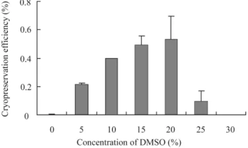
Assessment of ES cell growth after cryopreservation To evaluate the survival rate after cryopreservation, the effi-ciency of the slow-rate cooling method was assessed by counting the number of ES cell colonies, while that of the vitrification method Fig. 1. Cryopreservation efficiency of CMK6 ES cells by slow cooling
method. CMK6 ES cells were frozen in freezing media containing 0 to 30% DMSO. The number of ES cell colonies was counted at 6 days after thawing and compared to the control plate.
Fig. 2. Typical morphology of CMK6 ES cell colonies after subculturing, slow-cooling and vitrification.(A) A control plate prepared by passaging as described in Experimental Proce-dures. ES cell colonies at five days after cryopreservation by (B) the slow-cooling and (C) the vitrification method.
A
B
C
risks must be avoided for the potential clinical use of human ES cells. Therefore, we developed a novel cryopreservation method for primate ES cells that is simple and efficient on a large scale.
Experimental Procedures
ES cell culture
was assessed by counting ES cell numbers. The number of ES cells or ES cell colonies was counted and compared to that of the control plates prepared as described above.
Histochemical analysis of recultured ES cells
ES cells were fixed in 4% paraformaldehyde for 10 min. Alkaline phosphatase activity was detected with Vector Red Alkaline Phos-phatase Substrate Kit I (Vector laboratories, CA, USA). The expression of SSEA-4 and TRA1-60 was detected by immunostaining using specific antibodies (CHEMICON Interna-tional Inc., CA, USA) diluted at 1:100. After 2 hours of incubation, antibody staining was visualized with an HRP-labeled secondary antibody and diaminobenzidine.
Teratoma formation
Several passages after thawing, ES cells were dissociated. Approximately 1x10⁷ ES cells were subcutaneously injected into
SCID mice. Teratomas were formed within 5 to 8 weeks after injection. Teratomas were removed by dissection, fixed in 4% formalin, embedded in paraffin, sectioned at 5 µm and stained with hematoxylin and eosin.
Results and Discussion
Slow-rate freezing of monkey ES cells
For biological research and medical applications, it is necessary to develop an efficient cryopreservation method for the primate ES cell lines, CMK6 and CMK9. We therefore have attempted to establish an efficient and simple cryopreservation protocol appli-cable to primate ES cells. We first examined the efficiency of the slow-rate cooling method using freezing medium containing 10% DMSO for primate ES cells. It was necessary to determine the cell number after thawing to accurately evaluate the survival rates. However, this was impossible because of extremely poor recovery associated with this method (Fig. 2B). Thus, we estimate the survival rates by counting the number of colonies. After thawing, very few CMK6 cells remained alive, resulting in a cryopreservation efficiency of 0.4% compared to that for passaging (Fig. 1). To
optimize the cryopreservation medium, we tested several concen-trations of DMSO ranging from 0-30%. Although 20% DMSO proved to be most effective for slow cooling, the efficiency was only increased to 0.6%. We also examined a range of commercially available freezing media and tested the effect of additional compo-nents such as methylcellulose (Ohno et al., 1988) on the freezing efficiency. However the cell viability after thawing scarcely im-proved.
From these results, primate ES cells were found to exhibit low survival rates after cryopreservation using the slow-rate cooling method. Two weeks or more of expansion was required for undifferentiated stem cells to proliferate enough for subculturing. Thus, this time consuming and unreliable protocol is not practical for laboratory and clinical applications of primate ES cells.
Vitrification freezing of monkey ES cells
Vitrification is an alternative way to cryopreserve a variety of cell types, including mammalian fertilized eggs or embryos (Rall and Fahy, 1985; Kuleshova et al., 1999; Dobrinsky, 2002). Although vitrification of human ES cells has been previously attempted (Reubinoff et al., 2001), the potential contamination risks (Gorman and Patterson, 1995) and limited utility of this method has made it necessary to develop more efficient, reliable and simple vitrifica-tion methods.
As cryovials are the most commonly used cryopreservation containers for cultured cells, we replaced the OPSs used in this previously described method to include these vials. We also compared the efficacy of three freezing media, DES, DAP and EFS40, to optimize the vitrification protocol using cryovials. While DES (DMSO, Ethylene glycol and Sucrose) was used for the vitrification of human ES cells using OPS (Reubinoff et al., 2001), DAP (DMSO, Acetamide and Propylene glycol) is used for the cryopreservation of mammalian oocytes and embryos (Nakagata et al., 1993). EFS (Ethylene glycol, Ficoll and Sucrose) is also used for the cryopreservation of mammalian embryos in glass capillaries (Kasai et al., 1990; Nagy et al., 2003). The concentra-tion of ethylene glycol in EFS medium varies widely among
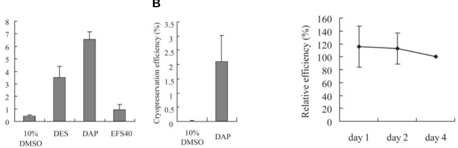
Fig. 3 (Left). Efficiency of cryopreservation by vitrification.(A) The average survival rate of CMK6 ES cells after vitrification using DES, DAP and EFS40 freezing media was estimated by counting cell numbers at four days after thawing. The efficiency of the slow-cooling method using 10% DMSO is also shown. (B) Cryopreservation efficiency for CMK9 cell lines was examined as in (A).
Fig. 4 (Right). Recovery of ES cells after cryopreservation. The survival rate of CMK6 ES cells was evaluated 1, 2 and 4 days after thawing from vitrification using DAP. The cryopreservation efficiencies for day 1 and day 2 relative to day 4 are shown.
protocols. We chose the EFS40 medium containing 40% v/v ethylene glycol, as this medium is widely used for mouse embryo cryopreservation. Since the vitrification methods yielded higher cell recovery rates than did the slow-rate freezing methods as shown in Fig. 2, we were able to accurately evaluate the survival rates by determining the cell number instead of the colony number. Among the vitrification freezing media examined, DAP exhibited the highest recovery rate of 6.5% (Fig. 3A), nearly twice
treatment with a less hypertonic medium than the freezing medium improved the efficiency of cryopreservation (Rall and Fahy, 1985). For the OPS vitrification method, ES cells were exposed to 1 M DMSO for 1 min before freez-ing. Therefore, we pretreated CMK6 cells with 1 M DMSO for 1 min before vitrification in DAP. This pretreatment, however, did not signifi-cantly alter the cryopreservation efficiency.
As vitrification freezing media contain high concentrations of cryoprotectants, these me-dia are toxic to ES cells. Therefore, we exam-ined the effect of the duration of time the cells spent suspended in the freezing medium be-fore soaking in liquid nitrogen on cell survival after thawing. The survival rates decreased with increased holding time before freezing (Fig. 5); when the holding time was longer than 1 min, cryopreservation efficiency was lower than 0.5 %. Therefore, EFS40 did not permit good recovery rates (Fig. 3A), probably be-cause it is too viscous to handle rapidly. A Fig. 5. Effect of
incuba-tion time in DAP during vitrification on the cryo-preservation efficiency of CMK6 ES cells. ES cells were incubated in DAP for 15, 30, 45 and 60 seconds before freezing. The cryopreservation ef-ficiency was then evalu-ated 4 days after thawing and it was found that the efficiency rapidly de-creased in accordance with the increase in incu-bation time.
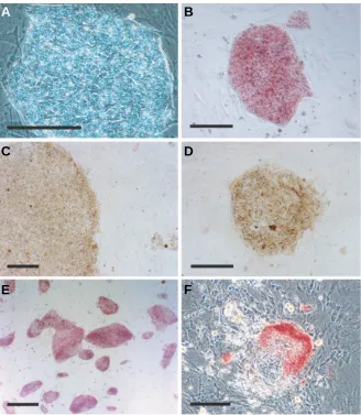
Fig. 6. Morphology and stem cell marker expres-sion in CMK6 ES cells after cryopreservation. A phase contrast microscopic image of vitrified CMK6 at 3 days after thawing (A) and vitrified CMK6 cells examined for alkaline phosphatase activity (B, E-F). Immunocytochemistry of vitrified CMK6 detected the expression of SSEA-4 (C)and TRA1-60 (D). A low power view of vitrified ES cells at 5 days stained for alkaline phosphatase activity showing that most colo-nies retained their undifferentiated state (E). A typical differentiating colony (F).Scale bars, 20 µm (A-D, F); 50 µm (E).
A
B
C
D
E
F
as high as the efficiency using DES (3.5%). EFS40 displayed a very low efficiency of 1.0%.
The cryopreservation efficiency was also examined using another cynomolgus monkey ES cell line, CMK9. The viability of vitrified CMK9 cells was about 2.1% (Fig. 3B). It was very low compared with that of CMK6 cells, which was about 6.5%. Such differences may represent a higher sensitivity of CMK9 cells to mechanical damage as suggested by previous experiments (Furuya et al., 2003).
We next tried to optimize this vitrification method using DAP. We examined a range of DAP concentrations from 0.25-1.5 fold of the initially tested concentration. The original concentration of DAP proved to be most effective. We attempted to improve the freezing efficiency by varying the ratios of the DAP components. Although we tested concentrations of DMSO ranging from 15-30% and propylene glycol ranging from 15-15-30%, we found little improvement in efficiency compared to the original DAP medium (data not shown).
We further analyzed the cryopreservation efficiency for CMK6 ES cells by vitrification using DAP immediately after thawing. As shown in Fig. 4, the cells exhibited rapid recovery from cryo-preservation and exhibited similar survival rates at 1, 2 and 4 days after thawing.
pre-holding time of approximately 15 s is thought to be the minimal handling time required for suspending cells, transferring to vials and freezing. Then, the greater time needed to manipulate the cells until freezing leads to increased cell death due to cryopro-tectant toxicity.
We also examined the effect of the cell concentration in the cryovials on the survival rate. CMK6 cells were subjected to our vitrification protocol over a wide rage of cell concentrations, from 5x10⁵ to 1x10⁷ cells/vial. Regardless of the cell density, ES cells
were successfully recovered at similar efficiencies. This result suggested that primate ES cells can be cryopreserved in cryovials for any culture scale, from 35 mm dishes to 100 mm or larger. The independence of the efficiency from culture size makes this proto-col advantageous for a variety of clinical and research applications using primate ES cells.
Reubinoff et al. demonstrated that 100% of the human ES cell colonies could be recovered after cryopreservation using the OPS vitrification method (Reubinoff et al., 2001). This difference in efficiency from our protocol was likely caused by differences in the vitrification protocols and assay systems. They evaluated the recovery efficiency by comparing colony numbers before and after vitrification, but he actual cell survival rate was not presented. In addition, more than 70% of the colonies exhibited a differentiated phenotype at 7 days after thawing by their OPS vitrification method. Using our culture system, spontaneous differentiation of primate ES cells was rarely observed (Fig. 6 E,F); less than 1% of the colonies exhibited differentiated morphology during routine matenance. The rate of occurrence of differentiated colonies in-creased to 3% after vitrification and this might have been caused by a relatively higher tolerance of differentiated cells to damage induced by the vitrification processes. Thus, our vitrification method induces differentiation at a lower rate.
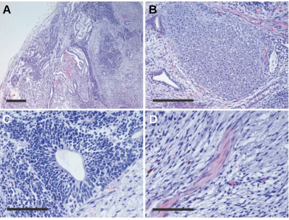
markers SSEA-4 and TRA1-60 (Fig. 6). We also analyzed the karyotype of CMK6 cells post-vitrification, demonstrating that these ES cells retained a normal karyotype of 40+ XY chromo-somes (data not shown). To examine differentiation potential of vitrified ES cells, we examined the formation of teratomas by injecting ES cells after vitrification into SCID mice. Teratoma formation was observed in two independent experiments. Histo-logical analysis of isolated teratomas revealed that the vitrified ES cells differentiated into a variety of cell types, including cartilage, neuroepithelium and muscle (Fig. 7). Thus, the vitrified ES cells retained their pluripotency.
Human ES cell cryopreservation
We examined the potential application of this vitrification method to cryopreservation of a human ES cell line, KhES-1. We could not recover any viable cells after cryopreservation by the slow-rate cooling method using 10% DMSO (Fig. 8). In contrast, human ES cells both survived and continued to grow after vitrification in DAP. The cryopreservation efficiency of KhES-1 was approximately 12.2%. As seen for monkey ES cells after vitrification, the vitrified human ES cells were indistinguishable from non-vitrified ES cells. Fig. 7. Teratoma formation by vitrified CMK6 ES cells. The gross appearance (A)
and higher magnification views (B-D) of a H & E-stained section of a teratoma produced from vitrified CMK6 ES cells. Cells expressing cartilage (B), neuroepithelium
(C) and muscle (D) phenotypes were identified. Scale bars, 20 µm (A,B); 10 µm (C, D).
Fig. 8. Cryopreservation efficiency of a human ES cell line. The survival rates of a human ES cell line, KhES-1, after cryo-preservation by the slow-cooling method using 10% DMSO and by the vitrification method using DAP.
Cell characteristics after cryopreservation
Our vitrification method using DAP freezing medium permitted the rapid recovery of ES cells after cryo-preservation. Usually, approximately two weeks are required following cryopreservation by slow-rate cooling for human ES cells to grow to the cell number present at the time of freezing. By our vitrification method, this number of cynomolgus monkey ES cells was recovered within about 4 days. The thawed ES cells tend to form small colonies compared with control ES cells probably because of additional dissociation caused by handling during freezing/thawing and damage induced by the preservation process (Fig. 2). Vitrified ES cells recov-ered very quickly and could be maintained for more than one month without any signs of differentiation or changes in their growth properties. Since human ES cells tend to differentiate after thawing (Reubinoff et al., 2001), our vitrification method may be an effective way to minimize this side effects. Such quick and safe recovery of ES cells also reduces the time and cost of experiments requiring cryopreservation from those using conven-tional slow-rate cooling methods.
Following the culture of vitrified ES cells, we as-sessed their stem cell marker expression (Suemori et al., 2001). Thawed cynomolgus monkey ES cells contin-ued to exhibit the alkaline phosphatase activity charac-teristic of ES cells and also expressed the stem cell
A
B
In conclusion, we have developed a novel cryopreservation method for primate ES cell lines, including human ES cell lines. This vitrification method provides the simple, efficient and reliable cryopreservation of primate ES cell lines. Such a system will be exceedingly helpful for experimental research and medical appli-cations.
Acknowledgements
This work was supported in part by the National Bioresource Project, Mext and the Japan Society for the Promotion of Science.
References
DOBRINSKY, J.R. (1996). Cellular approach to cryopreservation of embryos.
Theriogenology 45: 17–26.
DOBRINSKY, J.R. (2002). Advancements in cryopreservation of domestic animal embryos. Theriogenology. 57: 285-302.
FURUYA, M., YASUCHIKA, K., MIZUTANI, K., YOSHIMURA, Y., NAKATSUJI, N. and SUEMORI, H. (2003). Electroporation of cynomolgus monkey embryonic stem cells. Genesis. 37: 180-187.
GORMAN, A.M. and PATTERSON, K.G. (1995). Hepatitis B transmission from contaminated cryopreservation tank. Lancet 346: 137-140.
KASAI, M., KOMI, J.H., TAKAKAMO, A., TSUDERA, H., SAKURAI, T. and MACHIDA, T. (1990). A simple method for mouse embryo cryopreservation in a low toxicity vitrification solution, without appreciable loss of viability. J. Reprod. Fertil. 89: 91-97.
KELLER, G. and SNODGRASS, H.R. (1999). Human embryonic stem cells: the future is now. Nat. Med. 5: 151-152.
KULESHOVA, L., GIANAROLI, L., MAGLI, C., FERRARETTI, A. and TROUNSON, A. (1999). Birth following vitrification of a small number of human oocytes: case report. Hum. Reprod. 14: 3077-3079.
LUYET, B.J. (1937). The vitrification of organic colloids and of protoplasm.
Biodynamica. 1: 1-14
NAGY, A., GERTSENSTEIN, M., VINTERSTEN, K. and BEHRINGER, R. (2003) Embryo Cryopreservation by Rapid Cooling. In Manipulating the Mouse Em-bryo: A Laboratory Manual, Third Edition Cold spring Harbor Laboratory Press pp. 610-615
NAKAGATA, N. (1993). Survival of mouse morulae and blastocysts derived from in vitro fertilization after ultra rapid freezing. Jikken Dobutsu. 42: 229-231. OHNO, T., KURITA, K., ABE, S., EIMORI, N. and IKAWA, Y. (1988). A simple freezing
medium for serum–free cultured cells. Cryotechnology. 1: 257-260
RALL, W.F. and FAHY, G.M. (1985). Ice-free cryopreservation of mouse embryos at -196 degrees C by vitrification. Nature 313: 573-575.
REUBINOFF, B.E., PERA, M.F., FONG, C.Y., TROUNSON, A. and BONGSO, A. (2000). Embryonic stem cell lines from human blastocysts: somatic differentiation in vitro. Nat. Biotechnol. 18: 399-404.
REUBINOFF, B.E., PERA, M.F., VAJTA, G. and TROUNSON, A.O. (2001). Effective cryopreservation of human embryonic stem cells by the open pulled straw vitrification method. Hum. Reprod. 16: 2187-2194.
SUEMORI, H., TADA, T., TORII, R., HOSOI, Y., KOBAYASHI, K., IMAHIE, H., KONDO, Y., IRITANI, A. and NAKATSUJI, N. (2001). Establishment of embryonic stem cell lines from cynomolgus monkey blastocysts produced by IVF or ICSI.
Dev. Dyn. 222: 273-279.
THOMSON, J.A., KALISHMAN, J., GOLOS, T.G., DURNING, M., HARRIS, C.P., BECKER, R.A. and HEARN, J.P. (1995). Isolation of a primate embryonic stem cell line. Proc. Natl. Acad. Sci. USA 92: 7844-7848.
THOMSON, J.A., KALISHMAN, J., GOLOS, T.G., DURNING, M., HARRIS, C.P. and HEARN, J.P. (1996). Pluripotent cell lines derived from common marmoset (Callithrix jacchus) blastocysts. Biol. Reprod. 55: 254-259.
THOMSON, J.A., KALISHMAN, J., GOLOS, T.G., DURNING, M., HARRIS, C.P. and THOMSON, J.A., ITSKOVITZ-ELDOR, J., SHAPIRO, S.S., WAKNITZ, M.A., SWIERGIEL, J.J. MARSHALL, V.S. and JONES, J. (1998). Embryonic stem cell lines derived from human blastocysts. Science. 282:1145-1147.