1556-6811/09/$08.00
⫹
0
doi:10.1128/CVI.00148-09
Copyright © 2009, American Society for Microbiology. All Rights Reserved.
Development and Evaluation of an Enzyme-Linked Immunosorbent
Assay Based on Recombinant VP2 Capsids for the Detection of
Antibodies to Aleutian Mink Disease Virus
䌤
Anna Knuuttila,
1,2* Pirjo Aronen,
3Auli Saarinen,
2† and Olli Vapalahti
1,2Division of Microbiology and Epidemiology, Faculty of Veterinary Medicine, P.O. Box 66, 00014 University of Helsinki, Finland
1;
Department of Virology, Haartman Institute, P.O. Box 21, 00014 University of Helsinki, Finland
2; and
Finnish Fur Breeders’ Association (STKL), P.O. Box 92, 65101 Vaasa, Finland
3Received 26 March 2009/Returned for modification 11 May 2009/Accepted 22 July 2009
Aleutian disease (AD), a common infectious disease in farmed minks worldwide, is caused by Aleutian mink
disease virus (AMDV). Serodiagnosis of AD in minks has been based on detection of AMDV antibodies by
counterimmunoelectrophoresis (CIE) since the 1980s. The aim of this study was to develop and evaluate an
enzyme-linked immunosorbent assay (ELISA) based on recombinant virus-like particles (VLPs) for identifying
AMDV antibodies from mink sera. AMDV capsid protein (VP2) of a Finnish wild-type strain was expressed by
the baculovirus system in
Spodoptera frugiperda
9 insect cells and was shown to self-assemble to VLPs (with an
ultrastructure similar to that of the actual virion). A direct immunoglobulin G ELISA was established using
purified recombinant AMDV VP2 VLPs as an antigen. Sera from farmed minks were collected to evaluate the
AMDV VP2 ELISA (
n
ⴝ
316) and CIE (
n
ⴝ
209) based on AMDV VP2 recombinant antigen in parallel with
CIE performed using a commercially available traditional antigen. CIE performed with the recombinant
antigen had a sensitivity and specificity of 100% and ELISA a sensitivity of 99% and a specificity of 97%, with
reference to CIE performed with the commercial antigen. The results show that the recombinant AMDV VP2
VLPs are antigenic and that AMDV VP2 ELISA is sensitive and specific and encourage further development
of the method for high-throughput diagnostics, involving hundreds of thousands of samples in Finland
annually.
Aleutian mink disease virus (AMDV) is a member of the
genus
Amdovirus
, subfamily
Parvovirinae
, family
Parvoviridae
.
The icosahedral nonenveloped virion contains a 4.8-kb
single-stranded DNA genome and three nonstructural (NS1, NS2,
and putative NS3) and two structural (VP1 and VP2) proteins
(9, 10, 29). Several strains, ranging in pathogenicity from
non-pathogenic (G) to highly non-pathogenic (e.g.,
AMDV-Utah 1, -United, and -K), have been identified (6, 7, 18, 19).
AMDV causes an immune-mediated disease, called Aleutian
disease (AD), in minks (
Mustela vison
) and other mustelids
(16, 20, 22, 24, 28).
The disease manifestation varies, from mild nonprogressive
to fatal progressive disease in adult minks and acute fatal
pneumonia in mink kits, depending on the virus strain and host
factors (10). Nonpersistent infections have also been described
(10). The most serious form of AD, known as classical AD, is
associated with viremia, plasmacytosis,
hypergammaglobuline-mia, high AMDV antibody levels, formation of infectious
im-mune complexes, and glomerulonephritis (10, 19). Clinical
signs include lethargy, anemia, anorexia, cachexia, polydipsia,
poor pelt, infertility, renal failure, uremia, neurological
symp-toms, and clotting abnormalities (17). Minks with classical AD
die, depending on the mink genotype and the virus strain,
within 2 months to 4.5 years after infection (17).
AMDV can be found in all mink breeding countries, and it
causes considerable economic losses to farmers, e.g., in the
form of decreased production, loss of breeding animals, and
low-quality fur (17). The infection is usually persistent, often
fatal, and there is no effective vaccine or treatments against the
disease (3, 11, 26, 27). Transmission of AMDV occurs
hori-zontally by direct and indirect contact and vertically from dams
to kits (17). Furthermore, the virus is persistent in the
envi-ronment and resistant to various physical and chemical
treat-ments (17). Due to all of this, it is very challenging to sanitize
an infected farm. At the moment, the only effective means of
eradicating AD from farms is serological screening,
subse-quent identification, elimination of all antibody-positive
ani-mals, and pursuit of strict sanitary measures.
Diagnosis of AD in minks is based primarily on the clinical
signs and detection of AMDV antibodies. Several nonspecific
(iodine agglutination test, serum electrophoresis, and
glutar-aldehyde test) and specific (indirect immunofluorescence,
complement fixation, and counterimmunoelectrophoresis [CIE])
tests were developed in the late 1960s and 1970s (17). At the
moment, CIE (also abbreviated CCIE, CIEP, and CCE) is
used for routine detection of AMDV. CIE is based on the
formation and visual detection of immune complexes on an
agarose gel after electrophoresis. During the 1970s, the assay
has been carried out with organ-produced viral antigen (12).
Since the 1980s, an in vitro-grown antigen (strain AMDV-G;
Crandell feline kidney cells) has been applied (1). Other
im-* Corresponding author. Mailing address: Division of Microbiology
and Epidemiology, Faculty of Veterinary Medicine, P.O. Box 66, 00014
University of Helsinki, Finland. Phone: 358 9 191 57049. Fax: 358 9 191
57033. E-mail: [email protected].
† Present address: Department of Medical Genetics, Haartman
In-stitute, P.O. Box 63, 00014 University of Helsinki, Finland.
䌤
Published ahead of print on 29 July 2009.
1360
on August 17, 2020 by guest
http://cvi.asm.org/
(13, 14, 34, 35) and shown to be antigenic and able to form
virus-like particles (VLPs) (13, 14, 34). However, only a few
diagnostic applications have been described (3, 14, 35), and
published comparative data are scarce. Clemens et al. (14)
demonstrated that the recombinant VLPs are more sensitive
and give higher titers in CIE than the in vitro-produced
AMDV-G antigen (
n
⫽
10). Zeng et al. (35) expressed AMDV
VP2 protein in prokaryotic cells and used the purified antigen
in CIE. The detection results showed 94.3% identity with a
commercially available antigen in CIE (
n
⫽
54). Three
en-zyme-linked immunosorbent assay (ELISA)-based methods
have been described for diagnosis of AMDV infection from
mink serum samples (3, 11, 33). The only study comparing
ELISA and CIE test results was done more than 25 years ago
(33). In this study, fluorocarbon-activated AMDV (Guelph
strain) was used as an antigen in both tests (
n
⫽
1,329) and
the conclusion was that the ELISA method has a high rate
of false-negative reactions. Commercial applications of
ELISA assays for serodiagnosis of AD in minks are lacking.
To our knowledge, two ELISAs have been developed for
ferrets: one by Avecon Diagnostics (Bath, PA) and the other
by the University of Georgia (http://www.vet.uga.edu/VPP/clerk
/schuler/index.php). The former is commercially available.
In Finland, the Fur Animal Feed Laboratory started to test
farmed minks for AMDV by CIE in 1980. In 1981 and 1982,
the seroprevalence was approximately 50% to 60%. Since then,
it has decreased considerably due to control measures in
in-fected farms, varying from 3% to 11% in 1990 to 2008. In 2008,
almost 500,000 serum samples from minks were tested for
AMDV antibodies in Finland, and the number is increasing
each year.
In this study, a recombinant VP2 protein antigen based on a
wild-type Finnish AMDV strain and subsequently an
ELISA-based method for detecting AMDV antibodies in minks were
developed. The purified recombinant antigen was used in both
CIE and ELISA, and the results were evaluated in comparison
with those for the existing commercially available CIE antigen
and method.
MATERIALS AND METHODS
Serum samples.A total of 525 serum samples were collected from farmed minks in Finland. Blood was obtained by toenail cutting and collected into glass
capillary tubes. After centrifugation, the serum samples were stored at⫺20°C
until processed.
DNA extraction.DNA was extracted from the mesenteric lymph node of a Finnish mink, designated C8, in 2005 as previously described (21).
Construction of a recombinant plasmid and expression of the recombinant VP2 protein.The purified PCR product and the pGEM-T plasmid (Promega,
Madison, WI) were ligated and transformed intoEscherichia coliJM109
com-petent cells (Promega, Madison, WI). The bacteria were suspended in Luria
broth (LB), grown for 1 h in a shaker, plated on LB–ampicillin (250g/ml)–
IPTG (isopropyl--D-thiogalactopyranoside)–X-Gal
(5-bromo-4-chloro-3-indo-lyl--D-galactopyranoside) plates, and incubated overnight (o/n) at 37°C. White
bacterial colonies (generally containing the insert) were transferred in LB
con-taining ampicillin (100g/ml) and grown o/n at 37°C. Plasmids were purified
from the bacterial culture with a QIAprep Spin miniprep kit (Qiagen, Hilden, Germany). The purified plasmid DNA including the VP2 insert, the pGEM-T vector, and the baculovirus transfer plasmid pAcYML1 (kindly provided by
Johan Pera¨nen, Institute of Biotechnology, University of Helsinki; the plasmid
construct was modified from that reported by Matsuura et al. [23]) were digested with restriction enzymes BamHI and PstI (New England Biolabs, Ipswich, MA) and gel purified. The VP2 insert was ligated into baculovirus transfer plasmid
pAcYML1 o/n at 14°C (1l T4 DNA ligase [400 U/l], 1l 10⫻T4 DNA ligase
buffer [New England Biolabs, Ipswich, MA], 1l insert, 0.5l vector, and 10l
H2O). The transfer plasmid was further transformed intoE. coliand grown as
described above. The bacterial suspension was purified with an EndoFree plas-mid maxikit (Qiagen, Hilden, Germany). Two hundred fifty nanograms of
Bacu-loGold baculovirus DNA (Pharmingen, San Diego, CA) and 2g of purified
plasmid construct DNA were mixed in transfection buffer containing FuGENE transfection reagent (Roche Diagnostics, Indianapolis, IN) and Sf-900 medium
(Gibco, Paisley, United Kingdom). The mixture was then applied toSpodoptera
frugiperda9 (ATCC CRL-1711; American Type Culture Collection, Rockville,
MD) cells (3⫻106cells per 25-cm2flask) and incubated for 2 h at room
temperature (RT) and 1 h at 27°C. The transfection medium was then removed, and the cells were incubated in fresh growth medium (Sf-900 medium containing
10% fetal calf serum [Gibco, Paisley, United Kingdom], 1⫻
glutamine-penicillin-streptomycin [Haartbio, Helsinki, Finland], and 0.25 mg/ml Fungizone [Bristol-Myers Squibb, Rueil-Malmaison, France]) at 27°C for 4 days. The cells and supernatant were harvested by low-speed centrifugation. The supernatant was stored at 4°C and used for subsequent infections. One milliliter of the
superna-tant was used for 25-cm2, 2 ml for 50-cm2, and 4 ml for 75-cm2flasks. The
infectedS. frugiperda9 cells were incubated at 27°C for 48 to 72 h, until CPE was
evident. The cell paste was washed four times with phosphate-buffered saline (PBS) containing protease inhibitor (Roche Diagnostics, Indianapolis, IN) and
stored at⫺70°C until processed.
Sequencing. The gel-purified pGEM-T-VP2 construct was sequenced with commercial primers (M13, T7, and SP6) by the Haartman Institute core unit (Helsinki, Finland).
Extraction and purification of the recombinant VP2 protein.The recombinant proteins were extracted and purified from the cell paste in Tris-based buffer with heating as previously described (31), with slight modifications. The cell paste was sonicated and heated at 50°C, and after centrifugation, the supernatant was used to establish recombinant VP2 CIE and ELISA.
SDS-PAGE and Western blot analysis.The infected and noninfected cell pellets were diluted 1:100 and the purified antigen was diluted 1:10 in reducing Laemmli sample buffer, heated for 5 min at 95°C, and electrophoresed through a 10% sodium dodecyl sulfate-polyacrylamide gel electrophoresis (SDS-PAGE) gel. The proteins were visualized by Coomassie blue staining. For Western blot analysis, the proteins were transferred from the SDS-PAGE gel to a nitrocellu-lose membrane. Blocking of the membrane and both mink serum and conjugate dilutions were done with TEN (0.5 M Tris, 1.5 M sodium chloride, 0.05 M EDTA) containing 0.05% Tween 20 and 1% nonfat milk powder. The
on August 17, 2020 by guest
branes were blocked o/n at 4°C and incubated with pooled sera from AMDV-infected and nonAMDV-infected minks (1:300 dilution) for 1 h at RT. After being washed with TEN containing 0.05% Tween 20, the membranes were incubated with horseradish peroxidase-conjugated goat anti-cat immunoglobulin G (IgG) (1:5,000 dilution; Jackson ImmunoResearch, West Grove, PA) for 1 h at RT.
After the second wash, the reaction was visualized by 3,3⬘-diaminobenzidine
(DAB) (Sigma-Aldrich, Steinheim, Germany) staining.
Electron microscopy.The infected cell paste was studied after negative stain-ing with 2% tungstophosphoric acid, pH 6.0, with a Jeol JEM-100 CXII electron microscope.
CIE.The “gold standard,” which is CIE performed using a commercial antigen
(Antigen Laboratory of the Research Foundation of the Danish Fur Breeders’ Association, Glostrup, Denmark) by following the manufacturer’s instructions, was tested in parallel with CIE performed using recombinant AMDV VP2
antigen (n⫽209) and AMDV VP2 ELISA (n⫽316). The same CIE procedure
was used for both antigens. Before the recombinant antigen was used in CIE, it was diluted 1:16 in PBS containing 0.05% bovine serum albumin (BSA;
Sigma-Aldrich, Steinheim, Germany). Bromophenol blue (50g/ml; Merck Chemicals,
Darmstadt, Germany) was added to help the visualization of the antigen on the agarose gel.
AMDV VP2 ELISA.For all the ELISA procedures, 96-well Nunc
immuno-plates (Finnzymes, Espoo, Finland) and 100-l volumes of reagents were used.
All serum and reagent dilutions were done in PBS containing 0.05% Tween 20 and 0.5% BSA, all washes were done twice with PBS-T (PBS containing 0.05% Tween 20), and all incubations were done at RT. Plates were coated with the
purified recombinant AMDV VP2 antigen diluted 1:1,500 in 50 mM NaHCO3
buffer (pH 9.6). After incubation o/n, the plates were blocked with 1% BSA in PBS for 1 h. Serum samples were diluted (1:200), added in duplicate, and incubated for 1 h. The plates were washed and incubated for 1 h with peroxidase-conjugated goat anti-cat IgG (diluted according to the manufacturer’s
instruc-tions) and washed. The substrate reaction with 3,3⬘,5,5⬘-tetramethyl benzidine
(Sigma-Aldrich, Steinheim, Germany) was stopped with 0.5 M H2SO4after 15
min, and the optical density (OD) values were read with a Multiscan EX spec-trophotometer (Thermo Labsystems, Vantaa, Finland) at 450 nm. The mean OD for each sample was calculated. The mean OD for two blank wells (containing all reagents except serum) was subtracted from each result. Reference sera (nega-tive, low-posi(nega-tive, and positive) were run every time the assay was carried out. The assay cutoff was determined by counting the mean OD value of the
CIE-negative samples (n⫽211) plus 1 standard deviation (SD). In total, 316 serum
samples (105 positive and 211 negative as determined by CIE) were studied with both ELISA and CIE (commercial antigen).
Nucleotide sequence accession number.The VP2 nucleotide sequence of the Finnish AMDV strain C8 was deposited in GenBank under accession number GQ336866.
RESULTS
Expression and purification of recombinant AMDV VP2
protein.
The AMDV VP2 capsid gene, comprising nucleotides
2406 to 4349 of the full-length genome, was amplified by PCR
directly from an infected mink and cloned into the baculovirus
expression vector pAcYML1. The sequence and reading frame
of the VP2 gene were confirmed by DNA sequencing of the
recombinant plasmid used for subcloning. The insert had the
highest nucleotide (98%) and amino acid (97%) identities with
AMDV-G (GenBank accession no. M20036) (8) and -SL3
(GenBank accession no. X97629) (30) (BLAST search [http:
//blast.ncbi.nlm.nih.gov/Blast.cgi]). The AMDV VP2 protein
was extracted and purified from the cell paste by heating and
centrifugation. SDS-PAGE and Coomassie blue staining of the
purified recombinant protein revealed a protein band
corre-sponding to the expected size of the AMDV VP2 protein (75
kDa) (Fig. 1). The identity of the recombinant AMDV VP2
protein was further confirmed by a Western blot analysis (Fig. 1).
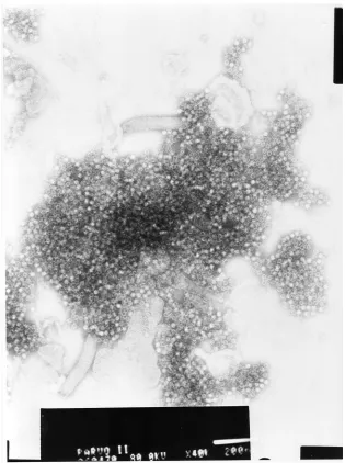
In electron microscopy, the recombinant VP2 proteins
spontane-ously formed empty VLPs, 23 to 25 nm in diameter, with a size
and ultrastructure typical of a parvovirus virion (Fig. 2).
Evaluation of recombinant antigen in comparison with the
commercially available antigen in CIE.
A serum panel (
n
⫽
209) was studied with CIE using both the recombinant antigen
FIG. 1. SDS-PAGE gel (A) and Western blot (B) of recombinant AMDV VP2 protein with sera from ADMV antibody-positive mink. Lanes:
1 and 5, molecular-mass marker; 2,
S. frugiperda
9 cells; 3, recombinant baculovirus-infected Sf9 cells; 4, purified recombinant antigen; 6, Western
blot of the purified recombinant antigen.
on August 17, 2020 by guest
http://cvi.asm.org/
and the commercially available antigen, with 100 AMDV
an-tibody-positive and 109 negative samples. CIE performed with
the recombinant antigen had a sensitivity and specificity of
100% (Table 1).
Evaluation of ELISA in comparison with CIE.
Three
hun-dred sixteen serum samples all together were studied with both
ELISA and CIE (commercial antigen). Two hundred eleven of
the samples were negative and 105 positive by CIE; 206 were
negative and 110 positive by ELISA. ELISA had a sensitivity of
99% and a specificity of 97% if CIE performed with a
com-mercial antigen was considered a “gold standard” method
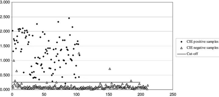
(Ta-ble 1). The cutoff was 0.099
⫹
0.127, or 0.255. The ODs of the
samples obtained by ELISA are shown in Fig. 3.
DISCUSSION
The aim of this study was to develop an ELISA method
based on recombinant antigen for detecting AMDV
anti-FIG. 2. Negative-stain electron micrograph of recombinant AMDV VP2 VLPs. The white bar at the bottom indicates 200 nm.
TABLE 1. Comparison between detection of AMDV antibodies
by CIE (commercial antigen) and that by CIE
(recombinant antigen) and ELISA
Assay and result
No. of samples with indicated result by CIE (commercial
antigen)
Correlation (%)
Sensitivity (%)
Specificity (%)
Positive Negative
ELISA
98
99
97
Positive
104
6
Negative
1
205
CIE (recombinant
antigen)
100
100
100
Positive
100
0
Negative
0
109
on August 17, 2020 by guest
bodies in minks. We cloned the full-length VP2 gene,
ex-pressed the protein in a baculovirus system, and demonstrated
that VLPs formed spontaneously. Using the purified
recombi-nant VP2 capsids as an antigen, we developed a direct IgG
ELISA for studying mink sera. We evaluated the AMDV VP2
ELISA and CIE using the purified recombinant antigen in
parallel with CIE using a commercially available traditional
antigen.
Different field strains of AMDV exhibit a considerable
de-gree of genetic variability (8, 18, 21, 25). The ELISA described
here could identify sera from minks infected with AMDV from
different genogroups as positive, indicating that this assay
should detect antibody-positive minks infected by any AMDV
strain.
The expression of AMDV VP2 protein in the baculovirus
system enables the production of large quantities of
conforma-tionally optimal antigen for diagnostic use. The results showed
that the recombinant protein has a good antigenicity and can
be used in both CIE and ELISA. Also, the results from both
assays were in good concordance with the commercial CIE
results. Previous studies have, however, indicated that the
sen-sitivity of CIE is not very good and that its role as a “gold
standard” is questionable (2). It is therefore possible that some
of the samples that were positive by ELISA and negative by
CIE could actually be true positives, and the ELISA might be
even more specific than these results indicate. This is also
supported by the fact that four out of six of the
ELISA-posi-tive-but-CIE-negative samples had rather high ODs, ranging
from 0.641 to 1.249 (Fig. 3).
The results of this study show that in CIE recombinant
antigen can replace the traditional antigen and that ELISA is
a suitable alternative to CIE for diagnosing AMDV infection
in minks. There are, however, a few requirements that need to
be met before ELISA can be used for mass screening. Both the
introduction of the sample from the glass capillary to the
ELISA plate and the dispensing of the various reagents are
laborious and time-consuming. Thus, a new method for
col-lecting samples should be created, and the whole assay should
be automated. If these requirements can be met, the ELISA
has the potential to become an economical, efficient, and
ac-curate test method. The ELISA would then have at least three
advantages over CIE. First, it requires less staff, as there are
fewer manual steps than in CIE. Second, the results are
ob-jective and independent of the experience of the reader,
whereas in CIE there is a possibility of false-positive results
due to errors in visualizing the results caused by nonspecific
precipitation lines. Third, the automated system allows for an
increase in the number of tested samples without recruitment
of additional staff. For example, in 2008 almost 500,000 serum
samples from minks were tested for AMDV antibodies in
Fin-land, and the number is increasing each year, which makes
using the traditional CIE test laborious. Thus, there is a need
for high-throughput assays, and this study, describing an
ELISA based on recombinant VLPs, should be taken as a
proof of concept for development of AMDV antibody testing
in this direction.
ACKNOWLEDGMENTS
This work was supported by Finnish Funding Agency for Technology
and Innovation (Tekes) grant 1596/31/05.
We thank Leena Kostamovaara (Haartman Institute) and Majvor
Eerola (Fur Animal Feed Laboratory) for excellent technical
assis-tance, Irja Luoto (HUSLAB) for electron micrographs, and Tarja
Hinkkanen (Finnish Fur Breeders’ Association) for sample collection.
REFERENCES
1.Aasted, B., and A. Cohn.1982. Inhibition of precipitation in counter current electrophoresis. A sensitive method for detection of mink antibodies to
Aleutian disease virus. APMIS Sect. C90:15–19.
2.Aasted, B., S. Alexandersen, A. Cohn, and M. Hansen.1986. Counter current line absorption immunoelectrophoresis in an alternative diagnostic screening test to counter current immunoelectrophoresis in Aleutian disease (AD)
eradication programs. Acta Vet. Scand.27:410–420.
3.Aasted, B., S. Alexandersen, and J. Christensen.1998. Vaccination with Aleutian mink disease parvovirus (AMDV) capsid proteins enhances dis-ease, while vaccination with the major non-structural protein causes partial
protection from disease. Vaccine16:1158–1165.
4.Alexandersen, S., and J. Hau.1985. Rocket line immunoelectrophoresis: an improved assay for simultaneous quantification of mink parvovirus (Aleutian
disease virus) antigen and antibody. J. Virol. Methods10:145–151.
5.Alexandersen, S., J. Hau, B. Aasted, and O. M. Poulsen.1985. Thin-layer counter current line absorption immunoelectrophoretic analysis of antigens and antibodies to Aleutian disease virus—a mink parvovirus. Electrophoresis
6:535–538.
6.Alexandersen, S.1990. Pathogenesis of disease caused by Aleutian mink disease parvovirus. Dissertation. The Royal Veterinary and Agricultural University of Copenhagen, Denmark.
7.Bloom, M. E., R. E. Race, and J. B. Wolfinbarger.1980. Characterization of
Aleutian disease virus as a parvovirus. J. Virol.35:836–843.
FIG. 3. Graph of the ELISA samples. The
x
axis shows the number of samples and the
y
axis the OD of the samples as obtained by
ELISA.
on August 17, 2020 by guest
http://cvi.asm.org/
14.Clemens, D. L., J. B. Wolfinbarger, S. Mori, B. D. Berry, S. F. Hayes, and M. E. Bloom.1992. Expression of Aleutian mink disease parvovirus capsid proteins by a recombinant vaccinia virus: self-assembly of capsid proteins
into particles. J. Virol.66:3077–3085.
15.Crawford, T. B., T. C. McGuire, D. D. Porter, and J. Cho.1977. A compar-ative study of detection methods for Aleutian disease viral antibody. J.
Im-munol.118:1249–1251.
16.Fournier-Chambrillon, C., B. Aasted, A. Perrot, D. Pontier, F. Sauvage, M. Artois, J.-M. Cassie`de, X. Chauby, A. Dal Molin, C. Simon, and P. Fournier.
2004. Antibodies to Aleutian mink disease parvovirus in free-ranging
Euro-pean mink (Mustela lutreola) and other small carnivores from southwestern
France. J. Wildl. Dis.40:394–402.
17.Gorham, J. R., J. B. Henson, T. B. Crawford, and G. A. Padgett.1976. The
epizootiology of Aleutian disease, p. 135–158.InR. H. Kimberlain (ed.),
Slow virus diseases of animals and man. Frontiers of Biology, North-Holland Publishing Co., Amsterdam, The Netherlands.
18.Gottschalck, E., S. Alexandersen, T. Storgaard, M. E. Bloom, and B. Aasted.
1994. Sequence comparison of the non-structural genes of four different types of Aleutian mink disease parvovirus indicates an unusual degree of
variability. Arch. Virol.138:213–231.
19.Hadlow, W. J., R. E. Race, and R. C. Kennedy.1983. Comparative patho-genicity of four strains of Aleutian disease virus for pastel and sapphire mink.
Infect. Immun.41:1016–1023.
20.Kenyon, A. J., B. J. Kenyon, and E. C. Hahn.1978. Protides of the Musteli-dae: immunoresponse of mustelids to Aleutian mink disease virus. Am. J.
Vet. Res.39:1011–1015.
21.Knuuttila, A., N. Uzca´tegui, J. Kankkonen, O. Vapalahti, and P. Kinnunen.
109:1–7.
27.Porter, D. D., A. E. Larsen, and H. G. Porter.1980. Aleutian disease of mink.
Adv. Immunol.29:261–286.
28.Porter, H. G., D. D. Porter, and A. E. Larsen.1982. Aleutian disease in
ferrets. Infect. Immun.36:379–386.
29.Qiu, J., F. Cheng, L. R. Burger, and D. Pintel.2006. The transcription profile Aleutian mink disease parvovirus in CRFK cells is generated by alternative
processing of pre-mRNAs produced from a single promoter. J. Virol.80:
654–662.
30.Schuierer, S., M. E. Bloom, O. R. Kaaden, and U. Truyen.1997. Sequence analysis of the lymphotropic Aleutian disease parvovirus ADV-SL3. Arch.
Virol.142:157–166.
31.Sico, C., S. White, E. Tsao, and A. Varma.2002. Enhanced kinetic extraction
of parvovirus B19 structural proteins. Biotechnol. Bioeng.80:250–256.
32.Uttenthal, Å.1992. Screening for antibodies against Aleutian disease virus (ADV) in mink. Elucidation of dubious results by additive
counterimmuno-electrophoresis. Appl. Theor. Electrophor.3:83–84.
33.Wright, P. F., and B. N. Wilkie.1982. Detection of antibody in Aleutian disease of mink: comparison of enzyme-linked immunosorbent assay and
counterimmunoelectrophoresis. Am. J. Vet. Res.43:865–868.
34.Wu, W.-H., M. E. Bloom, B. D. Berry, M. J. McGinley, and K. B. Platt.1994. Expression of Aleutian mink disease parvovirus capsid proteins in a bacu-lovirus expression system for potential diagnostic use. J. Vet. Diagn. Investig.
6:23–29.
35.Zeng, X. W., Y. P. Hua, and D. Y. Liang.2007. Prokaryotic expression and detective application of the main antigenic region of VP2 protein of Aleutian
mink disease parvovirus. Wei Sheng Wu Xue Bao47:1088–1090.