ASYNCHRONOUS .REPLICATION OF DNA IN A HETEROCHROMATIC SET OF CHROMOSOMES
IN
PSEUDOCOCCUS OBSCURUS'.DEE BAER
Biology Department> San Diego State College, San Diego, California
Received February 27, 1965
replication of chromosomal DNA has been shown to be asynchronous for T z o l e chromosomes or for parts of a single chromosome by LIMA-DE-FARIA (1959, 196l), MORISHIMA, GRUMBACH, and TAYLOR (1962), TAYLOR (1960), WIMBER ( 1961 ), WIMBER and PRENSKY (1963), and others. The chromosomes of the organisms studied generally contained heterochromatin. This heterochro- matin is genetically determined in the sense that certain parts of the genome, such as sex chromosomes, display heteropyknosis during all or some phases of the life cycle.
In a majority of the cases, heterochromatin has shown asynchrony of DNA synthesis with respect to the rest of the genome, although some parts of the eu- chromatin have also been found to replicate asynchronously.
This paper reports autoradiographic studies with tritiated thymidine on mealy bugs of the species Pseudococcus obscurus Essig. The results show that the magni- tude of genetic material involved in asynchronous replication may extend to an entire complement of heterochromatic chromosomes, for which a prior history
as euchromatin is known.
I n mealy bugs, both sexes are diploid and there are no distinguishable sex chromosomes. The sexes are, however, dramatically different: during embryo- genesis in the male the paternal set of chromosomes becomes heterochromatic and remains so throughout his life. The maternal chromosomes remain euchro- matic. The paternal chromosomes degenerate after spermatogenesis; the sperm contain and transmit only the maternal chromosomes. Thus the euchromatic set in the male of one generation becomes the heterochromatic set in the males of the next. This situation was recently reviewed by BROWN and NUR (1964).
Concurrent with this change to heterochromatin is a marked change in genetic function. The chromosomes become largely genetically inert as measured by radiation response (BROWN and NELSON-REES, 1961 ) and other means.
The present study was undertaken to determine if this newly acquired, o r facultative, heterochromatic state shows asynchronous replication similar to that of the genetically determined heterochromatin of other organisms. The results indicate that an asynchrony of the maternal, euchromatic ( E ) and paternal, heterochromatic (H) sets does indeed exist and that the E set precedes the H set
Fiom a thesis submitted In partial satisfaction of the requirements for the Ph D
Supported In part by Grant No G9772 from the National Science roundatlon to DR S IT BROWN
degree a t the Unixersitj of Cali forma, Berkeley
276 D. BAER
in replication. The transmission of chromosomes from father to son involves a shift to heterochromatin in association with a shift to late DNA synthesis.
MATERIALS A N D METHODS
Test organism: Mealy bugs of the species Pseudococcus obscurus, with 2N=10, were studied. These Homopteran insects feed on plant juices and are routinely cultivated, at room temperature, on sprouting potatoes in ventilated jars.
Testicular and abdominal diploid tissues of the second nymphal stage were investigated. Administration of HSTDR: Since mealy bugs suck plant juices, it was possible to administer the tritiated thymidine (H3TDR) in the following manner: ( 1 ) clean potatoes with apical sprouts less than '/8 inch high-but otherwise smoothly surfaced-were selected. All but about five apical sprouts were scraped off and these ex-sprout areas covered with liquefied paraffin. (2) Fifty young P. obscurus were then added to the potato in the culture jar and allowed to settle on the sprouts. (3) Four tunnels at 90 degrees to each other were made with a dissecting needle under each remaining sprout to a depth of % to '/e inch, at an angle to the surface of about 45 degrees. (4) H T D R , (New England Nuclear Corp., 575 Albany St., Boston 18, Mass.) as thymidine-methyl-H3, at a concentration of 1 mc/ml (maximum specific activity used was 5.15 c/mM) was injected into all tunnels. Each prepared potato received 40 to 60 X of H3TDR. This methcd allowed the mealy bugs to take in the H3TDR while feeding on the sprouts because the net movement of the injected HaTDR is upward through the phloem into the grow- ing sprouts. The presence of labeled nuclei in autoradiographs prepared from the young potato leaves showed that not all of the H3TDR reached the insects.
Collection times were 1.5, 3, 4.5 and 9 hours following administration of H3TDR.
Direct injection of H3TDR into the second nymphal stage animals was not feasible since they are small (less than 1 mm long), soft bodied, and covered with a waxy meal.
Autoradiography: The general procedure was fixation i n 4 parts chloroform : 3 parts absolute ethanol : 1 part glacial acetic acid (v/v/v) at various times after administration of H3TDR. The insects were then changed to 3:l (absolute ethanol : glacial acetic acid) for at least 6 hours, followed by distilled water for one day.
The tissues of interest were dissected out onto albuminized slides in 45% acetic acid, warmed briefly, and then the preparations were squashed, care being taken that the cells were well distributed and evenly flattened. The slide preparations were frozen on dry ice and the coverslips flicked off. After this the slides were set in absolute ethanol for 15 min, dried in air. then changed through 95%, 70%, and 30% ethanol to water before filming.
In sgme experiments, nucleases were used on comparable pieces of tissue from a given indi- vidual. Three slides were made per individual and carried through the procedures given above. The first slide received no enzyme treatment, but instead was incubated 3 hours in 0.1 % gelatin (w/v) in 0.003 M MgSO, at p H 6.5 and then returned to distilled water before filming. The second was treated for 3 hours with a 0.1% solution of DNase (Sigma Chemical Corp., 3500 DeKalb St., St. Louis, MO.) in 0.003 M MgSO, with 0.1% gelatin, adjusted to p H 6.5. The third slide was treated with a 0.02% solution of RNase (Sigma) at p H 5.5 for 3 hours. Following nuclease traatment, slides were immersed 5 min in 5% trichloroacetic acid at 4°C. All treatments were given at room temperature except as noted.
RNase did not remove the H3TDR label. When DNase was used on H3TDR treated tissues, no labeling of autographs was observed except in one series. In that series, following DNase treat-
ment, the label was not remned. However, it was concluded that the DNase preparation used in that series was inactive because the cytological appearance of the nuclei was no more diffuse than in the control slides.
D N A S Y N T H E S I S I N H E T E R O C H R O M A T I N 277 tried was a 2% aqueous solution of pyronin Y (E. Gum, London, England) extracted three times with equal volumes of chloroform. After 15 min in the stain, the autoradiographic slide prepara- tions were washed and air dried.
RESULTS
DifJerential nuclear labeling: Tissues were fixed at short intervals after ad- ministration of H3TDR to the potato on which the mealy bugs fed. I n autoradio- graphs prepared from somatic tissues of second nymphal stage males fixed after feeding for 1.5 hours, only a few large interphase nuclei were lightly labeled. Such large, endopolyploid nuclei typically contain no heterochromatin; they were never observed to be dividing or to be heavily labeled. In males fixed at 3 hours, interphase nuclei often showed label clustered exclusively over the E (euchromatic) or over the H (heterochromatic) complement; prometaphase nuclei with chromosomes individually distinguishable also often showed label only over the
H
orE
sets; metaphase nuclei, particularly in animals fixed after 3 to 4.5 hours feeding, showed label restricted to one side or to the middle of the plate of congressed chromosomes (Figure 1 ) . This last phenomenon reflects the nonrandom lateral distribution of the E andH
sets typically observed on the metaphase plate.Among labeled interphases from animals fixed at 3 hours, 54% of the somatic, H-containing nuclei showed only the H set labeled (Figure 1) whereas about
39% showed only the E complement labeled (Figure 2). The remainder, 7%, showed label over both E and H sets (Figure 3 ) .
These data suggest DNA asynchrony between the two complements. Asyn- chrony can be definitely established on the basis of these data if in the animals fixed at 3 hours, H3TDR was available for only a short part of the cell generation time.
Since few labeled nuclei were observed in animals fixed at 1.5 hours, H3TDR was available for a maximum of 1.5 hours in those animals fixed after feeding for 3 hours. As will be discussed later, it appears likely that 1.5 hours is only a small part of the cell generation time.
On the basis of the data from animals fixed after 3 hours it can therefore be concluded that the H and E sets synthesize DNA asynchronously with H-DNA synthesis occupying at least 54% of the S period (the time interval of DNA synthesis) and E-DNA synthesis at least 39%.
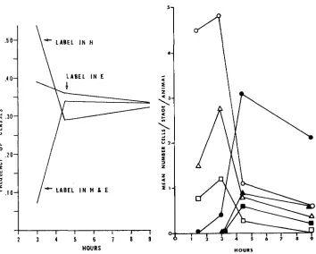
Among somatic interphases from individuals fixed at later times, up to 9 hours, the percentage of nuclei labeled exclusively in E changed very little, but the per- centage labeled in H decreased while the percentage labeled in both sets in- creased. The time sequenced data are presented in Figure 5. As the frequency of interphases labeled in both sets increased, the frequency of labeled metaphases increased (compare Figures 5 and 6 ) .
378
I- - -
D. R A E R
-
.
-l k u n m 1 - 3.-Srcon:I-iirstar nriilc uiri~atir rrlls. FIGURE I .-.Mrtaphaw ( M ) platr. lahrlcd to onr siclr. Intrrphiw with liilwl only ovrr tii~trrocliroririiti~~ ( H ) . Fixrtl nt ,+.5 hours. ~ 2 1 0 0 .
I;IC;WRF. 2.-Intrrphnsrs. I.iih:I only o v r r eurhrnrnirtin ( E ) nt hnttonr; Onlv ovcr H i i t top. l**ixr.tl ; I t 3 hours. x IMMH). l i i ( i u n ~ 3.--I.irhc.l ovrr h t h H and I< in crII nt top. Fixrd nt 4.5 hours. x 1750.
I;rr.unE +-Srrontl-instar ni-rlv sprrmatognniirl mitow.i. Fiirly prophasr (El') Inhrlrtl in H
D N A S Y N T H E S I S I N H E T E R O C H R O M A T I N 279
LABEL IN H
\
L A B E L I N E* LABEL I N H I E
I
I I I I I 1 I ' 1
2 3 4 5 6 i m 9
H O U R S
FIGURE 5.-Distribution of interphase label between the E and H complements. Collection times were at 1.5, 3, 4.5 and 9 hours after administration of HSTDR. At least 100 nuclei were counted for each collection point. Cells fall into three classes relative to labeling pat- tern: the class with label in H only, the class with label in E only, and the one with label in 110th E and H.
HOURS
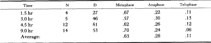
FIGURE 6.-Distribution of labeled and un- labeled somatic division figures (males plus females) at various times after administration of HSTDR given in terms of mean number of cells in a given stage per animal scored. Open symbols, unlabeled division figures; black sym- bols, labeled division figures.
0,
metaphases;A, anaphases; U, telophases. At least ten animals were scored for each collection point.
The question is whether the E set precedes or follows the H set in chromosome replication.
Since the testis is a parasynchronous tissue it was used to study this question. Early and late prophases and a prometaphase from the testis of a male fixed at
3 hours are shown in Figure 4. Only the early prophases are labeled; and they are labeled only in the H set. Later prophases and one prometaphase without label can be seen. Note that no E sets are labeled at all. No other stages of the spermatogonial mitotic cycle occurred in this testis.
280 D. BAER
the idea that E follows H in replication since in that case E labeling is later prophases in addition to the H labeling of early prophases should have been seen. This was not seen.
Spermatogonial interphases with label in E only, in
H
only, and in both sets, were found together in testes of other males fixed a few hours after administra- tion of H3TDR. However, none of these testes were analyzable as to the order of E and H in DNA synthesis.Autoradiographs of tissues from second nymphal stage females were made and examined under exactly the same conditions as obtained for males. These preparations of female cells often showed labeled interphases and mitotic stages. The interphases never showed clustering of label in any part of the nuclei, i.e., the label always appeared uniformly distributed.' Prometaphases with labeled, countable chromosomes showed all 10 chromosomes uniformly labeled. In meta- phases the label was also uniformly distributed across the plate.
Mitotic cycle: It is important to ascertain if the mitotic cycle in mealy bugs is sufficiently long so that the observations of differential labeling between
H
and E which led to the notion of H-E asynchrony can be regarded as valid.Mitotic cycles have been examined in many organisms and tissues by auto- radiographic methods by CRONKITE, BOND, FLIEDNER, and
RUBINI
(1959) HOWARD and DEWEY (1960), PELC and LACOUR (1960), and others. However, the condition of growth of mealy bugs are not conducive to such a study. Some of the difficulties encountered will be discussed later. In addition, labeled nuclei were scored from various abdominal tissues; the assumption was made that these tissues were comparable in mitotic activity. Mitotic cycle estimations presented here are only indicative.The interval from the end of S up to metaphase is easily estimated in this material. As shown in Figure 6, it is 1.5 hours (3.0 to 1.5). If cell division (D) is taken to consist of metaphase, anaphase, and telophase, the minimum D is then the interval from first labeled metaphase to first labeled telophase. Based on Figure 6, this would be 1.5 hours (4.5 - 3.0).
In the division figures seen metaphases comprised 63% of the total (Table 1 )
.
Assuming that the frequency of metaphases among all D's is proportional to the duration of metaphase, a second estimate of D can be obtained. It is 1.5 hoursTABLE 1
Frequencies of stages among the observed division figures.
Time N D Metaphase Anaphase Telophase
1.5 hr 4 27 .67 2 2 . l l
3.0 hr 5 46 .57 .30 .13
4.5 hr 12 SI .62 .26 . I 2
Average : .63 .26 . l l
9.0 hr 14 53 .70 .24 .06
D N A SYNTHESIS I N HETEROCHROMATIN 28 1 from first labeled metaphase to first labeled anaphase in Figure 6. If metaphase is 0.63D and lasts 1.5 hours, then D is 1.5/0.63 or about 2.4 hours.
D has been found by others to be in the ran.ge of 4 to 13% of the average cell generation time, G, for rapidly dividing tissues (see for instance CRONKITE, BOND, FLIEDNER, and RUBINI 1959; HOWARD and DEWEY 1960; WIMBER 1959). Lower and upper limits on G can therefore be estimated for the present system, and an indication of the duration of S derived from this estimate. If D is taken as 2.4 hours, the lower limit of G is 2.4/0.13 o r about 18 hours. The upper limit is about 60 hours.
S can be estimated as a fraction of G by the frequency of labeled, mitotically active interphases per animal after a short exposure to H3TDR. Four males fixed at 4.5 hours were used. Somatic cells containing an
H
set were scored; it was assumed they are all mitotically active. Females were not used because all nuclei are completely euchromatic.Approximately 200 interphases were scored per individual. The range of labeled interphases for these four individuals was 11% to 24%. The mean was 17%. This indicates, tenuously, the percent of cells in
S,
and thus S can be esti- mated as 17% of G. Therefore the minimum estimate of S is 3 hours, the maxi- mum is 10.In conclusion, the aspects of the mitotic cycle on which some information is available in abdominal, somatic tissues of mealy bugs as a result of these studies are (listed in decreasing order of certainty) : (1 ) the interval from S to D is a minimum of 1.5 hours, (2) D is 2.4 hours. or between 1.5 and 2.4 hours, (3) G is in the range of 18 to 60 hours, and (4) S takes 3 to 10 hours.
These estimates on the mitotic cycle are consistent with the notion that the differential labeling of H and E observed in 3 hours fixations is due to H-E asyn- chrony of DNA synthesis.
The search for chromosome breakage: In some organisms administration of H’TDR has been observed to increase chromosome breakage (e.g., WIMBER
1959).
In the present studies, labeled cells were carefully examined for chromosome breakage. Since the chromosomes of mealy bugs are holokinetic in regard to spindle activity, even small fragments can move normally in anaphase (BROWN and NUR 1964). Therefore, if any fragmentation had occurred it would have been readily observed in appropriate cells. None was detected. Furthermore, no other types of aberrant mitotic figures were observed.
DISCUSSION
Late. asynchronous H replication
in
terms of mitotic events: In the parasyn- chronous testicular tissue of one animal, only prophases through prometaphases were present. Among these, label was found only in the earliest prophases and then only in the H sets. Hence it was possible to say that the asynchrony of DNA synthesis between E and H is such that E precedes H.282 D. BAER
same sequence of replication. The large drop in the frequency of somatic inter- phases labeled in H only, coupled with the rise in the frequency of interphases labeled in both sets, with time (Figure 5), is understandable only if E precedes
H
in synthesis. Hence with longer exposure to H3TDR (3 to 4.5 hours fixations), more cells make the transition from the E to the H compartment of S while the isotope is available. Consequently, more nuclei show a label in both sets. This would be detrimental to the E-labeled class except that simultaneously more cells are entering the E compartment of S from the pre-S interphase period. This means that the E-labeled class is expected to stay constant, and, as seen in Figure 5, very little drop in this class was observed.
The class of cells labeled in H only decreases during the exposure period from 3 to 4.5 hours because more cells leave the
H
compartment of S and enter mitosis. Thus a rise in labeled metaphases is observed at this time (many of them clearly labeled nonuniformly in male cells) inversely proportional to the drop in the H-only class of labeled interphases (compare Figures 5 and 6).It is clear then that the DNA synthesis of heterochromatin here is out of phase with respect to that of euchromatin just as the condensation of heterochromatin, observed cytologically, is out of phase. Furthermore, when no heterochromatin is present, as in females, no out-of-phase DNA synthesis was detected.
Limitations of the experimental procedures: It seems reasonable to state that there is a late, asynchronous replication of H-DNA in P . obscurus. There are, however. a number of difficulties inherent in the procedures used which should be considered because they limit the precision that can be attained with auto- radiographic technics in this material.
Some of the difficulties encountered are: (1) No label was ever observed in autoradiographic preparations until 1.5 hours after application of H3TDR to the potato, and then only in a few of the large somatic interphases of a few (12%) individuals. (2) The mitotic index was variable but low. ( 3 ) The degree of synchrony of somatic cells could not be analyzed. (4) The proportion of non- dividing (endomitotic or permanent interphase) cells was high; the highest pro- portion of labeled cells recorded was at 4.5 hours fixation and even then less than 50% were labeled in a large sample of somatic, diploid abdominal cells. (5) The individual uptake of H3TDR was variable, depending on the feeding intensity of the insect and also on the proportion of cells in S when the precursor was avail- able, so that the length of the free pool time of the precursor (CRONKITE, BOND, FLIEDNER, and RUBINI 1959) could not be determined.
Cell-cycle estimations in relation to H-E asynchrony: In view of these diffi- culties, only rough measures of G, S, and D could be obtained. Nevertheless, it is not possible that the H-only and E-only labeled interphase classes which were observed are due to distribution of label to daughter nuclei during cell division following the application of H3TDR. A cogent reason for saying this is that H- only and E-only labeled classes were first observed in the 3 hour fixations, at which time labeled mitoses were almost completely absent.
D N A SYNTHESIS I N HETEROCHROMATIN 283 SWIFT 1961) and Secale
(LIMA-DE-FARIA
1959). In these cases, no differential genetic activity is known to be associated with the asynchrony.However, heterochromatization, differential thymidine labeling, and genetic function seem to be emerging as related phenomena in studies on grasshopper spermatogenesis (LIMA-DE-FARIA 1959, 1961 ), hamster cells (TAYLOR 1960), and human material ( BEUTLER, YEH, and FAIRBANKS 1962; MORISHIMA, GRUMBACH, and TAYLOR 1962). Similar relationships are probably operating in mice (LYON 1961).
The state of affairs in mealy bugs is straightforward once the salient features of the lecanoid genetic system are taken into account. This syndrome of chro- mosomal behavior is found in most mealy bugs and some related families (BROWN and NUR 1964). It is characterized by the fact that the paternal haploid set of chromosomes in the males becomes heterochromatic in early cleavage and persist in this condition throughout ontogeny. In males there is no pairing in meiotic prophase and the chromosomes separate equationally at metaphase I. The H set segregates from the maternal E set in anaphase 11, which is the re- duction division in the type of meiosis characteristic of the mealy bugs and their allies. Each H set degenerates post-meiotically, as a haploid nucleus, without undergoing spermiogenesis. Only the haploid E nuclei form mature sperm. All chromosomes in females and the maternal chromosomes in males are euchro- matic throughout ontogeny. Meiosis is normal in females.
In the lecanoid system heterochromatization is facultative; the set of euchro- matic chromosomes in a sperm either stays euchromatic or turns heterochromatic in the next generation in some unknown way dependent on the fate of the embryo, whether male or female.
In any case, males are characterized by the presence of a heterochromatic set of chromosomes. This H set is largely genetically inert. The evidence for this is threefold. (1 ) If a male has a normal allele on an H chromosome and a mutant “recessive” allele on the homologous E chromosome, his phenotype for that trait is mutant (BROWN and NUR 1964). (2) When X-ray induced dominant lethals are introduced into the heterochromatic set, via the sperm, no lethality of males ensues up to fairly high doses, but when dominant lethals are on the E set, the males die even at very much lower doses (BROWN and NELSON-REES 1961). (3) RNA synthesis in the H set is slow or absent ( BERLOWITZ 1965).
One evidence for activity of the H set is that the H set of one species will not substitute for that of a related species (NUR and CHANDRA 1963).
Males surviving high-dosage paternal irradiation (30,000-120,000 rep), in- terestingly enough, are sterile so that some role in male fertility must be at- tributed to the heterochromatic set ( NELSON-REES 1962). Furthermore these surviving males contain the entire bulk of H (95%) so that an approximately normal amount of H is necessary for male viability.
284 D. BAER
delian genes, dominant lethals, and RNA synthesis; activity at least with respect to male fertility; lack of transmission to the next generation; and heterochroma- tization.
I wish to express appreciation to DR. SPENCER W. BROWN, DR. WILLIAM A. JENSEN, DR. PATRICIA ST. LAWRENCE. DR. UZI NUR, and DR. H. SHARAT CHANDRA for their help and sug- gestions.
SUMMARY
DNA synthesis in mealy bugs was studied by tritiated thymidine autoradio- graphy, in order to determine whether there is a differential time of synthesis between the heterochromatic and euchromatic haploid sets of chromosomes. Cells labeled in only the heterochromatic and only the euchromatic sets were detected under various experimental conditions. On this evidence, it was concluded that the heterochromatic set synthesizes its DNA during roughly the last half of the synthetic period and the euchromatic set during roughly the first half. Uncertain- ties in the results of mitotic cycle experiments presented in the report and limi- tations in the methodology are discussed. The late, asynchronous replication of heterochromatin DNA in mealy bugs as associated with other genetic charac- teristics of heterochromatin in this system is discussed.
LITERATURE CITED
BERLOWITZ, L., 1965 Correlation of genetic activity, heterochromatization, and RNA metab- olism. Proc. Natl. Acad. Sci. U.S. 53: 68-73.
BEUTLER, E., M. YEH. and V. F. FAIRBANKS, 1962 The normal human female as a mosaic of X-chromosome activity: studies using the gene for g-6-pd as a marker. Proc. Natl. Acad. Sci. U.S. 48: 9-16.
Heterochromatic chromosomes in the Coccids. Science 145:
BROWN, S. W., and U. NUR, 1964
BROWN, S. W., and W. A. NELSON-REES, 1961 Radiation analysis of a lecanoid genetic system.
CRONKITE, E. P., V. P. BOND, T. M. FLIEDNER, and J. R. RUBINI, 1959 Use of H3-thymidine in the study of DNA synthesis and cell turnover in hemopoietic tissues. Lab. Invest. 8 :
263-275.
Variation in the period preceding DNA synthesis in bean root tips. pp. 155-160. The Cell Nucleus. Edited by J. S. MITCHELL. Academic Press, New York.
Differential uptake of tritiated thymidine into hetero- and euchromatin in Melanoplus and Secale. J. Biophys. Biochem. Cytol. 6: 457-466. ~ 1961 Initiation
of DNA synthesis at specific segments in the meiotic chromosomes of Melanoplus. Hereditas
47: 674-694.
LYON, M. F., 1961 Gene action in the X-chromosome of the mouse (Mus musculus L.). Nature
190: 372-373.
MORISHIMA, A., M. M. GRUMBACH, and J. H. TAYLOR, 1962 Asychronous duplication of human
The effects of radiation damaged heterochromatic chromosomes on 130-136.
Genetics 46: 983-1007.
HOWARD, A., and D. L. DEWEY, 1960
LIMA-DE-FARIA, A., 1959
chromosomes and the origin of sex chromatin. Proc. Natl. Acad. Sci. U.S. 48: 756763.
D N A S Y N T H E S I S IN HETEROCHROMATIN 285
NUR, U,, and H. S. CHANDRA, 1963 Am. Naturalist 97: 197-202. PELC, S. R., and L. F. LACOUR, 1960
TAYLOR, J. H., 1960
WIMBER, D. E., 1959
Interspecific hybridization and gynogenesis in mealy bugs.
Some aspects of replication in chromosomes. pp. 232-237. The Cell Nucleus. Edited by J. S. MITCHELL. Academic Press, New York.
Asynchronous duplication of chromosomes in cultivated cells of Chinese hamsters. J. Biophys. Biochem. Cytol. 7: 455-463.
Chromosomal breakage produced by tritiated thymidine in Tradescantia
paludosa. Proc. Natl. Acad. Sci. U.S. 45: 839-846. ~ 1960 Duration of the nuclear
cycle in Tradescantia paludosa root tips as measured with tritiated thymidine. Am. J. Botany
47: 828-834. ~ 1961 Asynchronous replication of deoxyribosenucleic acid in root tip
chromosomes of Tradescantia paludosa. Exptl. Cell Res. 23: 402-4.07.
Autoradiography with meiotic chromosomes of the male newt (Triturus uiridescens) using H3-thymidine. Genetics 48: 1731-1 738.
Nucleic acid and protein metabolism during the mitotic cycle in Vicia faba. J. Biophys. Biochem. Cytol. 9: 44.5-462.
WIMBER, D. E., and W. PRENSKY, 1963