Comparative Study of Sift & Surf Algorithm
in Alzheimer’s Disease Detection
Ameer Nisha1 , Shajun Nisha2
M.Phil. (PG Scholar), Dept. of Computer Science, Sadakathullah Appa College, India1
Prof & Head, P.G Dept. of Computer Science, Sadakathullah Appa College, India 2
ABSTRACT: Nowadays, automatic disease detection in MR images is very important in many diagnostic and therapeutic applications. Medical imaging is the technique and process of creating visual representations of the interior of a body for clinical analysis and medical intervention, as well as visual representation of the function of some organs or tissues. Magnetic resonance imaging of the brain is a safe and painless test that uses a magnetic field and radio waves to produce detailed images of the brain and the brain stem. Minimal structural variance of brain deliberately results in major disorders such as schizophrenia, epilepsy, inherited speech & language disorder Alzheimer’s dementia etc. Here the major pinpoint is given to diagnosticate the Alzheimer’s disease with the help of Image Registration Techniques. Alzheimer's disease is a neurological disorder in which the death of brain cells causes memory loss and cognitive decline. It affects a large proportion of the elderly population and will be one of the most challenging problems of public health in the future, as the population ages. This paper introduces a simple user friendly GUI based Image Registration application for detection of Alzheimer’s disease by processing images of brain. Image Registration is the process of overlaying images (two or more) of the same scene taken at different times, from different viewpoints and (or) different sensors. It is used to finding an optimal geometric transformation between corresponding image data. Two major algorithms such as Speeded-Up Robust Features (SURF) & Scale Invariant Feature Transform (SIFT) are used to detect the abnormality.
KEYWORDS: Brain MRI, Alzheimer’s disease, Image registration, Magnetic Resonance Imaging, SURF & SIFT
I.INTRODUCTION
Medical images are increasingly being used within healthcare for diagnosis, planning treatment, guiding treatment and monitoring disease progression. MRI of the brain can be useful in evaluating problems such as persistent headaches, dizziness, weakness, and blurry vision or seizures, and it can help to detect certain chronic diseases of the nervous system, such as multiple sclerosis. In some cases, MRI can provide clear images of parts of the brain that can't be seen as well with an X-ray, CAT scan, or ultrasound, making it particularly valuable for diagnosing problems with the pituitary gland and brain stem.MRI can detect a variety of conditions of the brain such as cysts, tumours, bleeding, swelling, developmental and structural abnormalities, infections, inflammatory conditions, or problems with the blood vessels. Brain is the kernel part of the body. Brain has a very complex structure. The brain is a soft, delicate, non-replaceable and spongy mass of tissue. It is a stable place for patterns to enter and stabilize among each other. One such brain abnormality may result in Alzheimer’s disease.
Two major techniques such as Scale-invariant feature transform (SIFT) is an algorithm in computer vision to detect and describe local features in images. SIFT's ability to find distinctive key points that are invariant to location, scale and rotation, and robust to affine transformations (changes in scale, rotation, shear, and position) and changes in illumination, they are usable for object recognition. Speeded Up Robust Features (SURF) is a local feature detector and descriptor. It that can be used for tasks such as object recognition, image registration, classification or 3D reconstruction. It is partly inspired by the scale-invariant feature transform (SIFT) descriptor.
II.RELATED WORK
A variety of feature detection algorithms have been proposed to compute reliable descriptors for image matching. SIFT and SURF descriptors are the most promising due to good performance and have now been used in many applications. Most common form of abnormality of brain is the deformation of cerebral [1, 2] cortex due to shrinking of brain. The sulci i.e. the spaces in folds of brain are grossly enlarged resulting to Alzheimer’s disease [3, 4]. In the paper [5] by Manjusha Deshmukh et.al has proposed about image registration and use of Mutual Information for image registration. In the paper [6] by YAO-MING YU had proposed an effective detecting system to distinguish the tumor from brain MRIs and to find the location and coarse contour of brain tumor.
In the paper [7] by Swapna et.al has proposed an improved version of SIFT, that is ISIFT. Which could overcome the limitation of conventional SIFT. By comparing the results of both matching algorithms, the ISIFT-based matching algorithm not only increases the number of correct matches, but also improves the matching accuracy. So ISIFT algorithm outstands than the SIFT algorithm. In the paper [8] by Reetika Verma et.al has proposed Neural Network (NN) and Surf technique plays an important role in performance of character recognition rate. In the paper [9] by Siddharth Saxena et.al makes genuine efforts to cover all possible techniques such as SIFT and SURF, and work done in the image registration field. In the paper [10] by Dr.PSJ Kumar et.al has proposed to detect the structured abnormality of the brain tissues. The task has almost being fulfilled & provides a better enhancement of images which is achieved by implementing bicubic interpolation in place of bilinear interpolation to get a better result.
In the paper [11] by Vivek Kumar Gupta et.al has proposed that the SIFT has detected more number of features compared to SURF but it is suffered with speed. The SURF is fast and has slightly less performance than SIFT. In the paper [12] by Peter Sykora et.al has compared the two feature extraction methods. SIFT as the first method and SURF method as second. From the obtained experimental results is evident that best result using SURF method with accuracy of 82.8% was achieved. In the paper [13] by Raju Anitha et.al have proposed to detect the structured abnormality of the brain tissues. The task has almost being fulfilled & provides a better enhancement of images. In the paper [14] by Sheena, Sheena Mathew have compared SIFT and SURF feature matching algorithm for the recognition of iris. It helps to found out matching keypoints much faster than using SIFT algorithm alone. In the paper [15] by Preeti Mandle et.al has compared the results of SIFT and SURF algorithms, performance of both the Algorithms are evaluated and find that SURF is fast and has good performance as the same as SIFT with better accuracy as compared to the existing system.
III.MOTIVATION AND JUSTIFICATION
Image registration techniques have gained popularity in recent times due to advancement of utilization in digital media and its storage. The main problem associated with image processing is when it is applied to fields like robotic vision and machine vision. The problem is due to clutter, i.e. the same frame with different objects has to be matched. Hence there has been need for efficient techniques of Image Registration. The two main algorithms in image registration, sift & surf has many advantages to match such different points.
IV. ORGANISATION OF THE PAPER
The remaining paper is organized as follows: - Section V includes proposed algorithm which includes outline of the framework, Section VI includes Experimental results and Section VII includes conclusion of the paper.
V.PROPOSED ALGORITHM
Image processing methods are able to visualize the objects and techniques developed for various image registration methods that enable to achieve possible solutions through the image registration process. It is pertinent to mention that most of the literature is summarized by keeping image registration in the domain of MIA in the view. Before proceeding towards various image registration methods we have to understand the terminology for better understanding.
Target image: The image which doesn’t change and is used as the basis of other images. Source image: The image which is geometrically aligned with the target image.
Transformation and Warping: The mapping function which is used to modify the source towards the target image
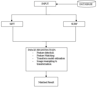
Fig. I Comparative study of SIFT & SURF algorithm
INPUT
SIFT SURF
DATABASE
IMAGE REGISTRATIOJN
– Feature detection
– Feature Matching
– Transform model estimation
– Image resampling & transformation
a) Image Registration Methodology
Image registration essentially consists of following steps as perZitova and Flusser .
i. Feature detection: Salient and distinctive objects (closed-boundary regions, edges, contours, line intersections, corners, etc) in both reference and sensed images are detected.
ii. Feature matching: The correspondence between the features in the reference and sensed image established.
iii. Transform model estimation: The type and parameters of the so-called mapping functions, aligning the sensed image with the reference image, are estimated.
iv. Image resampling and transformation: The sensed image is transformed by means of the mapping functions.
b)SIFT Detector
The algorithm was published by David Lowe in 1999. SIFT consists of four major stages: scale-space extreme detection, keypoint localization, orientation assignment and keypoint descriptor. The first stage used difference-of-Gaussian function to identify potential interest points, which were invariant to scale and orientation. DOG was used instead of Gaussian to improve the computation speed. In the keypoint localization step, they rejected the low contrast points and eliminated the edge response. Hessian matrix was used to compute the principal curvatures and eliminate the low contrast points. An orientation histogram was formed from the gradient orientations of sample points within a region around the keypoint in order to get an orientation assignment. According to the paper’s experiments, the best results were achieved with a 4 x 4 array of histograms with 8 orientation bins in each. So the descriptor of SIFT that was used is 4 x 4 x 8=128 descriptors.
Step: 1
Scale-space extrema detection, double size (width*=2, height*=2, size*=4) and use scale-space extrema in the
difference-of-Gaussian function convolved with image re-smooth the image with the Gaussian function. Step: 2
Scale-space extrema detection, double size (width*=2, height*=2, size*=4) and use scale-space extrema in the
difference-of-Gaussian function convolved with image pre-smooth the image with the Gaussian function. Step: 3
Key point localization, perform a detailed fit to the nearby data for location, scale, and ratio of principal curvatures. Determine the potential key points, accurate the location of them and remove the unstable points. Step: 4
Generate keypoint descriptor, find the orientation of key points and describe the local image region.
Image gradients Keypoint descriptor
c) SURF Detector
SURF (Speeded UP Robust Feature) is a local robust feature detector and firstly presented by Herbert BAY in 2006 that can be used in various computer vision tasks like object recognition, feature matching. SURF is Inspired by SIFT descriptor. The standard version of SURF is several times faster than SIFT and it is also more robust against SIFT. Surf Feature Extraction:
The purpose of feature point matching is to find up the feature point from the same location in two images and match a couple of feature points. SURF adopts nearest neighbour. A SURF descriptor is a 64-dimensional vector which need new data structure to place.
SURF Feature Matching:
Many approaches have been proposed to match corresponding features between two images. Low proposed the using of the ratio between the Euclidean distance to the nearest and the second nearest neighbours.
Step: 1
Detect interest points, use Hessian matrix approximation. Build the integral images and the scale space of image. Step: 2
Interest point description and matching, descriptor describes the distribution of the intensity content, similar to SIFT. Step: 3
Based on sum of Haar wavelet responses, construct a square region centred around the interest point and oriented along the orientation selected in previous section.
VI.EXPERIMENTAL RESULTS
Used input database of depth images and experimental results are described in this chapter.
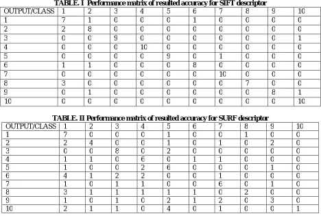
The experimental results in the Table. I shows the performance matrix of feature matching for SIFT and Table. II shows the performance matrix of feature matching for SURF. Database images of Brain MRI shown in Figure II.
Fig. II MRI DATABASE IMAGES
TABLE. I Performance matrix of resulted accuracy for SIFT descriptor
OUTPUT/CLASS 1 2 3 4 5 6 7 8 9 10
1 7 1 0 0 0 1 0 0 0 0
2 2 8 0 0 0 0 0 0 0 0
3 0 0 9 0 0 0 0 0 0 1
4 0 0 0 10 0 0 0 0 0 0
5 0 0 0 0 9 0 1 0 0 0
6 1 1 0 0 0 8 0 0 0 0
7 0 0 0 0 0 0 10 0 0 0
8 3 0 0 0 0 0 0 7 0 0
9 0 1 0 0 0 0 0 0 8 1
10 0 0 0 0 0 0 0 0 0 10
TABLE. II Performance matrix of resulted accuracy for SURF descriptor
OUTPUT/CLASS 1 2 3 4 5 6 7 8 9 10
1 7 0 0 0 1 0 0 1 0 0
2 2 4 0 0 1 0 1 0 2 0
3 0 0 8 0 2 0 0 0 0 0
4 1 1 0 6 0 1 1 0 0 0
5 1 0 0 2 6 0 0 0 1 0
6 4 1 2 2 0 0 1 0 0 0
7 1 0 1 1 0 0 6 0 1 0
8 3 1 1 1 1 1 0 2 0 0
9 1 0 1 0 2 1 2 0 3 0
10 2 1 1 0 4 0 1 0 0 1
A. PEFORMANCE ANALYSIS
a) Performance Metrices
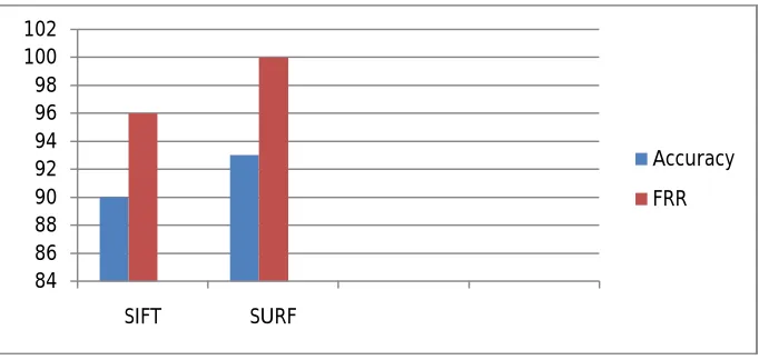
To evaluate the performance,FRR and accuracy are measured in this study. In order to check the proposed method increases the overall accuracy,calculates the accuracy of both algorithms.
Accuracy:
The accuracy of SURF is increased from 80% to 100%, but SIFT shows almost the same response with the given dataset. Results depend on the images taken.
Accuracy=Number of correct image matches x 100
Total number of images
FRR:
False Rejection Rate is also a way of investigating the best method for doing the image matching in any applications.FRR is basically used to find the capability of algorithm failing to identify the correct images.
FRR=Number of unrecognized images
Total no of images
b) Performance Evaluation
TABLE. III Performance Evaluation of SIFT & SURF descriptors
Algorithm Images Recognized
Images
Unrecognized Images
Accuracy FRR
SIFT
Class1
10
7 3 70 0.3
Class2 8 2 80 0.2
Class3 9 1 90 0.1
Class4 10 0 100 0
Class5 9 1 90 0.1
Class6 8 2 80 0.2
Class7 10 0 100 0
Class8 7 3 70 0.3
Class9 8 2 80 0.2
Class10 10 0 100 0
SURF
Class1
10
7 3 70 0.3
Class2 4 6 40 0.6
Class3 8 2 80 0.2
Class4 6 4 60 0.4
Class5 6 4 60 0.4
Class6 0 10 0 1
Class7 6 4 60 0.4
Class8 2 8 20 0.8
Class9 3 7 30 0.7
Class10 1 9 100 0.9
The Accuracy and False Rejection Rate of SIFT and SURF is shown in Figure II.
Fig. II ACCURACY & FRR
VII.CONCLUSION
In this paper the comparison between two feature detection methods for image matching was presented. SIFT as the first method and SURF method as second. Keypoints of brain were extracted and matching has been done using both SIFT and SURF. To compare the results of implementing algorithms, performance of both the algorithms are evaluated and find that SURF is fast and has good performance as the same as SIFT with better accuracy to the existing system. By combing both SIFT and SURF algorithm helps to found out matching keypoints much faster than using SIFT
84 86 88 90 92 94 96 98 100 102
SIFT SURF
Accuracy
algorithm alone. The future work will be based on developing algorithms to identify various other brain diseases, to improve the overall efficiency and also to further reduce the computational time.
REFERENCES
1. Sousa, David, “How the Special Needs Brain Learns, 2nded”, Crowin Press: USA. ISBN-10: 1-4129-4986-6 | ISBN-13: 978-1412949866, 2006. 2. Gilat , Amos, “Mat lab: An Introduction with Applications, 3rded”, Wiley: USA. ISBN-10: 0470108770 | ISBN-13: 978-0-470-10877-2, 2008. 3. Lashkari, AmirEhsan, “A Neural Network Based Method for Brain Abnormality Detect ion in MR Images Using Zernike Moments and Geometric Moments”, International Journal of Computer Applications, vol. 4, no. 7, ISSN: 0975-8887, July 2010.
4. Goyal, Soniya et.al, “Automatic Detect ion of Brain Abnormalities and Tumor Segmentation in MRI Sequences”, Image and Vision Computing New Zealand Conference, IVCNZ: New Zealand, 2011.
5. Dr. Manjusha et.al,” A Survey of Image Registration “, International Journal of Image Processing (IJIP), Volume (5) ,Issue (3), 2011.
6. YAO-MING YU, “Detecting and Locating of Brain Abnormality in MR Images Using Texture Feature Analysis and Improved Probabilistic Relaxation Methods”, WSEAS TRANSACTIONS On BIOLOGY And BIOMEDICINE, JULY 2013.
7. A. Swapna et.al, “A Comparison and Matching Point Extraction of SIFT and ISIFT”, ISSN: 2229-6093, DEC 2013.
8. Reetika Verma et.al, “Enhanced Character Recognition Using Surf Feature and Neural Network Technique”, (IJCSIT) International Journal of Computer Science and Information Technologies, Vol. 5 (4), 5565-5570, 2014.
9. Siddharth Saxena et.al, “A Survey of Recent and Classical Image Registration Methods”, International Journal of Signal Processing, Image Processing and Pattern Recognition, Vol.7, No.4, pp.167-176, 2014.
10. Dr. PSJ Kumar1 et.al, “Digital Image Processing based Detection of Brain Abnormality forAlzheime r’s disease”, International Journal of Engineering and Computer Science, ISSN: 2319-7242, Volume 3 Issue 12 December, Page No. 9479-9484, 2014.
11. Vivek Kumar Gupta et.al, “An Analytical Study of SIFT and SURF in Image Registration”, International Journal of Engineering and Innovative Technology (IJEIT), Volume 3, Issue 9, March 2014.
12. Peter Sykora et.al, “Comparison of SIFT and SURF Methods for Use on Hand Gesture Recognition Based on Depth Map”, AASRI Conference on Circuits and Signal Processing, 2014.
13. Raju Anitha et.al, “Detection of Brain Abnormality for Alzheimer’s Disease Using Image Processing Techniques”, ISSN (Online): 2347 - 2812, Volume-3, Issue -12, 2015.
14. Sheena S et.al, “A COMPARISON OF SIFT and SURF ALGORITHM FOR THE RECOGNITION OF AN EFFICIENT IRIS BIOMETRIC SYSTEM”, International Journal of Advanced Research in Computer and Communication Engineering, Vol. 5, Special Issue 1, FEB 2016.
15. Preeti Mandle et.al, “An Advanced Technique of Image Matching Using SIFT and SURF”, International Journal of Advanced Research in Computer and Communication Engineering, Vol. 5, Issue 5, MAY 2016.
BIOGRAPHY
Ameer Nisha.S is currently pursuing M.Phil degree in computer science in Sadakathullah Appa College, Tirunelveli. She has done her MSC degree in Computer Science from V.O.Chidambaram College,Thoothukudi and the B.Sc in Computer Science from St.Mary’s College(Autonomous),Thoothukudi.under Manonmaniam Sundaranar University,Tirunelveli.