ABSTRACT
KUHN, JEFFREY FRANCIS. Evolutionary Conservation of Eukaryotic and Archaeal Box C/D
Ribonucleoprotein Complex Structure. (Under the direction of E. Stuart Maxwell).
Ribosome biogenesis requires a large population of small nucleolar RNAs (snoRNAs) for
pre-rRNA processing and nucleotide modification. These snoRNAs can be classified into two major
families based on sequence and structural elements: the box C/D snoRNAs and the box H/ACA
snoRNAs. The box C/D snoRNAs possess conserved nucleotide boxes C and D contained within
a folded structural element defined as the box C/D core motif that is essential for snoRNA
biogenesis, nucleolar transport, and nucleotide modification. This dissertation describes the
purification, identification and characterization of box C/D snoRNA-associated proteins. These
proteins are conserved throughout evolution and homologs exist in Eukaryota and Archaea.
Homologs of the box C/D snoRNAs exist in Archaea as well, termed sRNAs. This work also
examines the structure of the box C/D core motif RNA and investigates the organization of the
box C/D RNP particle.
Eukaryotic snoRNA-associated proteins were initially identified by affinity chromatography
using the box C/D core motif RNA as the selection agent. The four proteins isolated consist of
two protein pairs, with members of each pair being highly related in sequence. One pair of
proteins, Nop56p and Nop58p/Nop5p, are essential nucleolar proteins associated with box C/D
snoRNAs, consistent with their designation as “core” snoRNP proteins. The second pair of
proteins, termed p50 and p55, are essential nucleoplasmic proteins and have been designated
“accessory” proteins. Immunoprecipitation experiments suggest that the eukaryotic proteins p50
and p55 are transiently associated with the box C/D snoRNP complex in the nucleoplasm,
consistent with a role in snoRNA biogenesis and/or snoRNA transport events. These results
have led to the designation of p50 and p55 as accessory proteins. Sequence analysis reveals
in archaeal genomes, where a single homolog for each of the Nop56p/Nop58p and p50/p55
protein pairs exists.
Thermal denaturation analysis of the box C/D core motif RNA demonstrates that it possesses
a well-defined structure in the absence of snoRNA binding proteins. Together with a mutational
analysis, the thermal denaturation experiments reveal that the box C/D core motif has an ordered
structure which is stable in solution and requires a set of critical G-A residues. These results are
consistent with the previous suggestion that the box C/D core motif forms a “kink-turn” (K-turn)
motif, a novel RNA fold defined by a highly kinked phosphodiester backbone and two base paired
stems flanking an asymmetric bulge region which contains tandem sheared G-A base pairs
critical to RNA folding.
Sequence analysis reveals that the archaeal homolog of the eukaryotic core protein termed
15.5kD is the archaeal ribosomal protein L7. The 15.5kD protein serves a dual function in
eukaryotes by binding both the spliceosomal U4 snRNA and the box C/D snoRNAs. Interestingly,
archaeal L7 also serves a dual function by binding the 23S rRNA as well as the box C/D sRNAs.
Our binding analyses reveal that L7 binds the box C/D core motif with high affinity. In addition,
we have cloned the remaining archaeal homologs of the box C/D-associated proteins and binding
analyses demonstrate that these proteins, together with the sRNAs, are able to form sRNP
complexes which closely resemble eukaryotic snoRNPs. Nuclease mapping experiments have
begun to establish the overall organization of the archaeal box C/D sRNP particle. The striking
similarity of the box C/D sRNP/snoRNP complex to the L7:KT15 RNP of the 50S ribosomal
subunit suggests that the archaeal sRNPs and eukaryotic snoRNPs could have their evolutionary
DEDICATION
For my parents, Joe and Jane, who always believed in me. And for my wife, Juliette, who supports me in all that I do.
BIOGRAPHY
ACKNOWLEDGEMENTS
I would like to express my deepest gratitude to Dr. E. Stuart Maxwell for being my mentor and guide throughout my graduate studies. His insightful scientific “discussions” will never go unremembered.
I would also like to thank the members of my advisory committee, Drs. Paul Wollenzien, Dennis Brown and James Brown, for their helpful advice and encouragement. They have been a wonderful committee and maintained an open door policy which I have found incredibly helpful. Additionally, Dr. James Brown has been an immense help in genetic analysis and RNA structural analysis, as well as providing materials for experiments.
I would like to express my appreciation to all of the faculty and staff of the Biochemistry department for their advice and stimulating scientific discussions. Especially Dr. Clay Clark who was an enormous help to me in both protein expression and protein binding studies. Also, Drs. Dick Guenther and Paul Agris who selflessly provided much time and energy in teaching me the techniques of thermal denaturation analysis. Additionally, I would like to thank Drs. Nicholas Watkins, Dima Moundus, Dalia Juzumiene, and Tatiana Shapkina for their friendship and guidance. They have taken much time to contribute to my training.
I would like to thank the graduate students of the Biochemistry department for their camaraderie over the years. Especially Elizabeth Tran and Donna Newman, who have worked alongside of me and contributed much of their talents to enriching both my work and my personal life.
TABLE OF CONTENTS
Page
LIST OF TABLES ... viii
LIST OF FIGURES ... ix
LITERATURE REVIEW ... 1
1. The Nucleolus ... 1
1.1 The Nucleolus and Ribosome Biogenesis ... 1
1.2 Ribosome Biogenesis and snoRNAs ... 1
2. The snoRNAs ... 3
2.1 Two Families of snoRNAs: Box C/D and H/ACA ... 3
2.2 Functions of snoRNAs ... 4
2.3 Biogenesis of snoRNAs ... 7
3. The snoRNP Proteins ... 9
3.1 Overview of snoRNP Proteins ... 9
3.2 Core Box C/D snoRNP Proteins ... 10
3.2.1 Fibrillarin or Nop1p ... 10
3.2.2 Nop58p (Nop5p) ... 11
3.2.3 Nop56p ... 12
3.2.4 15.5kD Protein ... 13
3.3 Accessory Proteins ... 14
3.3.1 Identification of the snoRNP Accessory Proteins p50 and p55 ... 14
3.3.2 The p50 and p55 Proteins are Linked to Important Cellular Functions ... 15
3.3.3 Functions of p50 and p55 in snoRNA Biogenesis ... 16
4. The Structure of Box C/D snoRNAs... 16
4.1 The “Kink Turn” Motif ... 16
4.2 Features of the Box C/D snoRNA Kink Turn ... 19
5. Archaeal sRNAs and sRNP Complexes ... 20
5.1 Archaea: A Prokaryotic Organism Related to Eukaryotes ... 20
5.2 Archaeal Homologs of Small Nucleolar RNAs: sRNAs ... 20
5.3 Archaeal Homologs of Box C/D snoRNP Proteins ... 21
5.5 Functions of Archaeal Box C/D sRNPs ... 26
6. Summary of Chapters ... 27
7. References ... 28
CHAPTER ONE ... 39
Foreword ... 40
Introduction ... 41
Results ... 42
In Vitro Assembly of the U14 snoRNP Core Complex ... 42
The U14 Box C/D Core Motif with Included Boxes C and D is Required for snoRNP Assembly ... 42
A Mouse 65-kDa Nuclear Protein Recognizes Boxes C/D and Crosslinks to the Box C/D Core Motif ... 44
Three Mouse Nuclear Proteins Co-fractionate with the U14 Terminal Core Motif in a Multiprotein snoRNP Complex ... 46
Discussion ... 48
Materials and Methods ... 50
References ... 51
CHAPTER TWO ... 53
Foreword ... 54
Introduction ... 55
Results ... 56
Affinity Chromatographic Isolation of the U14 Box C/D snoRNP Proteins ... 56
Mouse Nop58p and Nop56p are a Pair of Evolutionarily Conserved Nucleolar Proteins ... 57
P55 and p50 Constitute a Second Pair of Highly Conserved Nuclear Proteins ... 65
Mouse p55 and p50 are Nucleoplasmic Proteins ... 66
Immunoprecipitation of the Assembled U14 snoRNP Complex with p55 and p50 Antibodies ... 67
Discussion ... 67
Materials and Methods ... 70
References ... 72
CHAPTER THREE ... 74
Introduction ... 76
Materials and Methods ... 77
Results ... 78
Archaeal Ribosomal Protein L7 is a Functional Homolog of Eukaryotic 15.5kD snoRNP Protein and Binds the Box C/D snoRNA Core Motif ... 78
Archaeal L7 Protein Binds the Box C/D Core Motif with the Same Affinity as Eukaryotic 15.5kD Protein ... 79
L7 Requires the Same Box C/D Sequence and Structural Elements for Binding as does the 15.5kD Protein ... 80
Discussion ... 83
References ... 85
CHAPTER FOUR ... 87
Foreword ... 88
Introduction ... 89
Materials and Methods ... 90
Results ... 91
L7 Binds Both the Box C/D and C´/D´ Elements ... 91
Core sRNP Complex Formation Results in Conformational Change in the RNA ... 94
Protein binding is dependent on the conserved sequences in the box C/D and C´D´ elements ... 95
Discussion ... 97
References ... 99
SUMMARY AND FUTURE PERSPECTIVES ... 104
APPENDICES ... 109
A. Recombinant Proteins ... 110
B. Antibody Production ... 113
C. Chapter 3 Addendum: Immunoprecipitation of L7:Fibrillarin Complex ... 118
LIST OF TABLES
CHAPTER ONE
Table 1: Biochemical fractionation of the mouse U14 snoRNP core complex ... 48
CHAPTER THREE
Table 1: Relative binding affinity of L7 protein for selected box C/D snoRNA
mutants ... 80
APPENDIX A
Table A1: Eukaryotic and archaeal clone information ... 112
APPENDIX B
LIST OF FIGURES LITERATURE REVIEW
Figure 1. Processing pathway of the mammalian pre-ribosomal RNA transcript ... 2
Figure 2. Secondary structure of the box C/D and H/ACA snoRNAs ... 4
Figure 3. Proposed tertiary structure of the snoRNA:target RNA duplex ... 5
Figure 4. Processing of the intronic snoRNA precursor ... 8
Figure 5. Secondary structures of the U4 snRNA and proposed box C/D core motif RNA ... 17
Figure 6: Proposed tertiary structure of the box C/D core motif RNA ... 18
Figure 7. Comparison of the U4 K-turn and the KT-15 of archaeal 23S rRNA ... 23
Figure 8. Comparison of human 15.5kD, H. marismortui L7Ae and M. jannaschii L7 proteins ... 24
CHAPTER ONE Figure 1. Mouse U14 snoRNA and the box C/D core motif ... 43
Figure 2. In vitro assembly of the U14 snoRNP core complex ... 44
Figure 3. Requirement of nucleotide boxes C and D for snoRNP assembly ... 45
Figure 4. Requirement of phylogenetically conserved box C and D nucleotides for snoRNP assembly ... 45
Figure 5. Requirement of the U14 terminal stem structure for snoRNP assembly ... 46
Figure 6. UV crosslinking of mouse nuclear proteins to the U14 box C/D core motif ... 46
Figure 7. Biochemical fractionation of the in vitro-assembled U14 snoRNP complex and identification of U14 snoRNP core proteins ... 47
CHAPTER TWO Figure 1. Affinity chromatography isolation of box C/D snoRNA-associated proteins ... 56
Figure 2. Nuclear proteins of 65, 55 and 50 kDa are associated with the box C/D core motif ... 57 Figure 3. Alignment and sequence comparison of mouse Nop58 and Nop56
proteins with their eukaryotic homologs ... 58-59 Figure 4. Alignment and sequence comparison of mouse Nop58 and mouse
Nop56 proteins with the archaeal homologs and the phylogenetic relationship
Figure 5. Alignment and sequence comparison of mouse p50 and p55
proteins with their eukaryotic homologs ... 62-63 Figure 6. Alignment and sequence comparison of mouse p50 and p55 proteins
with the archaeal homologs and the phylogenetic relationship of eukaryotic
and archaeal p50/p55 proteins ... 64-65
Figure 7. Nuclear distribution of mouse p55 and p50 proteins ... 66
Figure 8. Immunoprecipitation of the assembled U14 snoRNP complex with p50 and p55 antibodies ... 67
Figure 9. Possible roles for the p55/p50 proteins in the eukaryotic nucleus ... 68
CHAPTER THREE Figure 1. Archaeal ribosomal protein L7 is a homolog of the eukaryotic 15.5kD snoRNP protein ... 78
Figure 2. Archaeal ribosomal protein L7 binds the box C/D snoRNA core motif ... 79
Figure 3. Folded structure of the box C/D snoRNA core motif ... 80
Figure 4. The box C/D core motif exhibits a highly ordered RNA structure ... 81
Figure 5. L7 requires specific sequence and structural elements of the box C/D core motif for RNA-protein interaction ... 82
Figure 6. Comparison of the U4 snRNA 5´ stem loop and the KT-15 K-turn of 23S rRNA ... 84
Figure 7. Comparison of human 15.5kD, H. marismortui L7Ae and M. jannaschii L7 proteins ... 85
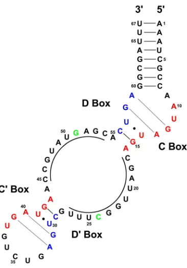
CHAPTER FOUR Figure 1. Proposed secondary structure of the M. jannaschii sR8 sRNA ... 92
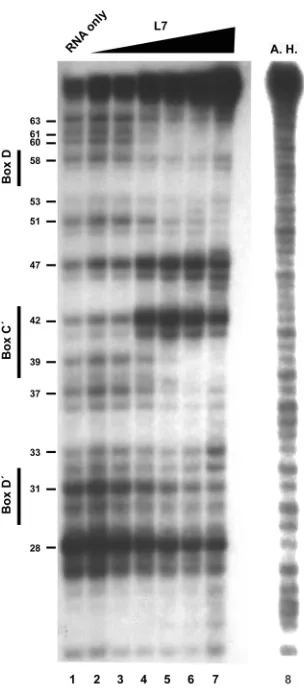
Figure 2. Nuclease mapping of sR8 RNA with titration of L7 ... 93
Figure 3. Nuclease mapping of sR8 RNA with all three sRNP core proteins ... 95
Figure 4. Nuclease mapping of the box D and box D´ mutant RNAs ... 96
Figure 5: Summary of nuclease protection experiments ... 98
LITERATURE REVIEW
1. The Nucleolus
1.1 The Nucleolus and Ribosome Biogenesis
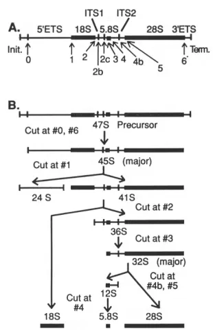
The nucleolus is a sub-nuclear structure present in eukaryotic cells and is the site of ribosome biogenesis. The nucleolus is a network of protein and RNA molecules organized around chromatin containing ribosomal RNA genes (rDNA) that are undergoing active transcription by RNA polymerase I. This chromosomal locus may contain as many as several thousand rDNA genes in tandem array and is known as a nucleolar organizer (1). The process of ribosome biogenesis is a series of coordinated steps including (not necessarily in this order): 1.) the transcription of rDNA genes, 2.) the modification of specific bases within the rRNA, 3.) the processing of the precursor ribosomal RNA (pre-rRNA) into separate 18S, 5.8S and 28S rRNA species, 4.) the import of 5S rRNA (transcribed separately in the nucleus by RNA pol III), 5.) the import of ribosomal proteins, 6.) the folding of the rRNA and assembly of the rRNA with the proteins (reviewed in 2). Ultimately, the nearly mature ribosome subunits are transported to the cytoplasm.
The nucleolus was first described approximately 150 years ago. As many as 100 years ago, it was noted that the nucleolus disappeared from cell nuclei during mitosis and subsequently reappeared (3). It would be 60 more years before the role of nucleoli in ribosome biogenesis became apparent. In the 1960’s, it was discovered that nucleoli contained the genes which code for 18S and 28S rRNA (4). The nucleolus possesses a distinct morphology and is comprised of dense regions known as the granular component (GC), the dense fibrillar component (DFC), and the fibrillar center (FC). The majority of the nucleolus is the DFC region (75%) in animal cells, however in plant cells the GC and DFC are roughly equivalent (5). The FC region of both animal and plant nucleoli is a very small portion, approximately 1-2% (5). Due to the variability in nucleoli, it is difficult to categorize these different regions by functions performed therein, but they do appear to have differences in protein makeup (reviewed in 6). Indeed, these dense regions are thought to be networks of interacting protein and RNA. The change in the appearance of nucleoli over time in a cell is likely due to a changing pattern of activities with respect to the synthesis and production of new ribosomes.
1.2 Ribosome Biogenesis and snoRNAs
external transcribed spacer regions (ETS1 and ETS2, see Fig. 1). Subsequent to transcription, the spacer regions and intervening sequences are removed by a host of processing and endonucleolytic factors which are too numerous to describe in detail here (for a complete review, see 2). Concomitant with this processing, the pre-rRNA is modified at selected nucleotide bases with either methylation of the ribose sugar at the 2´ position or conversion of a uridine to a pseudouridine as the mature forms of the 18S, 5.8S, and 28S species are produced. These RNAs are then folded, assembled together with protein and exported as essentially mature ribosomes.
Figure 1. Processing pathway of the mammalian pre-ribosomal RNA transcript.
(A). The 47S precursor rRNA with spacer regions and endonucleolytic processing points indicated. (B). The pre-rRNA processing pathway. Cleavage points correspond to the numbered sites in panel A. Adapted from D. Eichler and N. Craig (1994) Progress in Nucleic Acids Research and Molecular Biology, vol. 49, p.201.
through base pairing interactions. With over 200 modifications in the pre-rRNA (11), as well as sites of processing and regions chaperoned by snoRNAs during folding, it comes as no surprise that the snoRNAs are the most numerous small RNAs found in the eukaryotic cell.
2. The snoRNAs
2.1 Two Families of snoRNAs: Box C/D and H/ACA
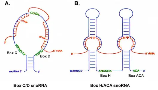
The snoRNAs are classified into two major families based on conserved sequence and structural elements (Fig. 2) (12-14). The conserved features are bound by common core proteins for each snoRNA family to form a ribonucleoprotein particle (15-17). The box C/D snoRNAs possess conserved nucleotide box C (UGAUGA) located near the 5´ end of the RNA and box D (CUGA) located near the 3´ end of the RNA. Box C/D snoRNAs are associated with the putative methylase fibrillarin, a conserved nucleolar protein, and are thus responsible for 2´-O-methylation of specific ribose sugars in the rRNA (18-20). The box C/D snoRNAs also contain internally positioned nucleotide boxes C´ and D´ which are similar to boxes C and D, although not as well conserved in sequence. Box C/D snoRNAs typically possess a terminal stem formed by complementary base pairing at the 5´ and 3´ ends of the RNA (Fig. 2A). They also possess an internal stem which forms from complementarities contained within boxes C and D that extend to the adjacent nucleotides (not shown, see Fig. 5). Those box C/D snoRNAs which do not possess a terminal stem have either extended internal stems or use sequences in the precursor snoRNA to form an exteRNAl stem during snoRNA biogenesis (21). The ultimate consequence of these base-paired structures is to bring box C and D together in close juxtaposition, forming an RNA motif that is essential for protein binding (14, 15, 22).
The box H/ACA snoRNAs possess a bipartite structure consisting of two base paired stems separated by the so-called “hinge” region containing conserved nucleotide box H (AnAnnA, where n is any base) and an ACA nucleotide triplet located at the 3´ end of the RNA (Fig. 2B) (13). Each of the two base paired stem regions contains a bulge within the base-paired stem which possesses short regions of complementarity that direct the snoRNA to the target RNAs (23, 24). H/ACA snoRNAs associate with the conserved putative psuedouridylase Cbf5p and are thus responsible for conversion of targeted uridines to pseudouridine(16, 17). The H/ACA snoRNAs also act as guide RNAs by directing the pseudouridine synthetase to the site of modification on the target RNA via base pairing.
common eight of the nine proteins that are found in each of the two RNP complexes (25). MRP, like several snoRNAs of both the box C/D and H/ACA families, is required for cleavage and processing of the 45S pre-rRNA. In particular, MRP catalyzes the cleavage in ITS1 which separates the 18S from the 5.8S and 28S species (26, 27). Although this cleavage site is not required for rRNA synthesis (an alternative site may be cleaved (28)), MRP is essential for cell viability (29). This raises the possibility that MRP has other targets or functions in addition to its role in pre-rRNA processing.
Figure 2. Secondary structure of the box C/D and H/ACA snoRNAs.
Conserved secondary structures of the box C/D snoRNAs (A.) and box H/ACA snoRNAs (B.) In each panel, the blue line drawing represents the snoRNA as indicated, the conserved box elements are highlighted in green, and the red line represents the substrate RNA as indicated. Base pairing is represented by dashes. Positions of the modified nucleotide in the snoRNA:rRNA duplex are shown for both 2´-O-methylation (2´-O-Me) and pseudouridylation (Ψ). Adapted from Smith and Steitz (1997) Cell, vol. 89, p. 670.
2.2 Functions of snoRNAs
the pre-rRNA by base pairing interactions between the snoRNA and the target RNA. Also, base pairing interactions can be envisioned to occupy internal base pairing sites during transcription of the nascent target RNA, delaying RNA folding until the mature configuration can be achieved. In this manner, the snoRNA can serve as a chaperone during RNA folding reactions, although this function has not yet been widely demonstrated.
The snoRNAs function in nucleotide modification by using regions of complementarity to guide their respective associated protein enzymes to the target site of modification, a common feature of both box C/D and H/ACA snoRNAs (Fig. 2) (8). The snoRNAs form base-pairing interactions with their respective target RNAs such that the nucleotide to be modified is positioned at precise distances from conserved sequences in the snoRNAs which serve as protein binding sites. Experiments have confirmed these potential guide functions by examining sites within rRNA which contain sequences complementary to snoRNA guides (24, 31). Mutation analysis has demonstrated that destroying the complementarity between substrate and snoRNA results in a loss of modification of the target nucleotide (23, 32). Furthermore, compensatory changes which restore base pairing in this region result in the restoration of the correct modification. Experiments have gone on to show that novel modifications can be introduced into target RNAs by introducing artificial snoRNA:target RNA base pairing interactions (33).
The complementary region of the box C/D snoRNAs is typically long (11-20 nucleotides) and is located just upstream of box D or D´, guiding the methylation of the RNA substrate to the base-paired nucleotide five residues upstream of box D (or D´) (Fig. 2A). Interestingly, it has been noted this position on the RNA substrate occurs on the same side of the snoRNA:RNA substrate duplex as box D (or D´) (Fig. 3) (30). Thus, the site of modification is positioned close to box D (or D´), which is presumably bound by protein.
The green strand of model is the target RNA and the blue strand of model is the snoRNA. Nucleotides of box D are indicated by a bracket and shaded dark blue. The residue targeted for 2´-O methylation modification is shown in gold. Adapted from D. Tollervey (1996) Science, vol. 273, p. 1057.
The H/ACA snoRNAs possess a conserved hairpin-hinge-hairpin structure with the region of complementarity located in the internal bulge region of the hairpins (Fig. 2B). The uridine residue to be converted to pseudouridine in the target RNA is an unpaired nucleotide in the snoRNA:target duplex, guided to the bulge region by base pairing interactions. The residue in the duplex is positioned 15-16 nucleotides upstream from the box H and ACA region(s), in the so-called “pseudouridylation pocket” (24). The targeted nucleotide is directed by base-pairing interactions to the bulge region of the snoRNA stems. As a result, the RNA substrate is positioned in a “pseudouridylation pocket” that is formed by short regions (4-5 nucleotides) of complementarity between the snoRNA and the substrate, flanking the target nucleotide (Fig. 2B).
Although the eukaryotic rRNA is extensively modified (approximately 200 modifications in humans (11)), the functional significance of these nucleotide modifications remains unclear. Individual snoRNAs can be genetically deleted with no apparent adverse effect to the organism (34), with the exception of those few indispensable snoRNAs that are involved in pre-rRNA processing (35-40). It has not been possible to abolish global methylation, because mutations in fibrillarin which eliminate methylation are lethal (41). However, it is not possible to conclude from these experiments whether the lethality is due to defects in snoRNA biogenesis or loss of methylation. Interestingly, while the individual modifications are not conserved, they typically fall in the most highly conserved regions of rRNA and are situated near the catalytic core (11).
Modified nucleotides could enhance the activity of the ribosome, a confirmed ribozyme (42), or increase the stability of protein:RNA interactions within the ribosome. It has recently been shown that some 2´-O-methylation modifications at select sites do affect the rate of translation in vivo (M. Fournier, personal communication). Another possibility is that the modifications are necessary for structural reasons. For example, in the case of 2´-O-methylation, addition of a methyl group may add a desirable hydrophobic interaction which stabilizes a protein:RNA contact. Additionally, 2´-O-methylation may prevent RNA degradation at vulnerable sites by blocking the hydrolysis of the phosphodiester bond, which requires formation of a cyclic bond between the 2´ and 3´ hydroxyl groups. Pseudouridylation changes the nature of the base-pairing ability of the modified nucleotide. Such modifications may serve to enhance the activity or stabilize the overall folded structure of the mature ribosome.
protein enzyme to a site of endonucleolytic cleavage that releases the mature rRNA species from the pre-rRNA transcript. It was shown by genetic manipulation in yeast that these snoRNAs include the box C/D U3, U8, U14 and U22 snoRNAs, as well as the H/ACA snoRNAs U17, snR10 and snR30 (35-40). Deletion of these snoRNA species disrupts cleavage of the pre-rRNA and blocks accumulation of mature rRNA species. Although it has not been demonstrated, the snoRNA could prevent the pre-rRNA from incorrect or premature folding which blocks the site of cleavage by using its complementary region to form a competing snoRNA:pre-rRNA base pairing interaction.
The snoRNAs may serve as chaperones for rRNA folding interactions as well. Base pairing between the nascent pre-rRNA and snoRNA could inhibit undesirable internal base pairing interactions within the pre-rRNA as the transcript is made by RNA polymerase, thereby promoting the correct base pairing in the mature molecule. Several snoRNAs from both the box C/D and H/ACA families may possibly participate in these activities, as well as MRP.
In addition, snoRNAs have been shown to modify other cellular RNAs in addition to ribosomal RNA, such as the splicing snRNA U6 (10, 44, 45). A number of snoRNAs found exclusively in the brain, termed “small non-messenger RNAs“ (snmRNA) have recently been discovered (46-48). These RNAs possess the hallmarks of bona fide snoRNAs, but are not involved in modification or processing of either rRNA or snRNAs. It is not clear what function these snmRNAs serve. The antisense elements of these snmRNAs do not correspond to any known cellular RNA, with the exception of one snmRNA which possesses the potential to guide modification of the serotonin receptor mRNA. Clearly though, these snmRNAs represent an entirely new set of potential functions for snoRNAs, possibly regulating gene expression.
2.3 Biogenesis of snoRNAs
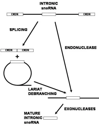
generating the mature ends of the snoRNA (57, 59). In the absence of splicing and lariat formation, it has been shown that the U16 and U18 snoRNAs can be excised from the intron by endonucleolytic cleavage as a minor pathway (60, 61). Exonucleolytic trimming then removes the flanking sequence from the precursor snoRNA. In both pathways, proteins which bind the conserved sequences within the mature snoRNA before it is fully processed serve to block complete degradation of the RNA by exonucleases and thereby define the mature ends of the molecule.
Figure 4. Processing of the intronic snoRNA precursor.
Correct processing requires the evolutionarily conserved sequence elements present in the snoRNA, which serve as protein binding recognition signals (14, 23, 62). It has been shown that mutation of these sequence elements results in a failure to accumulate stable, steady state snoRNA populations. It is assumed that abolishing protein binding results in full degradation of the precursor snoRNA by exonucleases. Thus, protein binding is essential for the processing of the snoRNA from the intron.
transcribed exclusively from classical promoters, and are either mono- or, more often, polycistronic (52). Biogenesis of polycistronic snoRNAs does not involve splicing and no polycistronic snoRNAs have yet been found in metazoans. Despite this fact, these snoRNA precursors have the same requirements for processing as their monocistronic counterparts and must bind proteins to accumulate the mature form of the snoRNA (52).
The protein-coding genes which serve to “host” intronic snoRNA genes are typically proteins involved in translation or ribosome biogenesis (7, 54, 63, 64). These include ribosomal proteins, initiation factors, and nucleolar proteins. The reason for this biased distribution is not clear, but could provide a means for coordinating snoRNA biogenesis with production of the translation machinery. Other host genes, such as the heat shock cognate gene, are constitutively transcribed and could thus serve to supply the cell with a steady level of new snoRNAs (53). Additionally, the same snoRNA coding sequence can be found within different introns of the same host gene. Interestingly, the host gene of a particular snoRNA coding sequence can differ among evolutionarily related organisms, suggesting that the snoRNA was a highly mobile element throughout evolution (55).
In addition to protein coding genes, several host genes serve only to produce intronic snoRNAs, as their exons do not code for proteins (65-67). How these genes arose and what purpose they serve, other than production of snoRNAs, is not clear. One such snoRNA host gene identified was gas5 (growth arrest-specific transcript 5) (66). With the discovery of gas5, it was speculated that the pyrimidine rich 5´ terminal oligopyrimidine (5´TOP) sequences that served as promoters for such non-protein coding host genes had some regulatory role in snoRNA biogenesis. This was most likely to occur through coordinating transcription with mRNA processing. It is noteworthy that all known snoRNA host genes have 5´TOP-like regions, suggesting that a common mechanism for regulating such a diverse group of host genes might exist (65). This raises the possibility that ribosome biogenesis could be coordinated with snoRNA biogenesis through transcriptional regulation via the 5´TOP elements.
3. SnoRNP Proteins
3.1 Overview of snoRNP Proteins
may include regulatory and processing factors that may not be absolutely essential for snoRNP function or stability, but are crucial to coordinating the biogenesis of both snoRNAs and ribosomes.
The snoRNAs themselves are not assumed to be the catalytic agents in the pre-rRNA modification and cleavage reactions. Proteins which associate directly with the RNA or through protein:protein interactions are therefore responsible for carrying out specific reactions being mediated by the snoRNA. The box C/D snoRNAs associate with four core proteins: fibrillarin, Nop56p, Nop58p and 15.5kD protein (15, 19, 68, 69, 71-73, 75). The box H/ACA snoRNAs associate with four core proteins as well: Nhp2p, Gar1p, Cbf5p and Nop10p (16, 17, 70, 74). In addition, the putative helicases termed p50 and p55 associate transiently with box C/D snoRNAs and are involved in the biogenesis of both box C/D and H/ACA snoRNAs (15, 76). To date, p50 and p55 are the only known examples of accessory proteins. SnoRNAs are believed to associate with many other proteins such as Rat1p, an endonuclease that is involved in pre-rRNA processing (28). Interactions with many as yet unidentified proteins will be key to understanding the biogenesis and function of the snoRNAs.
An exhaustive survey of all proteins associated with snoRNAs and ribosome biogenesis is beyond the scope of this manuscript (for reviews see, 2). I will instead focus on the box C/D snoRNA-associated proteins.
3.2 Core Box C/D snoRNP Proteins
Box C/D snoRNA-associated proteins have been identified through genetic screening, immunoprecipitation experiments, and chromatographic isolation. The four core proteins which have been defined by these various techniques (described in detail below) are highly conserved throughout evolution and are essential for the function and biogenesis of the mature box C/D snoRNP particle. Several of the eukaryotic core proteins have not yet proven amenable to recombinant protein expression techniques, therefore structural information is scarce. In the following sections, I will highlight what is known about the core box C/D-associated proteins fibrillarin, Nop56p, Nop58p and the 15.5kD protein.
3.2.1 Fibrillarin or Nop1p
of the mature 18S rRNA (41, 79). In addition, global methylation of the ribosomal RNA is also inhibited when fibrillarin expression is disrupted (41). However, it is difficult to determine from these experiments whether the loss of methylation is due to fibrillarin depletion or if some other nucleolar component is being affected by fibrillarin depletion. Interestingly, thermosensitive (ts) conditional mutants were discovered in which either pre-rRNA processing or methylation was affected, indicating that these functions are dependent upon distinct regions of the protein (41).
Fibrillarin is a highly conserved 34kD protein that contains two characteristic domains: an “RNA recognition motif” (RRM) and a “glycine-arginine rich” (GAR) domain, also called an RGG motif (68, 80). The RRM has been well characterized and contains two short, highly conserved core regions. The positively charged GAR domain has, not surprisingly, been shown to function in nucleic acid binding. However, this binding is nonspecific in nature and it is postulated that the GAR domain functions in nucleolar localization. Interestingly, an obvious sequence for nuclear localization is absent in fibrillarin.
More recently, the fibrillarin gene has been cloned and it is apparent that the recombinant protein has a very weak RNA binding affinity, suggesting that it does not interact directly with the snoRNA but requires other proteins to remain stably bound to the RNA (81). However, there are reports of specific binding of fibrillarin to the box C/D core motif, which requires conserved box C and D sequences (82). This weak binding interaction may seem surprising in light of the sequence elements such as the RRM contained in the fibrillarin sequence. However, it is consistent with the well-documented fact that fibrillarin binding activity is very sensitive to salt concentrations. The crystal structure of fibrillarin suggests that the protein contains a putative S-adenosyl-L-methionine (SAM) binding site, indicating that it is the methylase enzyme (20). This is consistent with the finding that ts mutants defective in methylation are found clustered in the putative SAM motif.
3.2.2 Nop58p (Nop5p)
Nop58p was the second box C/D snoRNP core protein to be identified (69). Nop58p was originally isolated by immunoprecipitation experiments directed against fibrillarin which demonstrated that Nop58p was associated with box C/D snoRNAs. Further experiments using antibodies against Nop58p and tagged versions of the Nop58 protein in yeast also demonstrated that it was a box C/D snoRNP core protein (71, 72, 75). In other experiments, screens for lethal mutations linked to mutations in the fibrillarin gene (synthetic lethals, sl) in yeast identified Nop58p and its partner Nop56p (described below) as associated with fibrillarin (73).
motif”). In mammals, this domain is less well defined and consists of long stretches of basic residues (the “K-tail”). The KKE/D family of yeast nucleolar proteins includes both Nop58p and Nop56p, as well as the putative pseudouridylase Cbf5p and the putative RNA helicase Dbp3p (73, 83, 84). Nop58p is an essential protein and gene deletion experiments have demonstrated the requirement of Nop58p for cell growth in yeast (71, 73). Specifically, the processing of 18S rRNA is inhibited, resulting in a depletion of the 40S ribosomal subunit (69). The processing defect appears to be in the cleavage of the 5´ETS. Experiments have shown that loss of the box C/D snoRNAs U3 and U14 also disrupt the processing of pre-rRNA in the 5´ETS (36, 40). These results are consistent with the definition of Nop58p as a core box C/D snoRNP protein. Interestingly, genetic manipulations have shown that the “K-tail” or KKE/D motif is not essential for Nop58p accumulation or cell growth in yeast as it can be deleted from the gene with no apparent growth phenotype (73).
Immunoprecipitation experiments have demonstrated that Nop58p is associated with box C/D snoRNAs, although these experiments do not indicate if the protein is able to bind the snoRNA directly (71, 72, 75). Crosslinking experiments incorporating the photoreactive 4-thiouridine nucleotide in place of native uridines in the box C/D core motif RNA substrate demonstrated that a mouse 65kD protein specifically associates with the box C/D snoRNAs and is dependent on boxes C and D for association (detailed in chapter 1). The identity of the 65kD protein is likely Nop58p due to the fact that, although the calculated molecular weight of mouse Nop58p is approximately 58kD, the apparent mobility of purified Nop58p in SDS polyacrylamide gel electrophoresis is 65kD. However, it should be noted that the same case can be made for Nop56p (see below), so it is difficult to determine if the identity of the 65kD protein is Nop56p, Nop58p, or both. The crosslinking results indicate that the 65kD protein is in close proximity to the RNA, since crosslinks based on 4-thioU have a range of approximately 5 Å.
In addition, affinity chromatographic isolation of the box C/D snoRNP demonstrates that Nop58p co-purifies with an snoRNA construct (detailed in Chapter 2). The box C/D core motif was covalently linked to Sepharose resin beads and a nuclear extract rich in nuclear and nucleolar proteins was passed over this matrix. Among the proteins isolated from this extract was a polypeptide of 65kD apparent molecular weight. Microsequencing revealed that the polypeptide corresponded to two proteins, Nop58p and Nop56p.
3.2.3 Nop56p
indicating the presence of an unidentified but conserved motif. Nop56p was first implicated as a snoRNA-associated protein in synthetic lethal (sl) screens of fibrillarin mutants (73). The results of the sl screen indicated that Nop56p was associated with fibrillarin, a known box C/D snoRNA-associated protein.
Nop56p is highly conserved from yeast to man, but unlike Nop58p, it is not an essential gene in yeast (85). Despite the sequence similarity between Nop56p and Nop58p, overexpression of either gene cannot compensate for deletion of the other (73). This suggests that Nop58p and Nop56p have independent functions. It has been shown that Nop56p is required for several steps of pre-rRNA processing (73, 85). Like Nop58p, deletion of the Nop56 gene led to the accumulation of processing intermediates of the 18S rRNA species. However, there was also an accumulation of large subunit rRNA processing intermediates.
Affinity purification using a box C/D snoRNA construct demonstrated that Nop56p, like Nop58p, is associated with box C/D snoRNP (see Chapters 1 and 2). However, it was not possible to determine if Nop56p and Nop58p are binding the same snoRNA molecule or whether the co-purification is a result of isolating a mixed population of snoRNP complexes. Crosslinking experiments indicated that a 65kD protein associates specifically with a box C/D snoRNA construct, consistent with the apparent mobility of both Nop56p and Nop58p in SDS-PAGE analysis. Pulldown of affinity-tagged snoRNP particles (described in detail in section 3.2.4) from yeast cell lysates indicated that Nop56p interacts with both fibrillarin and Nop58p, suggesting that all three proteins are present in the same complex. However, these experiments cannot determine which, if any, of the proteins interact with one another. The possibility remains that all three proteins only interact with the RNA and co-purify when any one of the three is isolated. Therefore, it is not possible to say whether the protein is bound to the snoRNP particle through protein:protein or protein:RNA interactions. To date, it has not been determined which sequence or structural element of the box C/D snoRNA or snoRNP core protein is responsible for the recruitment of Nop56p to the snoRNP particle.
3.2.4 15.5kD Protein
Nop58p, Nop56p and fibrillarin were thus co-isolated, as well as an unidentified 13kD protein. The identity of the protein was determined by sequencing to be Snu13p, the yeast counterpart to the 15.5kD protein. The 15.5kD protein binds the box C/D core motif with high affinity and specificity (see Chapter 3), serving as the base upon which the snoRNP core complex is formed.
The 15.5kD protein is well conserved in eukaryotes, with nearly 80% sequence similarity between human 15.5kD and yeast Snu13p. It also shares a weaker homology with eukaryotic ribosomal proteins RPL7 (yeast and human), S12 (human), and L30 (yeast), as well as the RNA binding protein Nhp2p (an H/ACA snoRNP core protein) (72). The region of similarity in these proteins has been postulated to be an as yet uncharacterized RNA binding motif (87, 88). As expected, the putative RNA binding motif seems to be essential for the RNA binding capability of 15.5kD protein, as mutations within this region either abolish or greatly reduce the binding affinity of the protein for U4 snRNA (86). However, residues outside this presumed binding motif, most notably the C-terminal portion, are also required for RNA binding (89).
Strikingly, the 15.5kD protein binds both the U4 snRNA and the box C/D core motif RNA. This dual role suggests that 15.5kD could provide a regulatory link between the important cellular processes of mRNA maturation and ribosome biogenesis. While it has not yet been demonstrated, the rate of transcription and translation could be coordinated through regulation of the 15.5kD protein. The evolutionary implication of this dual role is interesting. The fact that 15.5kD plays a part in these two RNA metabolic processes indicates that they likely were adapted from a common process.
3.3 SnoRNP Accessory Proteins
3.3.1 Identification of the snoRNP Accessory Proteins p50 and p55
P50 and p55 are both approximately 50kD molecular weight proteins and are so named due to their apparent mobility in SDS-PAGE analysis. Both proteins possess the combination of Walker A (GxxxxGKT) and B (DExH) motifs, which are thought to function in nucleotide binding and hydrolysis (90). This combination of motifs is a hallmark of proteins which function as helicases. Both proteins are highly conserved in eukaryotes as well as some species of Archaea, where a single homolog equivalently similar to both p50 and p55 is found (15). Interestingly, a search of genomic databases reveals that the presence of a p50/55 homolog is not obvious throughout the Archaeal Kingdom.
3.3.2 The p50 and p55 Proteins are Linked to Important Cellular Functions
Many independent investigators have described this pair of proteins with various nomenclatures. P55 has also been designated Pontin52 (from the Latin pons, meaning bridge, indicating that this protein “bridges” the interaction between β-catenin and TBP) (91), ECP-54 (erythrocyte cytosol protein 54) (92), TIP49 (trans-activation domain-interacting protein 49) (93), NMP238 (nuclear matrix protein 238) (94), RUVBL1 (RuvB-like 1) (95), TAP54-α (Tip60 associated protein 54) (96) and Tih1 (Tip49 homology protein 1) (97). Similarly, p50 has also been designated Tip49b (TBP-interacting protein 49b) (98), ECP-51 (erythrocyte cytosol protein 51) (92), TIP48 (trans-activation domain-interacting protein 48) (99), Reptin52 (repressor of pontin52) (100), TAP54-β (Tip60 associated protein 54) (96), P47 (47kD protein) (101), RUVBL2 (RuvB-like 2) (102) and Tih2 (Tip49 homology protein 2) (97). These alternative designations indicate the roles that p50 and p55 play in essential cellular functions such as transcription and DNA replication.
P50 and p55 have links to the process of transcription. P55 was originally designated TIP49a (TBP-interacting protein 49a) and was identified in a screen for proteins which interact with the TATA-box binding protein (TBP), a general transcription factor required for activity of all eukaryotic RNA polymerases (93). In addition, p55 and p50 have been suggested to function as transcriptional activator and repressor, respectively, in the wnt signaling pathway (100). Finally, p55 was isolated in a preparation of the nuclear matrix (94), a superstructure comprised mainly of protein which is thought to provide a means to coordinate and facilitate complex nuclear processes (103). The nuclear matrix is purported to play an important regulatory role in transcription by providing a structural framework for this process. Collectively, these observations are consistent with a role for p50 and p55 in the regulation of transcription or in transcriptional activation/repression.
motifs), suggests that p50 and p55 might serve a role similar to RuvB during DNA replication in eukaryotes. Consistent with this idea, p55 has been shown to interact with replication protein A (RPA), the eukaryotic homolog of the bacterial single stranded DNA-binding protein (SSB) (95). SSB functions in DNA strand exchange during homologous crossover events and interacts with RuvB. In vitro analysis of p55 has indeed revealed both ATP binding and DNA helicase activity. These results indicate that p50 and p55 are involved in the process of DNA replication (98, 105).
3.3.3 Functions of p50 and p55 in snoRNA biogenesis
To characterize the role of p50 and p55 in snoRNA biogenesis and/or function, gene disruption experiments were performed in yeast (76). Loss of p50 resulted in a decrease in the accumulation of mature box C/D snoRNA. Unexpectedly, accumulation of H/ACA snoRNAs was also decreased. However, accumulation of RNAs such as 5S ribosomal RNA, an unrelated RNA not dependent upon snoRNA for production, was unaffected by p50 depletion. Depletion of p50 also resulted in the aberrant localization of snoRNP associated proteins. Fibrillarin, an exclusively nucleolar protein associated with box C/D snoRNAs, became delocalized and accumulated throughout the nucleus and even the cytoplasm after depletion of p50. Strikingly, a nucleolar protein associated with H/ACA snoRNAs, Gar1p, also became delocalized upon depletion of p50. Since various research groups have indicated that p50 and p55 are transcription factors, a role in transcriptional regulation of snoRNA genes could explain the observed effect. However, it was found that levels of host mRNA that contain intronic snoRNAs were not reduced by p50 depletion, indicating that p50 does not act as transcriptional activator of these genes. Taken together, these results suggest that p50 and p55 function during snoRNA biogenesis at a point subsequent to transcription of snoRNA-containing genes. At this time, it is not known how p50 and p55 influence the biogenesis of snoRNAs. However, considering the possibility that p50 and p55 are involved in transcription and replication, these proteins could provide a link between ribosome biogenesis and these other important nuclear cellular functions.
4. The Structure of Box C/D snoRNAs
4.1 The “Kink Turn” RNA Motif
severely kinked configuration. The K-turn motif was described in greater detail in the atomic resolution structure of the Haloarcula marismortui 50S ribosomal subunit (42). The 23S rRNA component of the subunit was found to contain six K-turns which all served as protein binding sites (106). Therefore, these motifs serve as important sites of protein recognition, as well as mediate RNA tertiary structure.
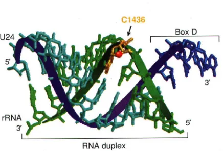
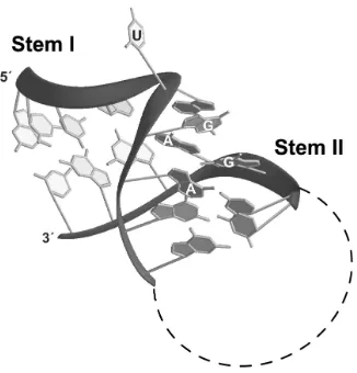
The 5´ stem loop structure of U4 snRNA which serves as the binding site for the 15.5kD protein in the U4:15.5kD co-crystal also exhibits a K-turn motif (89). Interestingly, the box C/D core motif possesses sequence elements similar to those found in the U4 site, such that the two RNAs can be folded into similar secondary structures (Fig. 5) (72). The demonstration that the 15.5kD protein recognizes both the U4 and box C/D core motif, along with the sequence similarity of the two RNAs, strongly suggests that U4 and the box C/D RNAs adopt a similar tertiary structure. This motif is characterized by two stem structures, termed stems I and II, which flank an internal asymmetric bulge, termed the 5+2 bulge due to the arrangement of five nucleotides on one side of the bulge and two on the opposite side. In tertiary space, the bulge region adopts a dramatic bend which positions the two stems at an acute angle between the helical axes. The severely kinked structure is stabilized by base stacking interactions.
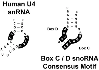
Figure 5. Secondary structures of the U4 snRNA and proposed box C/D core motif RNA.
U4 snRNA is shown on the left with those nucleotides required for binding the 15.5kD protein shaded in black. The box C/D core motif RNA is shown on the right with conserved nucleotide boxes C and D shaded black (N stands for any base and R stands for any purine). Base paired nucleotides are indicated by dashed lines.
of the “GA motif” (109). The K-turn motif folds into a kinked RNA helix, with a bend of approximately 65° between the helical axes of stems I and II. Most often, the base of stem I at the internal loop side ends in a Watson-Crick G-C pair, while stem II begins by definition at the bulge region with the G-A pairs (106). Therefore, the 5+2 asymmetric bulge secondary structure element actually leaves three unpaired nucleotides on the “long side” of the bulge. Of these three nucleotides, the first two nucleotides are most often purines and stack upon the base of the two stems. The third nucleotide is rotated out and away from the interior of the helix, exposing it to the surrounding solvent. It was noted in the case of the U4/15.5kD structure, that purines in the first two positions of the bulge were required to maintain the stacking interactions within the bulge (89). These arrangements serve to severely bend the backbone in the bulge region, which results in the “flipped-out” nucleotide rotating away from the kinked helix formed by stems I and II.
Figure 6: Proposed tertiary structure of the box C/D core motif RNA.
RNA is represented in a stick configuration. Positions of the “flipped-out” uridine nucleotide and the G-A base pairs are indicated.
from in vitro SELEX (Systematic Evolution of Ligands by EXponential Enrichment) for L30 binding exclusively possessed the GA motif that is a hallmark of the K-turn RNAs (110). In higher eukaryotes, U4 snRNA possesses a K-turn motif (89). In Eukaryota and Archaea, the box C/D RNAs adopt the K-turn fold (detailed in Chapter 3). Given the similarity of Nhp2p to other K-turn RNA binding proteins (see section 5.3), it is possible that the box H/ACA snoRNAs may include a K-turn motif not previously predicted in their secondary structures. In addition, RNase MRP, an snoRNA which functions during pre-rRNA processing has been predicted to contain a K-turn motif based on sequence analysis (106).
K-turn motifs serve as protein recognition motifs. The features of the K-turn provide an abundance of sequence and structural information which may be utilized for interaction with proteins. These features include the wide major groove of stem I, the shallow minor groove of stem II, the kinked backbone and the protruding nucleotide of the asymmetric bulge. Proteins which bind the K-turns are able to contact a relatively large hydrophobic region, as well as make many base-specific contacts with the exposed base edges in each groove. In addition, the identity of the protruding nucleotide can be crucial, as a pocket within the protein must be made to accommodate the deep insinuation of the nucleotide into the interior of the protein. These aspects of the K-turn allow the RNA to form the base for a large RNP complex.
4.2 Features of the Box C/D snoRNA Kink Turn
Before the U4:15.5kD co-crystal shed light on the structure of the box C/D core motif RNA, it was thought that the box C/D motif consisted of an external stem (equivalent to stem I) flanked by nucleotide boxes C and D (see Fig. 2A) (14, 62). Mutational analyses provided insight as to what residues were important to the box C/D core motif for protein binding and snoRNA biogenesis. Early evidence suggested that the box C/D snoRNA required some type of external stem, as well as the nearly universally conserved nucleotides in box C and box D, for complex formation in vitro and accumulation of mature snoRNP particles in vivo. These requirements overlap with the requirements of the Kink turn motif, consistent with the idea that the box C/D snoRNA adopts a K-turn motif (72, 106). Furthermore, the box C/D core motif and the K-turn motif have both been shown to be important protein recognition elements.
snoRNA precursor flanking the termini of the mature snoRNA sequence (21). The stem I requirement suggests that the conserved boxes C and D must be brought together in close juxtaposition. This is completely consistent with the idea that the box C/D core motif RNA adopts a K-turn structure, because boxes C and D are partially base paired in the K-turn motif.
The nucleotide sequences of boxes C and D are nearly 100% conserved throughout eukaryotes, suggesting that this region is particularly important for protein binding (14). The positioning of boxes C and D in the K-turn motif forms most of the internal stem (stem II, see Fig. 5B). The first two unpaired nucleotides in the bulge region that occur just upstream of box C are most often purines in the box C/D consensus motif, which is consistent with the stacking requirements of stems I and II in the K-turn motif. The initial U in the box C sequence is the nucleotide which rotates out into solution to contact the protein. This U is the preferred nucleotide for the 15.5kD protein as seen in binding analyses using RNA mutants in vitro. The G and A which follow form the tandem G-A base pairs with the GA nucleotides of box D. The positions that these nucleotides occupy in the tertiary space of the K-turn motif are critical for binding the 15.5kD protein (89). However, the remainder of boxes C and D do not seem to be essential for binding the protein, even though they are highly conserved in eukaryotes (detailed in Chapter 3). It has been suggested that these nucleotides function in binding other proteins in the snoRNP core complex.
5. Archaeal sRNAs and sRNP Complexes
5.1 Archaea: A Prokaryotic Organism Related to Eukaryotes
Archaea are single celled organisms that possess many cellular components more closely resembling those found in Eukarya than Bacteria (reviewed in 111). Examples include chromatin structures with DNA wrapped around histones, TATA box sequences in promoter regions, and most of the protein components of transcriptional, translational and splicing machinery. However, despite the resemblance to eukaryotes, a membrane-delimited nucleus has not been discovered in Archaea. The close relationship between the two has prompted evolutionary biologists to place the Kingdom of Archaea close to Eukarya. It is likely that an early divergence took place during evolution in which the ancestor of modern Bacteria was separated from the common ancestor of Archaea and Eukarya.
5.2 Archaeal Homologs of Small Nucleolar RNAs: sRNAs
sequences within many of the identified sRNAs correspond to sites of modification within rRNA. Ribosomal RNA in Archaea is highly modified with both 2´-O-methylation and psuedouridylation, although not to the extent of eukaryotic rRNA. Interestingly, the sRNAs also contain guide sequences which target other small cellular RNAs for modification. The presence of guide RNAs in Archaea indicates that this is an ancient mechanism for the nucleotide modification of cellular RNAs.
The sRNAs were initially identified in archaeal genomes via a computational method that screened sequentially for the presence of a box D sequence (CUGA), a box C sequence (UGAUGA), a region of complementarity to ribosomal RNA, box D´, and a terminal stem (if present) (112). These features were constrained to be within a certain distance of one another and the presence of these sRNAs in the cell was then confirmed by primer extension analysis. Target RNAs were also analyzed to determine if they indeed possessed the modification at the expected target site. In most cases, the target RNA did carry the expected modification, consistent with the postulated function of sRNAs in nucleotide modification.
Primary sequence analysis of the archaeal sRNAs indicates that they closely resemble eukaryotic snoRNAs. All box C/D sRNAs sequences identified to date have a well conserved box C and D region and an internally positioned box C´ and D´. In addition, many sRNAs possess an external stem which serves to bring the boxes C and D into close juxtaposition. Notable exceptions are the sRNAs from S. acidocaldarius, which contain no apparent stem I. It is interesting that none of the sRNAs identified in this one organism possess an external stem and it is not clear if sequences upstream or downstream can base pair and bring boxes C and D into juxtaposition. This lack of a stem seems to violate the rules governing snoRNA biogenesis and assembly. However, in vitro binding reactions have shown that these sRNAs are competent for binding the sRNP core proteins (81).
5.3 Archaeal Homologs of Box C/D snoRNP Proteins
similar to p55. A single homolog in Archaea for each of these related protein pairs suggests that there has been a gene duplication event that gave rise to both eukaryotic homologs.
Not readily evident in the archaeal genomic sequence database is a homolog for the eukaryotic snoRNP core protein 15.5kD. The archaeal protein most similar to 15.5kD in sequence is the previously designated ribosomal protein L7 (114). Given the sequence similarity between the two proteins, it seemed likely that L7 should bind a K-turn motif in a similar manner as the 15.5kD protein. Indeed, as detailed in Chapter 3, the L7 protein binds the box C/D core motif with the same specificity and affinity as the 15.5kD protein, requiring the same structural and sequence elements in the RNA necessary for protein recognition. Like 15.5kD, L7 binds the box C/D core motif with a Kd estimated in the low nanomolar range. Mutational analysis of the box C/D core motif demonstrated that binding requires highly conserved residues and structures in the RNA. Furthermore, immunoprecipitation experiments using antibodies specific for the archaeal core sRNP protein fibrillarin are able to co-immunoprecipitate the archaeal L7 protein (detailed in Appendix C). These results are consistent with the idea that L7 is the archaeal sRNP homolog of the eukaryotic snoRNP protein 15.5kD.
The similarity of archaeal ribosomal protein L7 and the eukaryotic 15.5kD protein is noteworthy. The two proteins are 60% similar and belong to a distantly related family of proteins that were first identified as a new class of RNA binding proteins (87). Included in this collection are the eukaryotic and archaeal ribosomal protein L7, eukaryotic ribosomal proteins L30 and S12, as well as 15.5kD and the eukaryotic H/ACA snoRNP core protein Nhp2p. The relatedness of these proteins suggests that they arose from a common ancestor early in evolution. Inclusion of both the box H/ACA and box C/D snoRNP proteins in this group suggests that the eukaryotic snoRNP complexes are also of ancient origin.
5.4 Conservation of Archaeal and Eukaryotic Box C/D sRNP/snoRNP Structure
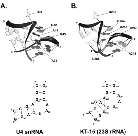
The structure of the large ribosomal subunit of H. marismortui has been solved, revealing that L7 also binds a K-turn RNA within the 23S rRNA (designated KT-15) (42). Interestingly, the structure of the KT-15:L7 RNP superimposes very well on that of the U4:15.5kD structure (114). The conserved residues of both the protein and RNA reflect the similarity evident between the two structures. However, while similar in overall structure, distinct differences between the KT-15 K-turn and the U4/snoRNA K-turn are evident (Fig. 7). These similarities and differences may provide insight as to how the sRNP/snoRNP complexes evolved, as well as how the variety of snoRNA sequences might lead to the formation of structurally similar snoRNP complexes.
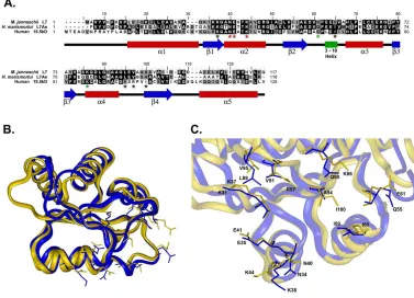
present in both proteins between sheet β2 and helix α3. Conserved residues in the L7 and 15.5kD proteins which contact the RNA overlap very closely in three-dimensional space. Residues near the C-terminus of both proteins form a large hydrophobic pocket that contacts the G-A pairs of the asymmetric bulge which are critical for the K-turn motif. In addition, this hydrophobic pocket is shaped to accept a pyrimidine nucleotide. This nucleotide corresponds to the "flipped-out" residue in the asymmetric bulge. In U4 snRNA, the box C/D motif and the 23S rRNA, this nucleotide is a uridine and mutation analysis has indicated that a pyrimidine but not a purine is tolerated at this position. Thus the shape and size of the hydrophobic pocket are important for recognition of RNA sequence as well as RNP stability.
Figure 7. Comparison of the U4 K-turn and the KT-15 of archaeal 23S rRNA.
The U4 K-turn (A.) and KT-15 (B.) are shown in both tertiary structure (top panels) and secondary structure (bottom panels). Base pairs are indicated by short, thick dashes in the secondary structures. The “flipped-out” nucleotide and residues contributing to the G-A base pairs are indicated (42, 89).
helix. Interestingly, the amino acids which make contact with the G-A pairs in helix α2 are identical in L7 and 15.5kD, whereas the residues which form the hydrophobic pocket are not identical but similar in nature, each one being a hydrophobic amino acid. The 3-10 helix, although only 3 amino acids long, appears to be important as it falls near the binding site and carries conserved residues which make contact with RNA. A residue internal to the 3-10 helix (shown in Fig. 8C and marked with a black asterisk in Fig. 8A) makes hydrophobic contact with the flipped out nucleotide of the asymmetric bulge, while an amino acid just outside the 3-10 helix (shown in Fig. 8C and marked with a green asterisk in Fig. 8A) makes a base-specific hydrogen bonding interaction with this same nucleotide.
sequence specific contacts with nucleotide bases in the bound RNA according to the U4:15.5kD crystal structure. Black asterisks indicate residues making hydrophobic contacts, red asterisks indicates residues contacting the conserved G:A pairs in the kink-turn motif, with green asterisks indicating residues contacting the protruding nucleotide. (B.) Overlay of the 15.5kD protein (gold) and H. marismortui L7Ae protein (blue) tertiary structures. Coordinates for the 15.5kD and L7Ae proteins were obtained from the PDB (entries 1E7K and 1JJF, respectively). The side chains of amino acid residues denoted by asterisks in Panel A are shown. (C.) Closeup view of the RNA binding pocket from the overlayed proteins shown in Panel B. Amino acids are numbered according to their respective sequences and colors correspond to Panel B. (The presented graphic images were generated using the InsightII software module of Molecular Simulations, Inc. release 2000.)
There are, however, slight differences between the two RNP complexes which may provide a clue as to how the core sRNP/snoRNP proteins are able to bind snoRNAs which differ in sequence. Two amino acids which make base-specific hydrogen bond interactions with the RNA in both RNP complexes possess different geometries and biochemical properties. The ASN34/LYS38 residues in L7 and ASN40/LYS44 residues in 15.5kD (in helix α2 of both proteins, shown in Fig. 8C and marked with a red asterisk in Fig. 8A) are involved in specific contacts with the RNA substrate. However, while they occupy nearly the same position in three dimensional space, they are rotated in different positions in each of the two crystal structures (see Fig. 8C). As a result, ASN34 makes a base-specific hydrogen bond with the second of the tandem base G-A base pair (from the 5´ end) in the L7/23S structure, while G-ASN40 contacts the first G-G-A base pair in the 15.5kD/U4 structure. The LYS44 residue makes a base-specific hydrogen bond with the second G-A pair in the 15.5kD U4 structure, while the LYS38 makes a hydrophobic contact away from the G-A pair and an ionic interaction with the backbone. The net result of these differences is that the L7 protein binds slightly farther downstream of the bulge in the stem II region of the K-turn than the 15.5kD protein. Since these proteins must be able to recognize all of the various snoRNAs/sRNAs within the organism, this seemingly small difference in binding geometry could be a clue as to how these proteins might deal with slight differences in each sRNA/snoRNA.
relative to the U4 K-turn. In terms of three-dimensional space, however, the two G-A base pairs of KT-15 are in tandem and occupy nearly the same position in both K-turn structures (see fig 7). This situation illustrates the difficulty in predicting such complicated tertiary structures solely on the basis of primary sequence data.
5.5 Functions of Archaeal Box C/D sRNAs
The presence of snoRNP protein homologs indicates that Archaea possess a mechanism similar to eukaryotes for ribosome processing and/or nucleotide modification. To examine this possibility, researchers have cloned the archaeal box C/D sRNP core proteins and in vitro methylation assays have been performed using full length Archaeal sRNAs (81, E. Tran, unpublished results). These experiments demonstrate that, like their eukaryotic counterparts, archaeal sRNP complexes act as guides to direct methylation of target RNA substrates. These results indicate that the function of the box C/D sRNP/snoRNPs is conserved.
The structure of the fibrillarin homolog in M. jannaschii has revealed that the C-terminal portion of the protein adopts a Rossmann fold topology which is common to most methyltransferases. A short sequence within the protein was identified as a potential consensus sequence for binding S-adenosyl-L-methionine and the crystal structure shows a potential SAM binding pocket in the core of the Rossmann fold which contains the consensus sequence. Archaeal fibrillarin proteins differ from their eukaryotic counterparts in that the N-terminal GAR domains are absent. This domain has been implicated in targeting the protein to the nucleolus, a structure presumably absent from Archaeal cells. However, the SAM-binding region of the protein is conserved, suggesting that fibrillarin does indeed serve as the methylase enzyme during nucleotide modification guided by box C/D RNAs.
6. Summary of Chapters
7. REFERENCES
1. Long, E.O. and I.B. Dawid, Repeated genes in eukaryotes. Annu Rev Biochem, 1980. 49: 727-64.
2. Venema, J. and D. Tollervey, Ribosome synthesis in Saccharomyces cerevisiae. Annu Rev Genet, 1999. 33: 261-311.
3. Montgomery, T.H., Comparative cytological studies, with especial regard to the morphology of the nucleolus. J Morphol, 1898. 15: 265-565.
4. Ritossa, F.M., K.C. Atwood, D.L. Lindsley, and S. Spiegelman, On the chromosomal distribution of DNA complementary to ribosomal and soluble RNA. Natl Cancer Inst Monogr, 1966. 23: 449-72.
5. Jordan, E.G. and J.H. McGovern, The quantitative relationship of the fibrillar centres and other nucleolar components to changes in growth conditions, serum deprivation and low doses of actinomycin D in cultured diploid human fibroblasts (strain MRC-5). J Cell Sci, 1981. 52: 373-89.
6. Shaw, P.J. and E.G. Jordan, The nucleolus. Annu Rev Cell Dev Biol, 1995. 11: 93-121.
7. Maxwell, E.S. and M.J. Fournier, The small nucleolar RNAs. Annu Rev Biochem, 1995. 64: 897-934.
8. Smith, C.M. and J.A. Steitz, Sno storm in the nucleolus: new roles for myriad small RNPs. Cell, 1997. 89(5): 669-72.
9. Bachellerie, J.P. and J. Cavaille, Guiding ribose methylation of rRNA. Trends Biochem Sci, 1997. 22(7): 257-61.
10. Kiss, T., Small nucleolar RNA-guided post-transcriptional modification of cellular RNAs. EMBO J, 2001. 20(14): 3617-22.
11. Maden, B.E., Locations of methyl groups in 28 S rRNA of Xenopus laevis and man. Clustering in the conserved core of molecule. J Mol Biol, 1988. 201(2): 289-314.
13. Ganot, P., M. Caizergues-Ferrer, and T. Kiss, The family of box ACA small nucleolar RNAs is defined by an evolutionarily conserved secondary structure and ubiquitous sequence elements essential for RNA accumulation. Genes Dev, 1997. 11(7): 941-56.
14. Xia, L., N.J. Watkins, and E.S. Maxwell, Identification of specific nucleotide sequences and structural elements required for intronic U14 snoRNA processing. RNA, 1997. 3(1): 17-26.
15. Newman, D.R., J.F. Kuhn, G.M. Shanab, and E.S. Maxwell, Box C/D snoRNA-associated proteins: two pairs of evolutionarily ancient proteins and possible links to replication and transcription. RNA, 2000. 6(6): 861-79.
16. Watkins, N.J., A. Gottschalk, G. Neubauer, B. Kastner, P. Fabrizio, M. Mann, and R. Luhrmann, Cbf5p, a potential pseudouridine synthase, and Nhp2p, a putative RNA-binding protein, are present together with Gar1p in all H BOX/ACA-motif snoRNPs and constitute a common bipartite structure. RNA, 1998. 4(12): 1549-68.
17. Henras, A., Y. Henry, C. Bousquet-Antonelli, J. Noaillac-Depeyre, J.P. Gelugne, and M. Caizergues-Ferrer, Nhp2p and Nop10p are essential for the function of H/ACA snoRNPs. EMBO J, 1998. 17(23): 7078-90.
18. Baserga, S.J., X.D. Yang, and J.A. Steitz, An intact Box C sequence in the U3 snRNA is required for binding of fibrillarin, the protein common to the major family of nucleolar snRNPs. EMBO J, 1991. 10(9): 2645-51.
19. Tyc, K. and J.A. Steitz, U3, U8 and U13 comprise a new class of mammalian snRNPs localized in the cell nucleolus. EMBO J, 1989. 8(10): 3113-9.
20. Wang, H., D. Boisvert, K.K. Kim, R. Kim, and S.H. Kim, Crystal structure of a fibrillarin homologue from Methanococcus jannaschii, a hyperthermophile, at 1.6 A resolution. EMBO J, 2000. 19(3): 317-23.
21. Darzacq, X. and T. Kiss, Processing of intron-encoded box C/D small nucleolar RNAs lacking a 5',3'-terminal stem structure. Mol Cell Biol, 2000. 20(13): 4522-31.
22. Watkins, N.J., D.R. Newman, J.F. Kuhn, and E.S. Maxwell, In vitro assembly of the mouse U14 snoRNP core complex and identification of a 65-kDa box C/D-binding protein. RNA, 1998. 4(5): 582-93.