C A S E R E P O R T
Open Access
Metastasis of distal esophageal carcinoma to the
thyroid with presentation simulating primary
thyroid carcinoma: a case report and review of
the literature
En-dong Chen
1†, Pu Cheng
1†, Xing-qiang Yan
1, Yun-liang Ye
1, Cheng-ze Chen
1, Xiu-huan Ji
2and Xiao-hua Zhang
1*Abstract
Metastasis to the thyroid is extremely rare. There is a lack of awareness of and adequate preparation for this situation, especially in an individual without a past history of malignancy. We describe a rare case of a 61-year-old man in whom a primary distal esophageal carcinoma gave rise to a metastatic palpable mass in the thyroid gland. Palliative bilateral near-total thyroidectomy was performed with pathology showing squamous cell carcinoma and tracheostomy was carried out simultaneously due to airway compression with related symptoms. A review of the literature only reveals 4 similar cases. Secondary neoplasm of the thyroid mimicking a primary malignant lesion is seldom encountered, however, in order to make appropriate treatment, the most critical problem is to distinguish the difference between the above two and the final diagnosis can only be confirmed on pathologic examination. Although the prognosis of thyroid metastasis is commonly felt to be poor, improvement of living quality and prolongation of survival may be obtained in such patients through correct diagnosis and treatment.
Keywords:Thyroid metastasis, esophageal neoplasms, squamous cell carcinoma
Background
Despite being a highly vascularized organ, the thyroid gland is an unusual site of clinically detectable cancer metastasis. According to reports, the incidence of intra-thyroid metastases (ITM) in autopsy series varies from 1.25% to 24.0% in cancer patients [1,2].The most fre-quently noted primary sites are the kidney, breast, and lung [3-6]. The metastatic spread of gastrointestinal ma-lignancies to the thyroid gland is relatively rare, and the majority come from the colo-rectum [7]. ITM originat-ing from the alimentary tract are quite unusual based on our previous experience. In clinical practice, metastatic involvement of the thyroid is usually characterized by an indolent growth pattern, which results in few visible in-dications of the illness and difficulty in making a differ-ential diagnosis from other primary thyroid neoplasms
[8]. Thyroid metastasis from the esophagus has only been reported in four cases in the English literature [9-12]. Herein, a further case is reported of metastasis of esophageal carcinoma to the thyroid, which was mis-diagnosed in the first place as primary thyroid carcinoma with unilateral lymph node metastasis, and a systematic literature review shows that this is the first case of this rare condition to be described in eastern China.
Case presentation
In July 2011, a 61-year-old man presented to our depart-ment complaining of dyspnea, mild dysphagia and hoar-seness for a few months with exacerbation for 10 days. There was no remarkable past medical, surgical, or fam-ily history. On admission, a large nodular mass on the left lobe of the thyroid was readily recognizable and was irregular, hard, immobile and painless on palpation. Thy-roid sonogram findings demonstrated a 6.1 cm × 3.9 cm mass in the left lobe with low and heterogeneous echo and an enlarged cervical lymph node measuring 2.5 cm × 1.8 cm on the left side of the neck in the level-III region * Correspondence:zxh.surgeryoncology@gmail.com
†Equal contributors 1
Department of Surgical Oncology, The First Affiliated Hospital of Wenzhou Medical University, Wenzhou, Zhejiang, People’s Republic of China Full list of author information is available at the end of the article
with hypoechogenicity. Fine-needle aspiration cytology of the thyroid disclosed diffuse infiltration of atypical cells with a high suspicion of malignancy, therefore the patient was scheduled for surgery.
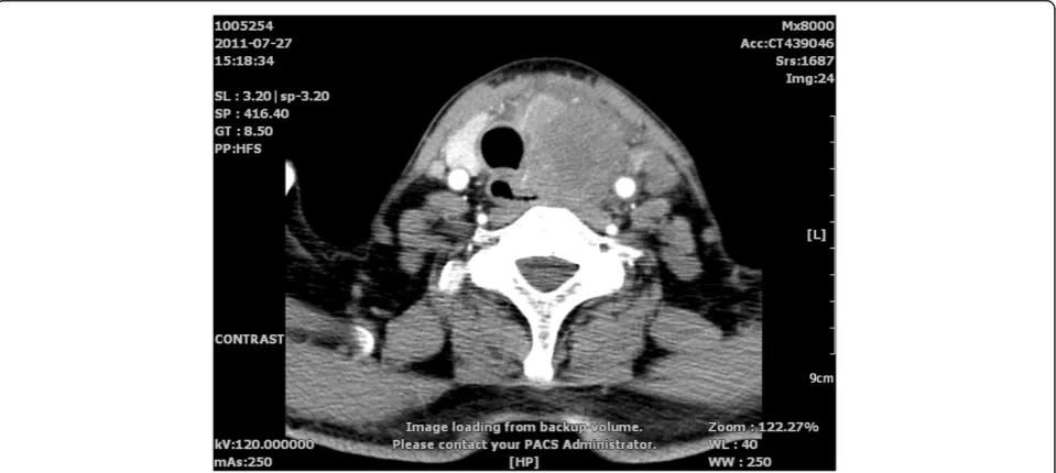
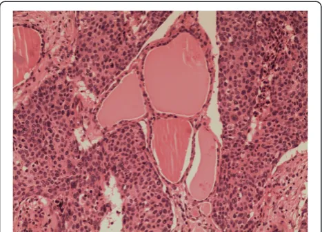
Chest radiography and laboratory examinations inclu-ding thyroid hormone showed no abnormal fininclu-dings. Blood calcium level was also checked for the possibility of medullary thyroid cancer, and was within normal limits. Neck contrast-enhanced computed tomography revealed a mass encasing the left carotid sheath vessels, esophagus, and trachea in the left thyroid lobe (Figure 1). The man underwent bilateral near-total thyroidectomy and trache-ostomy for palliative purposes because adhesions to adja-cent structures and the aggravation of dyspnea were observed pre- and intra-operatively. Histological examin-ation from the resected specimen verified moderate differ-entiated squamous cell carcinoma (SCC) of the thyroid gland (Figure 2) with metastasis to the level-III region of the left cervical lymph nodes and characteristics similar to the esophageal lesions. Further workup with introscopy biopsy revealed a squamous cell carcinoma of the distal esophagus (Figure 3). One month post surgery, the patient started chemotherapy with a docetaxel and cisplatin (DC) regimen, and radiotherapy according to the 2011 Natio-nal Comprehensive Cancer Network (NCCN) Esophageal Cancer Guidelines. Although the patient’s condition was kept under control initially, after five cycles of chemo-therapy, his illness grew worse as the cancer disseminated to the peritoneal and mediastinal lymph nodes. Finally, eleven months after the diagnosis of metastasis, the pa-tient died due to advanced esophageal carcinoma compli-cated by pneumonia and sepsis.
Discussion
The incidence of metastatic spread of gastrointestinal malignancies to the thyroid gland is relatively low and most of them are from the colo-rectum [7]. ITM ori-ginating from the gastro-esophagus is poorly docu-mented. A review of the English-language literature, searching for patients with secondary cancerous growth in the thyroid formed by transmission of tumor cells from primary esophageal and gastric carcinoma dis-closed a total of four [9-12] and five reported cases [7,13-16], respectively. This article presents an addi-tional case of thyroid metastasis from esophageal car-cinoma occurring in a Chinese man and reviews the related references.
Table 1 summarizes the clinical circumstances for the nine cases previously published plus our report of thy-roid metastasis from esophageal cancer. The age of the five female and five male patients at presentation was variable, ranging from 32 to 74 years with an average age of 61.5 years. The majority of patients underwent thyroidectomy (specific types of surgery are presented in Table 1: this was unknown in four cases). In the postop-erative histopathological specimen, there were four pa-tients whose thyroid cancer seeded from squamous cell carcinoma, two originated from the undifferentiated car-cinoma with signet-ring cells, two were derived from poorly differentiated carcinoma with characteristics simi-lar to the gastric lesions, and the remaining cases were from poorly differentiated adenocarcinomas. Of the pa-tients with thyroid metastasis, most had a poor outcome and died shortly after the original diagnosis (specific sur-vival times are presented in Table 1).
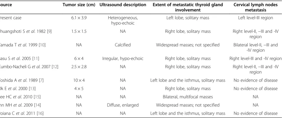
Table 2 summarizes the diagnosis of thyroid cancer for reported cases. Three patients had multifocal or wide-spread masses and all the others had only a solitary mass. Five patients were confirmed with unilateral or bilateral cervical lymph nodes metastases and two patients had no evidence of cervical lymphadenopathy (there was no refer-ence to this for the remaining cases. Several patients re-ceived additional examinations (specific investigations are presented in Table 2).
Generally, despite being second only to the adrenal glands as the most vascular perfused organ in the body [17], the thyroid is rarely considered to be the sole site of metastases in the clinical setting and is usually asymp-tomatic [8,18]. Cichonet al. reported that metastasis to the thyroid only accounts for 2% to 3% of all thyroid car-cinomas identified in the clinical setting [19]. The most common primary sites are the kidney, breast, and lung
(see related references [3-6]). To the best of our know-ledge there is little information to date in the English lit-erature about thyroid metastasis from the esophagus, except for four published cases [9-12].
One question is raised by our case: how to prove that the SCC of the thyroid in our patient originated from a primary esophageal carcinoma? In general, it is difficult and challenging to accurately distinguish between pri-mary and secondary neoplasm in the thyroid. Pripri-mary SCC rarely arise from the thyroid gland, especially in older patients with a long-standing history of goître [20,21]. The etiology remains mysterious and unclear, and presumed to originate from the metaplastic glandu-lar epithelium. Fine-needle aspiration cytology is often used to obtain tissues for diagnosis. Nevertheless, its value in the discrimination between primary and meta-static thyroid malignancies is still uncertain when highly anaplastic cells are observed microscopically [2]. Our pa-tient had no particular history of thyroid disease and the thyroid mass grew rapidly in a few months. What is more, the patient suffered an exacerbation of dyspnea. An urgent thyroidectomy was necessary to relieve threa-tening airway obstruction caused by tracheal compres-sion. Postoperative histopathological examination revealed multi-focal lesions with similar pathological profiles to the esophageal lesions. Thorough work up reavealed a squa-mous carcinoma of the distal esophagus on esophago-gastroduodenoscopy biopsy. Other findings included: dysphagia, dyspnea, hoarseness, vascular infiltration of SCC under the microscope, and some imaging observa-tions. It was therefore confirmed as a primary esphageal cancer with metastasis to the thyroid.
Direct extension of adjacent primaries, a hematoge-nous pathway and lymphatic route for metastatic spread to the thyroid have been suggested [11,22]. Czechet al. suggested that the vertebral vein plexus may play an im-portant role in the process of metastases from other or-gans to the thyroid [5]. Unfortunately, according to a review of the related literature, there is no reported case of careful imaging and pathologic evaluation of the most likely route of metastasis in the thyroid. Our patient was supposed to have lymphogenous metastasis in view of unilateral lymphadenopathy and the obvious peritoneal and mediastinal lymphatic vessel infiltration.
The main method of treatment for metastatic thy-roid cancers usually involves radiotherapy and surgery [8,11,17,23]. The role of radiation therapy is still con-troversial because thyroid metastases are mainly re-vealed as highly anaplastic carcinomas and are usually radiation-resistant, and are often rapidly fatal. Further-more, until recently there has been no clear consensus on the election of surgical means for metastatic thyroid can-cers [5,7,24]. Sadly, thyroidectomy had no remarkably beneficial effects on the outcome of our patient. On the Figure 2Multi-focal nests of tumors cells are distributed
nearby the follicles.Hematoxylin and eosin staining, ×100.
whole, thyroid metastasis from esophageal cancer shows a poor prognosis, with reported 9-month survival after diag-nosis [25].
Conclusion
This case highlights the need for an awareness of the possibility of potential metastatic deposits in unexpected sites. A new thyroid mass with dysphagia appearing in a patient, however remote, should be evaluated for the possibility of metastasis. Whenever the histology is un-usual for a thyroid primary, metastasis should be strongly considered. Although the prognosis of metastasis in the
thyroid is commonly poor, patients with a single infiltrat-ing thyroid mass may have improved quality of life and longer survival time after accurate diagnosis and proper treatment.
Consent
Written informed consent was obtained from the son of the patient for publication of this Case report and any accompanying images. A copy of the written consent is available for review by the Editor-in-Chief of this journal.
Table 2 The diagnosis of thyroid cancer for reported cases
Source Tumor size (cm) Ultrasound description Extent of metastatic thyroid gland involvement
Cervical lymph nodes metastasis
Present case 6.1 × 3.9 Heterogeneous,
hypo-echoic
Left lobe, solitary mass Left level-III region
Shuangshoti Set al. 1982 [9] 1.5 × 1.5 NA Right lobe, solitary mass Right level-II,−III and -IV region
Yamada Tet al. 1999 [10] NA Calcified Widespread masses; not specified Bilateral level-II,−III and -IV region
Basu Set al. 2005 [11] 6 × 4 Irregular, hypo-echoic Right lobe, solitary mass Right level-III and -IV region Cumbo-Nacheli Get al. 2007 [12] 2.5 × 2.8 NA Right lobe, solitary mass Right level-II,−III and -IV
region
Yoshida Aet al. 1989 [7] 10 × 4 NA Left lobe and the isthmus, solitary mass No evidence of disease Ok Eet al. 2000 [13] 4 × 5 NA Right lobe, solitary mass No evidence of disease
Lee HCet al. 2010 [15] NA NA Bilateral, multifocal masses NA
Ihn MHet al. 2009 [14] NA Diffuse, enlarged Widespread masses; not specified NA
Poiana Cet al. 2011 [16] NA NA Left lobe and the isthmus, solitary mass No evidence of disease NA, no data available.
Table 1 Clinical data of patients with metastatic involvement of the thyroid
Source Age (yr) Sex Site of
primary lesion
Type of secondary thyroid surgery
Pathology results from thyroid specimen
Outcomes (months)*
Present case 61 M Esophagus Palliative bilateral
NT + tracheostomy
SCC 11
Shuangshoti Set al. 1982 [9] 58 M Esophagus TT + ipsilateral CL SCC 5
Yamada Tet al. 1999 [10] 74 F Esophagus ST + Bilateral CL SCC /
Basu Set al. 2005 [11] 55 F Esophagus NA SCC NA
Cumbo-Nacheli Get al. 2007 [12] 32 M Esophagus NA Poorly differentiated adenocarcinoma NA
Yoshida Aet al. 1989 [7] 71 M Stomach ST Poorly differentiated adenocarcinoma 7
Ok Eet al. 2000 [13] 60 F Stomach Bilateral ST Undifferentiated carcinoma (with signet-ring cells)
1.5
Lee HCet al. 2010 [15] 71 M Stomach Bilateral NT Poorly differentiated carcinoma 4
Ihn MHet al. 2009 [14] 63 F Stomach NA Undifferentiated carcinoma (with
signet-ring cells)
6
Poiana Cet al. 2011 [16] 70 F Stomach NA Poorly differentiated neuroendocrine
carcinoma (with small cells)
NA
Abbreviations
ITM:intrathyroid metastases; SCC: squamous cell carcinoma.
Competing interests
There is no financial relationship that might lead to a conflict of interest in relation to the manuscript.
Authors’contributions
ZXH performed the surgery and carried out the initial conception; CED and CP wrote the manuscript; YXQ, YYL and CCZ helped to revise the manuscript; JXH helped collecting references. All authors read and approved the final manuscript.
Acknowledgements
We thank all of the pathologists at the First Affiliated Hospital of Wenzhou Medical University for their assistance with the pathologic analysis. Without their efforts, this article would not be possible. This work was supported by a grant from the National High Technology Research and Development Program of 863 project of China (NO.2012AA02A210).
Author details 1
Department of Surgical Oncology, The First Affiliated Hospital of Wenzhou Medical University, Wenzhou, Zhejiang, People’s Republic of China. 2Department of Pathology, The First Affiliated Hospital of Wenzhou Medical
University, Wenzhou, Zhejiang, People’s Republic of China.
Received: 28 July 2013 Accepted: 9 April 2014 Published: 23 April 2014
References
1. Berge T, Lundberg S:Cancer in Malmo 1958–1969. An autopsy study.
Acta Pathol Microbiol Scand Suppl1977,1:235.
2. Nakhjavani MK, Gharib H, Goellner JR, Van Heerden JA:Metastasis to the thyroid gland. A report of 43 cases.Cancer1997,79:574–578. 3. Pillay SP, Angorn IB, Baker LW:Tumour metastasis to the thyroid gland.
S Afr Med J1977,51:509–512.
4. Ericsson M, Biorklund A, Cederquist E, Ingemansson S, Akerman M:Surgical treatment of metastatic disease in the thyroid gland.J Surg Oncol1981,
17:15–23.
5. Czech JM, Lichtor TR, Carney JA, Van Heerden JA:Neoplasms metastatic to the thyroid gland.Surg Gynecol Obstet1982,155:503–505.
6. McCabe DP, Farrar WB, Petkov TM, Finkelmeier W, O'Dwyer P, James A:
Clinical and pathologic correlations in disease metastatic to the thyroid gland.Am J Surg1985,150:519–523.
7. Yoshida A, Imamura A, Tanaka H, Hirano M, Kamma H, Ueno E, Ushio H, Aiyoshi Y, Soeda S:A case of metastasis from gastric cancer to the thyroid gland.Jpn J Surg1989,19:480–484.
8. Chen H, Nicol TL, Udelsman R:Clinically significant, isolated metastatic disease to the thyroid gland.World J Surg1999,23:177–180. discussion 181.
9. Shuangshoti S:Primary carcinomas of esophagus and bronchus with presentation simulating primary carcinoma of thyroid gland.J Med Assoc Thai1982,65:38–44.
10. Yamada T, Tatsuzawa Y, Yagi S, Fujioka S, Kitagawa S, Nakagawa M, Minato H, Kurumaya H, Matsunou H:Lymphoepithelioma-like esophageal carcinoma: report of a case.Surg Today1999,29:542–544. 11. Basu S, Nair N, Borges AM:Squamous cell carcinoma of esophagus
masquerading as solitary thyroid nodule.Indian J Cancer2005,
42:205–207.
12. Cumbo-Nacheli G, De Sanctis JT, Chung MH:Proximal esophageal adenocarcinoma presenting as a thyroid mass: case report and review of the literature.Thyroid2007,17:267–269.
13. Ok E, Sozuer E:Thyroid metastasis from gastric carcinoma: report of a case.Surg Today2000,30:1005–1007.
14. Ihn MH, Kim YJ, Kim JJ, Cho JY, Jin SY:A case of thyroid metastasis originating from early gastric cancer.J Korean Med Sci2009,
24:1230–1233.
15. Lee HC, Chen FF, Lo CC, Wang CJ, Lo WC, Luh SP:Metastasis of gastric carcinoma to the thyroid and lung: a case report and review of literature.J Zhejiang Univ Sci B2010,11:542–546.
16. Poiana C, Carsote M, Ardeleanu C, Terzea D, Avramescu ET, Neamtu MC, Miulescu RD:The value of the immunohistochemistry in a case of gastric neuroendocrine tumor and thyroid metastasis.Rom J Morphol Embryol 2011,52:187–192.
17. Wychulis AR, Beahrs OH, Woolner LB:Metastasis of Carcinoma to the Thyroid Gland.Ann Surg1964,160:169–177.
18. Kihara M, Yokomise H, Yamauchi A:Metastasis of renal cell carcinoma to the thyroid gland 19 years after nephrectomy: a case report.Auris Nasus Larynx2004,31:95–100.
19. Cichon S, Anielski R, Konturek A, Barczynski M, Cichon W:Metastases to the thyroid gland: seventeen cases operated on in a single clinical center.
Langenbecks Arch Surg2006,391:581–587.
20. Huang TY, Assor D:Primary squamous cell carcinoma of the thyroid gland: a report of four cases.Am J Clin Pathol1971,55:93–98.
21. Simpson WJ, Carruthers J:Squamous cell carcinoma of the thyroid gland.
Am J Surg1988,156:44–46.
22. Fujita T, Ogasawara Y, Doihara H, Shimizu N:Rectal adenocarcinoma metastatic to the thyroid gland.Int J Clin Oncol2004,9:515–519. 23. Mirallie E, Rigaud J, Mathonnet M, Gibelin H, Regenet N, Hamy A, Bretagnol
F, de Calan L, Le Neel JC, Kraimps JL:Management and prognosis of metastases to the thyroid gland.J Am Coll Surg2005,200:203–207. 24. Murakami S, Yashuda S, Nakamura T, Mishima Y, Iida H, Okano H, Nakano M:
A case of renal cell carcinoma with metastasis to the thyroid gland and concomitant early gastric cancer.Surg Today1993,23:153–158. 25. Lin JD, Weng HF, Ho YS:Clinical and pathological characteristics of
secondary thyroid cancer.Thyroid1998,8:149–153.
doi:10.1186/1477-7819-12-106
Cite this article as:Chenet al.:Metastasis of distal esophageal carcinoma to the thyroid with presentation simulating primary thyroid
carcinoma: a case report and review of the literature.World Journal of
Surgical Oncology201412:106.
Submit your next manuscript to BioMed Central and take full advantage of:
• Convenient online submission
• Thorough peer review
• No space constraints or color figure charges
• Immediate publication on acceptance
• Inclusion in PubMed, CAS, Scopus and Google Scholar
• Research which is freely available for redistribution