R E S E A R C H
Open Access
Combined epithelial-mesenchymal transition
with cancer stem cell-like marker as predictors of
recurrence after radical resection for gastric cancer
Gui-fang Xu
1†, Wei-jie Zhang
2,3*†, Qi Sun
4, Xinyun Xu
4, Xiaoping Zou
3*and Wenxian Guan
2,3*Abstract
Background:The aim of the study was to identify the incidence and the predictors of recurrence after curative resection and the clinical significance of epithelial-mesenchymal transition (EMT) and stem cell-like phenotypes in gastric cancer.
Methods:In a total of 1,463 patients that underwent curative resection for gastric cancer between January 2001 and January 2008 at Drum Tower Hospital, 402 (27.5%) experienced recurrence. They were divided into early recurrence (within two years) and late recurrence (more than two years). The clinicopathological characteristics, including five EMT-related proteins (Snail-1, ZEB-1, E-cadherin, vimentin, andβ-catenin) and the gastric cancer stem cell markers CD44 and CD54, therapeutic modalities, survival time after recurrence, and recurrence patterns were compared between the two groups.
Results:Loss of E-cadherin expression and aberrant expression of vimentin and the known gastric cancer stem cell maker CD44 were significantly associated with aggressive clinicopathologic features. Multivariate analysis showed that stage III gastric cancer patients with early recurrence had larger tumors and more lymph node metastasis, coupled with aberrant expression EMT and cancer stem cell marker, than patients with late recurrence. Early recurrence was associated with more distant metastasis than late recurrence and patients tended to die within two years of recurrence.
Conclusions:Combined EMT with cancer stem cell-like marker is a predictor of recurrence after radical resection for gastric cancer. Advanced TNM stage was associated with early cancer death after recurrence.
Background
Gastric cancer is the fourth most common cancer in the world, and surgery remains the main treatment, with curative intent. Even after gastrectomy and lymphade-nectomy, every year many patients die of recurrence. The epithelial-mesenchymal transition (EMT), a devel-opmental process in which epithelial cells show reduced intercellular adhesion and acquire migratory fibroblastic properties, is considered to be critical for invasive and metastatic progression in cancer [1]. The process of
EMT is associated with the downregulation of epithelial markers, abnormal translocation of β-catenin, and aber-rant upregulation of mesenchymal markers. Cancer stem cells are cells within a tumor that possess self-renewal and tumor-initiating capacities. In gastric cancer, CD44-positive tumor cells were shown to have cancer stem cell properties, including the tumor-initiating ability [2], and Chenet al. [3] showed that CD44 + CD54+ cells exhibit cancer stem cell capabilities in gastric cancer tissues. These tumor-initiating cells provide a reservoir that can cause tumor recurrence after therapy [4].
Previously, Ryuet al. [5] showed that a combination of EMT and stem cell-like phenotypes is an important pre-dictor of aggressive biologic behavior of gastric cancer; however, few studies currently have been investigating EMT with cancer stem cell-like marker in gastric cancer with recurrence and prognosis. The aim of the current * Correspondence:zhangweijie1616@163.com;zouxiaoping795@hotmail.com;
guan-wx@163.com
†Equal contributors
2
Department of General Surgery, Drum Tower Clinical College of Nanjing Medical University, 321 Zhongshan Road, Nanjing 210008, China
3
Department of General Surgery, Affiliated Drum Tower Hospital of Nanjing University Medical School, 321 Zhongshan Road, Nanjing 210008, China Full list of author information is available at the end of the article
study was to characterize recurrence patterns, identify the predictors of time of recurrence and initial recur-rence patterns of gastric cancer with early and late recurrence, and find risk factors to predicate early recur-rence after radical resection for gastric cancer. This could help in the preoperative assessment of appropriate therapeutic strategies and in providing more intensive and tailored follow-up strategies for high-risk patients.
Methods Patient selection
Between January 2001 and January 2008, excluding pa-tients who were lost to follow-up or poor compliance, a total of 1,463 patients underwent curative resection for gastric cancer at Drum Tower Hospital. A total of 402 (27.5%) of these patients experienced recurrence and were subsequently included in the study; thus the recur-rence rate in our study was 27.5%. The mean age of the 402 patients was 64.7 years (range: 26 to 91 years), with a male:female ratio of 1.87:1. Medical charts and path-ology reports were reviewed to record clinical and pathological data. To evaluate whether the expression profiles of the various EMT markers were different be-tween primary and metastatic gastric cancers, cases with regional lymph node metastasis were also evaluated. In terms of the timing of recurrence, some tumors recur within the first year after resection, and in these cases, inadequate radical surgical approach or systematic me-tastasis at operation may be suspected. In our study, we defined early recurrence as recurrence within two years after surgery. Late recurrence was defined as recurrence more than two years after surgery. These 402 patients were divided into two groups, namely the early recur-rence group, which contained 248 patients, and the late recurrence group, which contained 154 patients. Exclu-sion criteria included synchronous gastric double cancer, a previous history of surgery for gastric cancer, gastric stump cancer, or cancer anatomically classified as esophageal cancer according to the American Joint Committee on Cancer (AJCC) seventh edition [6].
The study was approved by the clinical research ethics committees of the Drum Tower Hospital and informed consent was obtained from all patients.
Tissue array and immunohistochemistry
All samples in the study were removed from formalin-fixed paraffin-embedded gastric cancer tissues. For all of the arrays, three cores from different areas of the tumor in each case were removed and placed into a new blank recipient paraffin block as previously described by Hsu et al. [7], and 4-μm thick sections were obtained for im-munohistochemistry. The specimens were deparaffinized and dehydrated by a graded series of ethanol solutions (Nanjing Chemical Reagent Co., Ltd., Nanjing, China).
The immunohistochemical staining for EMT-related proteins Snail-1, zinc finger E-box-binding homeobox 1 (ZEB-1), vimentin, E-cadherin, β-catenin, and cancer stem cell-like markers CD44 and CD54, were performed and evaluated according to the previous method [3,5].
As for statistical analysis of multiple markers, the spe-cimen was recorded as having positive immunoreactivity for each antibody, excluding E-cadherin, if more than 5% of the cancer cells were immunoreactive, and tumor cells with less than 5% were considered as negative. The scoring criteria for E-cadherin were defined as ‘positive (intact)’ when immunoreactivity on the cell membrane was present in more than 25% of the gastric cancer cells.
Patterns of recurrence
Patterns of recurrence reported represent the first sites of documented recurring disease after curative resection. Re-currence was documented from the first clinical or radio-logical signs of disease with an unrelenting course leading to tumor progression and/or death. Confirmation through biopsy was recommended for any evidence of recurrent disease or distant metastases. Recurrence patterns were classified as locoregional, peritoneal, or hematogenous. Locoregional recurrence was defined as any cancer recur-rence at the resection margin or lymph nodes (including regional nodes as well as retropancreatic and para-aortic nodes) or operation bed within the region of the resection (below the diaphragm and liver, and above the pancreas and abdominal wound). Peritoneal recurrence was defined as any cancer recurrence within the abdominal cavity due to intraperitoneal distribution including visceral metasta-sis, rectal shelf, pericholedochal, and periureteral infiltra-tion. Hematogenous recurrence was defined as any metastatic lesion detected in liver, lung, bone, and other distant organs.
Follow-up
When metastasis was suspected, further techniques were utilized, such as bone scan, positron emission tomog-raphy (PET), and biopsy sampling.
Statistical analysis
All values are expressed as the mean ± standard devi-ation. Categorical variables were analyzed using the χ2 test and continuous variables using the independent-samples t-test. Logistic regression multivariate analysis was conducted to determine the differences between the two groups. Analysis of survival was performed by the Kaplan-Meier method, and differences between the curves were tested using a two-tailed log-rank test. P <0.05 was considered to be statistically significant. Statistical analysis was performed using SPSS (SPSS 13.0 for Windows; SPSS Inc., Chicago, Illinois, United States) software.
Results
Expression of epithelial-mesenchymal transition and cancer stem cell-like marker association with tumor recurrence
Table 1 shows the clinicopathologic characteristics of the patients with early recurrence and late recurrence. Among the perioperative variables recorded for univari-ate analysis were: presence of CD44, CD54, and vimen-tin, loss of E-cadherin and β-catenin expression, with a tumor size of 5 cm or more, more advanced T-stage, and a more advanced TNM stage with early recurrence to late recurrence Multivariate analysis was employed to identify the independent risk factors for overall recur-rence. The analysis showed that patients presenting with a tumor size of 5 cm or more, and a more advanced TNM stage, coupled with EMT expression and expres-sion of cancer stem cell marker CD44, were prominent in early recurrence (Table 2).
Patterns of recurrence
The median time to recurrence was 17.0 months (range: 2.0 to 89.0). Most recurrences in our study group (248 out of 402, 61.7%) occurred within two years after cura-tive surgery (Table 3). In the early recurrence group, the most common pattern was hematogenous metastasis, followed by locoregional and peritoneal recurrence. In-cluding patterns that developed concurrently, locoregional and hematogenous recurrence was the most common pat-tern, followed by peritoneal recurrence. In the late recur-rence group, the most common pattern was locoregional and peritoneal recurrence, followed by hematogenous recurrence as single patterns, and when patterns that developed simultaneously were included, locoregional re-currence was still the most common pattern. Distant metastasis is more common in early recurrence, but peri-toneal dissemination is not significant. Among patients
with hematogenous metastasis, liver metastasis is more common in early recurrence.
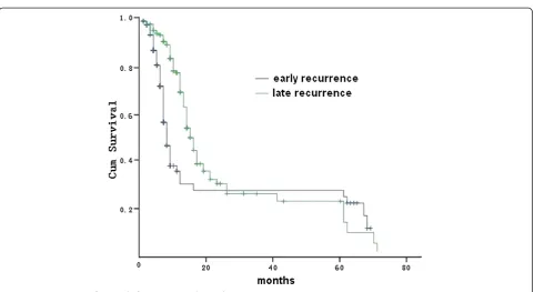
Survival time after recurrence
The majority of patients with recurrence of gastric can-cer (321 out of 402, 79.9%) died within two years after recurrence. The median survival time after recurrence was 8.1 months in early recurrence and 13.3 months in late recurrence, respectively. A total of 16 patients sur-vived more than five years after recurrence: 10 with early recurrence and six with late recurrence. Among the 10 patients with early recurrence, the recurrence pattern in-cluded one liver metastasis, two lung metastasis, three anastomosis recurrences, two hepatoduodenal ligament recurrences, one abdominal wall recurrence, and one peritoneal seeding. Among the six patients with late recurrence, the recurrence patterns included one ab-dominal incision recurrence, two liver metastases, one remnant stomach relapse, one hepatoduodenal ligament recurrence, and one anastomosis recurrence with liver inva-sion. Survival time after recurrence was significantly shorter in the early group (P<0.001, log-rank test) (Figure 1).
Discussion
Gastric cancer represents the fourth leading cause of can-cer mortality worldwide. Locoregional relapse may occur after curative resection, including adjacent lymph nodes resection and multidisciplinary treatment, possibly in the form of peritoneal carcinosis and/or distant metastases, which is one reason for the high rate of recurrence. Car-cinoma relapse is essentially lethal, and so far there are no specific treatments to avoid recurrence [8-10].
The clinical significance of EMT has been reported in various human cancers, and specific mechanisms in-volved in cancer progression, such as the evasion of apoptosis, resistance to chemotherapy, and the acquisi-tion of stem cell-like properties and their influence on patient survival have been suggested [1,5,11,12]. CD44 is a transmembrane glycoprotein that is well known as a cancer stem cell marker in gastric cancer [2]. CD44 is positively and significantly associated with tumor metas-tasis, recurrence, and mortality in gastric cancer [13,14].
There have been several studies [1,5] on the complex-ity of the interactions between EMT-related proteins and their effects on cancer recurrence. The present study determined that the combination of alterations in the protein expression of vimentin, E-cadherin, and CD44 was the most effective prognostic factor in gastric cancer. In addition, this combination was an independ-ent predictive factor of recurrence, together with a con-ventional clinicopathologic parameter such as pTNM stage and histologic differentiation.
In our series, there is a large difference in cancer size, gross appearance, lymph node metastasis, lymphovascular invasion, depth of invasion, and stage between the early and late recurrence groups, and patients with CD44 and vimentin, and loss of E-cadherin andβ-catenin expression, have a higher tendency towards early recurrence. Our re-sults showed that patients presenting with a tumor size of 5 cm or more, and a more advanced TNM stage, coupled with EMT expression and expression of cancer stem cell
Table 1 Clinicopathological characteristics of recurrent gastric cancer patients
Early recurrence (n = 248) n (%)
Late recurrence (n = 154) n (%) P
value
Age, years 0.392
<65 109 61
≥65 139 93
Gender, male/female 162/86 100/54 0.937
Tumor size, cm 0.013
<5 76 68
≥5 172 86
Gastrectomy 0.061
Total 91 71
Subtotal 157 83
Location 0.232
Upper 46 27
Middle 49 20
Lower 130 95
Diffuse 23 12
Gross appearance 0.001
Superficial tumor 18 25
Bormann type I and II 25 40
Bormann type III and IV 205 89
Histology type 0.315
Differentiated 76 40
Undifferentiated 172 114
Lauren’s classification 0.572
Intestinal type 104 69
Diffuse type 144 85
Lymph node metastasis 0.002
Negative 52 54
Positive 196 100
Lymphovascular invasion 0.003
Absent 57 56
Present 191 96
Depth of invasion <0.001
T1 and T2 46 53
T3 and T4 202 101
TNM stage <0.001
I 14 27
II 43 59
III 191 68
Lymph node dissection 0.414
D1 109 75
D2 86 54
D3 53 25
Table 1 Clinicopathological characteristics of recurrent gastric cancer patients(Continued)
EMT expression (%)
Aberrant expression of mesenchymal marker
Snail-1 117(47.2) 64(41.6) 0.271
ZEB-1 168(67.7) 109(70.8) 0.522
Vimentin 87(35.1) 21(13.6) <0.001
Expression loss of epithelial marker
E-cadherin 112(45.2) 35 (22.7) <0.001
β-catenin 29 (11.7) 19(12.3) 0.846
Expression of cancer stem cell marker (%)
CD44 163(65.7) 71(46.1) 0.001
CD54 120(48.3) 63(40.9) 0.037
Adjuvant chemotherapy, yes/no
221/27 142/12 0.308
Table 2 Multivariate analysis of factors independently associated with the timing of recurrence (early versus late)
Early Late Pvalue Odds ratio
95.0% CI for experiment B
Tumor size≥5 cm 172(69.4) 86(55.8) 0.001 8.750 (2.265 - 33.799)
TNM stage III 191(77.0) 68(44.2) 0.040 0.493 (0.251 - 0.970)
EMT expression (%)
Vimentin 87(35.1) 21(13.6) 0.031 2.359 (1.082 - 5.145)
E-cadherin 112(45.2) 35(22.7) <0.001 0.125 (0.046 - 0.338)
Expression of cancer stem cell marker (%)
marker CD44, were prominent in early recurrence in the multivariate analysis.
As for recurrence patterns, in general, there are three main recurrence patterns of gastric cancer after curative surgery: locoregional recurrence, peritoneal dissemination, and hematogenous metastasis. Our data showed that pa-tients with early recurrence had more distant metastasis, but not peritoneal dissemination, than those with late re-currence. Hematogenous metastasis was more common in early recurrence, which was similar to some studies [9,10]. There is no significant difference between early and late recurrence with respect to locoregional recurrence. We found that liver, lung, and bone are the most frequently hematogenous metastatic organs. This result concurs with others [10,15]. Liver metastases show a tendency to occur earlier, whereas lung and bone metastases occur later. The liver is the first filter of cancer cells through the portal venous blood flow of the stomach; the lung functions as a secondary filter. This result suggests that patients should be monitored carefully for hematogenous metastasis dur-ing the first two years of follow-up, and for locoregional or peritoneal recurrence subsequently.
Survival times after recurrence have rarely been docu-mented in previous studies, although a new study per-formed by Wu et al. [16] reported that most patients succumbed within one year of receiving a diagnosis of re-currence, with the median survival time after recurrence being only 6.7 months. In our results, the median survival time of the early recurrence group was significantly lower
Table 3 The pattern of initial recurrence of gastric cancer
Early recurrence (n = 248) n (%)
Late recurrence (n = 154) n (%) P
value
Locoregional recurrence 161(64.9) 85(55.2) 0.052
Hepatoduodenal ligament 57(23.0) 37(24) 0.810
Perigastric area 25(10.1) 24(15.6) 0.101
Anastomosis 23(9.3) 15(9.7) 0.877
Remnant stomach 15(6) 12(7.8) 0.601
Peripancreatic area 12(4.8) 9(5.8) 0.660
Abdominal wall 10(4) 7(4.5) 0.804
Lymph node 51(20.6) 27(17.5) 0.455
Mixed 10(4) 3(1.9) 0.251
Distant metastasis 207(83.5) 109(70.8) 0.003
Peritoneal dissemination 106(42.7) 62(40.3) 0.624
Hematogenous metastasis 112(45.2) 49(31.8) 0.008
Liver 64(25.8) 23(14.9) 0.010
Lung 25(10.1) 17(11) 0.760
Bone 9(3.6) 4(2.6) 0.570
Adrenal gland 7(2.8) 3(1.9) 0.584
Brain 2(0.8) 0 0.264
Skin 2(0.8) 0 0.264
Mixed 3(1.2) 2(1.3) 0.938
(8.06 versus 14.97 months,P<0.001). Despite poor survival after recurrence of cancer and an unfavorable response to the chemotherapy in some patients, 16 patients had long-term survival of more than five years after recurrence. We suggest that close follow-up with B-ultrasound and endos-copies are important, especially in the first two years, in order to prolong survival.
Conclusions
Hematogenous metastasis was found to be common in patients that experienced recurrence within two years of curative resection, and patients with large tumor size (≥5 cm) and advanced TNM stage (stage III) were found to be more prone to early recurrence. EMT and stem cell-like phenotypes are correlated with aggressive clinical features in gastric cancer, and the three proteins, E-cadherin, vimentin, and CD44, may be the best combin-ation for predicting patient recurrence.
Abbreviations
EMT:Epithelial-mesenchymal transition; ZEB-1: Zinc finger E-box-binding protein 1; CD44: Cluster of differentiation 44; CD54: Cluster of differentiation 54; AJCC: American Joint Committee on Cancer; CT: Computed tomography; PET: Positron emission tomography; SPSS: Statistical product and service solutions; TNM:‘Tumor, node, metastases’.
Competing interests
The authors declare that they have no competing interests.
Authors’contributions
GFX and WJZ participated in the study design, clinical data and specimen collection, and data analysis. QS and XYX as pathologists re-classified all the adenocarcinoma specimens. GFX and WJZ wrote the draft paper. XPZ and WXG contributed equally: both conceived and conducted the study, and revised the whole manuscript. All authors read and approved the final manuscript.
Acknowledgments
This work was supported by National Natural Science Foundation of China (grant number: 81201909), and Nanjing Medical Science and Technology Development program (grant numbers: QYK11166, YKK12072).
Author details
1
Department of Gastroenterology, Affiliated Drum Tower Hospital of Nanjing University Medical School, 321 Zhongshan Road, Nanjing 210008, China.
2
Department of General Surgery, Drum Tower Clinical College of Nanjing Medical University, 321 Zhongshan Road, Nanjing 210008, China.
3
Department of General Surgery, Affiliated Drum Tower Hospital of Nanjing University Medical School, 321 Zhongshan Road, Nanjing 210008, China.
4
Department of Pathology, Affiliated Drum Tower Hospital of Nanjing University Medical School, 321 Zhongshan Road, Nanjing 210008, China.
Received: 13 June 2014 Accepted: 18 November 2014 Published: 2 December 2014
References
1. Steinestel K, Eder S, Schrader AJ, Steinestel J:Clinical significance of epithelial-mesenchymal transition.Clin Transl Med2014,3:17.
2. Takaishi S, Okumura T, Tu S, Wang SS, Shibata W, Vigneshwaran R, Gordon SA, Shimada Y, Wang TC:Identification of gastric cancer stem cells using the cell surface marker CD44.Stem Cells2009,27:1006–1020.
3. Chen T, Yang K, Yu J, Meng W, Yuan D, Bi F, Meng W, Yuan D, Bi F, Liu F, Liu J, Dai B, Chen X, Wang F, Zeng F, Xu H, Hu J, Mo X:Identification and expansion of cancer stem cells in tumor tissues and peripheral blood derived from gastric adenocarcinoma patients.Cell Res2012,22:248–258. 4. Stojnev S, Krstic M, Ristic-Petrovic A, Stefanovic V, Hattori T:Gastric cancer
stem cells: therapeutic targets.Gastric Cancer2014,17:13–25.
5. Ryu HS, Park Do J, Kim HH, Kim WH, Lee HS:Combination of epithelial-mesenchymal transition and cancer stem cell-like phenotypes has independent prognostic value in gastric cancer.Hum Pathol2012, 43:520–528.
6. Edge SB, Byrd DR, Carolyn CC, Fritz AG, Greene FL, Trotti A (Eds):AJCC Cancer Staging Manual.7th edition. Chicago, IL: Springer; 2010:117–126. 7. Hsu FD, Nielsen TO, Alkushi A, Dupuis B, Huntsman D, Liu CL, van de Rijn M,
Gilks CB:Tissue microarrays are an effective quality assurance tool for diagnostic immunohistochemistry.Mod Pathol2002,15:1374–1380. 8. Li F, Zhang R, Liang H, Liu H, Quan J:The pattern and risk factors of recurrence
of proximal gastric cancer after curative resection.J Surg Oncol2013, 107:130–135.
9. Otsuji E, Kuriu Y, Ichikawa D, Okamoto K, Ochiai T, Hagiwara A, Yamagishi H: Time to death and pattern of death in recurrence following curative resection of gastric carcinoma: analysis based on depth of invasion.
World J Surg2004,28:866–869.
10. Eom BW, Yoon H, Ryu KW, Lee JH, Cho SJ, Lee JY, Kim CG, Choi IJ, Lee JS, Kook MC, Park SR, Nam BH, Kim YW:Predictors of timing and patterns of recurrence after curative resection for gastric cancer.Dig Surg2010, 27:481–486.
11. Fan QM, Jing YY, Yu GF, Kou XR, Ye F, Gao L, Li R, Zhao QD, Yang Y, Lu ZH, Wei LX:Tumor-associated macrophages promote cancer stem cell-like properties via transforming growth factor-beta1-induced epithelial-mesenchymal transition in hepatocellular carcinoma.Cancer Lett2014, 352:160–168.
12. Wellner U, Schubert J, Burk UC, Schmalhofer O, Zhu F, Sonntag A, Waldvogel B, Vannier C, Darling D, zur Hausen A, Brunton VG, Morton J, Sansom O, Schüler J, Stemmler MP, Herzberger C, Hopt U, Keck T, Brabletz S, Brabletz T: The EMT-activator ZEB1 promotes tumorigenicity by repressing stemness-inhibiting micro-RNAs.Nat Cell Biol2009,11:1487–1195. 13. Chen S, Hou JH, Feng XY, Zhang XS, Zhou ZW, Yun JP, Chen YB, Cai MY:
Clinicopathologic significance of putative stem cell marker, CD44 and CD133, in human gastric carcinoma.J Surg Oncol2013,107:799–806. 14. Wakamatsu Y, Sakamoto N, Oo HZ, Naito Y, Uraoka N, Anami K, Sentani K,
Oue N, Yasui W:Expression of cancer stem cell markers ALDH1, CD44 and CD133 in primary tumor and lymph node metastasis of gastric cancer.Pathol Int2012,62:112–119.
15. Chiang CY, Huang KH, Fang WL, Wu CW, Chen JH, Lo SS, Hsieh MC, Shen KH, Li AF, Niu DM, Chiou SH:Factors associated with recurrence within 2 years after curative surgery for gastric adenocarcinoma.World J Surg2011, 35:2472–2478.
16. Wu J, Liu X, Cai H, Wang Y:Prediction of tumor recurrence after curative resection in gastric carcinoma based on bcl-2 expression.World J Surg Oncol2014,12:40.
doi:10.1186/1477-7819-12-368
Cite this article as:Xuet al.:Combined epithelial-mesenchymal transition with cancer stem cell-like marker as predictors of recurrence after radical resection for gastric cancer.World Journal of Surgical Oncology201412:368.
Submit your next manuscript to BioMed Central and take full advantage of:
• Convenient online submission
• Thorough peer review
• No space constraints or color figure charges
• Immediate publication on acceptance
• Inclusion in PubMed, CAS, Scopus and Google Scholar
• Research which is freely available for redistribution