R E S E A R C H
Open Access
An immunohistochemical study of
cyclin-dependent kinase 5 (CDK5) expression in
non-small cell lung cancer (NSCLC) and
small cell lung cancer (SCLC): a possible
prognostic biomarker
Kanglai Wei
1†, Zhihua Ye
1†, Zuyun Li
1, Yiwu Dang
1, Xin Chen
1, Na Huang
1, Chongxi Bao
1, Tingqing Gan
2,
Lihua Yang
2*and Gang Chen
1*Abstract
Background:Cyclin-dependent kinase 5 (CDK5) is an atypical CDK which plays a vital role in several cancers via regulating migration and motility of cancer cells. However, the clinicopathological impact and function of CDK5 in lung cancer remain poorly understood. The present study was aimed at exploring expression and clinicopathological significance of CDK5 in lung cancer.
Methods:There were 395 samples of lung tissue including 365 lung tumors (339 non-small cell lung cancers and 26 small cell lung cancers) and 30 samples of normal lung. CDK5 expression was detected by immunohistochemistry on lung tissue microarrays.
Results:Over expression was detected in lung cancer compared with normal lung tissues (P= 0.001). Furthermore, area under curve (AUC) of receiver operating characteristic (ROC) of CDK5 was 0.685 (95 % CI 0.564~0.751,P= 0.004). In lung cancer, we also discovered close correlations between CDK5 and pathological grading (r= 0.310,P< 0.001), TNM stage (r= 0.155, P= 0.003), and lymph node metastasis (r= 0.279, P< 0.001) by using Spearman analysis. In two subgroups of non-small cell lung cancer (NSCLC) and small cell lung cancer (SCLC), the expression of CDK5 was also higher than that of normal lung tissue, respectively (P= 0.001 and P= 0.004). Moreover, in NSCLCs, Spearman analysis revealed that expression of CDK5 was correlated with TNM stages (r= 0.129, P= 0.017), lymph node metastasis (r= 0.365, P< 0.001), and pathological grading (r= 0.307, P< 0.001), respectively. The significant correlation was also found between CDK5 expression and TNM stages (r= 0.415, P= 0.049) and lymphatic metastasis (r= 0.469, P= 0.024) in SCLCs.
Conclusions: The results of this present study suggest that the CDK5 expression is associated with several clinicopathological factors linked with poorer prognosis.
Keywords: Lung neoplasms, Cyclin-dependent kinase 5, Immunohistochemistry, Tissue array analysis, Neoplasm metastasis
* Correspondence:150871746@qq.com;chen_gang_triones@163.com
†Equal contributors
2Department of Medical Oncology, First Affiliated Hospital of Guangxi
Medical University, No.6 Shuangyong Road, Nanning, Guangxi Zhuang Autonomous Region 530021, People’s Republic of China
1Department of Pathology, First Affiliated Hospital of Guangxi Medical
University, No.6 Shuangyong Road, Nanning, Guangxi Zhuang Autonomous Region 530021, People’s Republic of China
Background
Lung cancer is the most common type of cancer and the leading cause of cancer-related deaths in the world [1, 2]. In China, the incidence and the mortal-ity of lung cancer increase rapidly, and now, lung cancer is the first dominating cancer [3]. Non-small cell lung cancer (NSCLC) is the most frequent (ap-proximately 85 %) class of lung cancers [4, 5]. As a result of the insufficiency of efficacious biomarkers for early diagnosis, the majority of lung cancer pa-tients are diagnosed in an advanced stage [6]. Al-though there are increasing proofs of therapeutic targets like EGFR, HER2, ALK, ROS1, BRAF, MET, VEGF, and FGFR1 and perpetual endeavor in clinic, the prognosis for patients with NSCLC still remains poor, with only a 5-year survival rate of 15 % with the normal therapy [7–9]. Therefore, there is an ur-gent requirement to discover new stable and inde-pendent biomarkers for prognosis and molecular therapy for lung cancer.
Cyclin-dependent kinases (CDKs) are serine/threo-nine kinases activated by cyclins [10]. CDK5 is a mem-ber of CDKs and the investigation of CDK5 in cancer is increasing. In addition to western blot analysis, im-munohistochemistry has also been performed to de-tect expression of CDK5 in cancer tissue [11]. CDK5 has been reported to be upregulated in prostate cancer, breast cancer, medullary thyroid carcinoma, pituitary adenoma, and hepatocellular carcinoma, and CDK5 gene amplification was found in lung cancer [11–16]. However, decreased expression of CDK5 was detected in gastric cancer [17]. The results of the stud-ies showed that CDK5 was greatly related to prolifera-tion, migraprolifera-tion, and motility of cancer cells [13–17]. Moreover, downregulation of CDK5 indicated higher overall survival in multiple myeloma [18]. With regard to prognostic implications, decreased expression of CDK5 was associated with advanced clinical stage and poor survival in gastric cancer patients and increased CDK5 expression was correlated to high pathological grading in breast cancer [11, 17]. So far, several arti-cles have studied the potential role of CDK5 in lung cancer in vitro [19–22]. However, only one paper mentioned the clinical contribution of CDK5 in lung cancer with only 95 NSCLC patients and without small cell lung cancer (SCLC) cases by Liu et al. [23]. In the current study, we set up a larger sample size of 365 lung cancers, 3.8 times bigger than the previous study performed by Liu et al. [23]. Hence, the object-ive of this study was to explore the expression and clinicopathological significance of CDK5 in lung can-cers and investigate its potential role of CDK5 as a biomarker for diagnosis and prognosis prediction for lung cancer patients.
Methods Tissue samples
This study was conducted with 395 samples including 365 lung cancers and 30 normal lung tissues. The fix-ation was performed shorter than 15 min after surgical removal of the tissue with neutral-buffered formalin (10 %), and fixation time was 24–48 h according to the tissue size. Two pathologists (Kanglai Wei and Gang Chen) screened all the collected hematoxylin- and eosin-stained sections and selected areas of the paraffin-embedded tissue specimens that contained representa-tive tumor or non-tumorous cells. Two tissue cores of 0.6 mm in diameter were taken from each donor block sample and arrayed into a new blank recipient paraffin block (35 mm × 22 mm × 5 mm) with a commercially available microarray instrument (Beecher Instruments, USA). Two TMA blocks included 150 cases (300 tissue cores) and the third one comprised of 95 cases (190 tissue cores), respectively. The total lung material was mounted into three blocks. The age range of lung can-cer patients was from 19 to 84 years and was from 19 to 73 years of normal lung tissue. The mean age was 57.67 and 54.03 years for cancer and normal controls, respectively. When lung cancer was separated into two subgroups, there were 339 samples of NSCLCs and 26 samples of SCLCs. Furthermore, NSCLCs were composed of 127 samples of adenocarcinomas, which included four subtypes of 83 cases of acinar adenocarcinoma, 19 cases of papillary adenocarcinomas,18 cases of bronchioloalveolar cell carcinomas, and 7 cases of mucinous carcinomas. NSCLCs also included 175 squamous cell carcinomas, 28 adenosquamous carcinomas, 8 undifferentiated carcinomas, and 1 large cell carcinoma (Table 1). Various clinicopathological factors of patients were collected, including gender, age, pathological grading, TNM stage, lymph node metastasis, tumor size, and distal metastasis. The samples were obtained by random selection of the lung cancer patients by surgery without cancer-related treatment in the First Affiliated Hospital of Guangxi Medical University from January 2010 to Decem-ber 2012. Approval of this study was achieved from the Ethical Committee of the First Affiliated Hospital of Guangxi Medical University, clinical doctors and patients. Moreover, two pathologists were responsible for the diagnosis, independently.
Immunohistochemistry
tumor cells and the corresponding score were assigned as follows: 0 (0 %), 1 (1–25 %), 2 (26–50 %), 3 (51– 75 %), and 4 (76–100 %). The intensity of CDK5 staining was scored from 0 to 3, and the detailed standard was as follows: 0 (no staining), 1 (weak staining), 2 (moderate staining), and 3 (strong staining). Samples were scored by the summation of the percentage of CDK5-positive cells and staining intensity. The total score of immuno-staining was more than two which was considered as positive expression of CDK5. Immunostaining was assessed and graded independently by two pathologists (Kanglai Wei and Gang Chen).
Statistical analysis
The statistical analysis was conducted by SPSS 20.0 completely, andP values less than 0.05 were considered statistically significant. The chi-square test was used in the analysis of contrast of two groups, and when it exceeded two groups, Kruskal-Wallis H test was per-formed. Further, Spearman analysis was performed to study the relationship between CDK5 expression and clinicopathological characteristics. Moreover, we con-ducted ROC curve to evaluate the diagnostic significance of CDK5 in lung cancer, and the area under curve (AUC) of CDK5 more than 0.5 was considered significant.
Results
(CDK5) expression in lung cancer
CDK5-positive signaling located in the cytoplasm of the tumor cells. Significantly increased expression of CDK5 in lung cancer tissues (51.5 %, 188/365) was found as com-pared to that in normal lung tissues (20 %, 6/30,P= 0.001, Table 2, Fig. 1). Furthermore, we conducted ROC curve to evaluate the diagnostic significance of CDK5 in lung cancer. The AUC of CDK5 was 0.685 (95 % CI 0.564~0.751, P= 0.004). However, there was no statistical significance be-tween expression of CDK5 in NSCLC and SCLC. Concern-ing the correlation between CDK5 expression and clinical features, CDK5 was found to be related to several clinico-pathological parameters (Table 3). The positive rate of CDK5 expression was higher (68.3 %, 43/63) in advanced stages (III and IV) than in early stages (I and II) (47.8 %, 143/299, P= 0.003, Table 3). Higher expression of CDK5 was found in lung cancer patients with lymphatic metasta-sis (76.6 %, 98/128) compared to those without lymphatic metastasis (37.6 %, 88/234, P< 0.001, Table 3). In
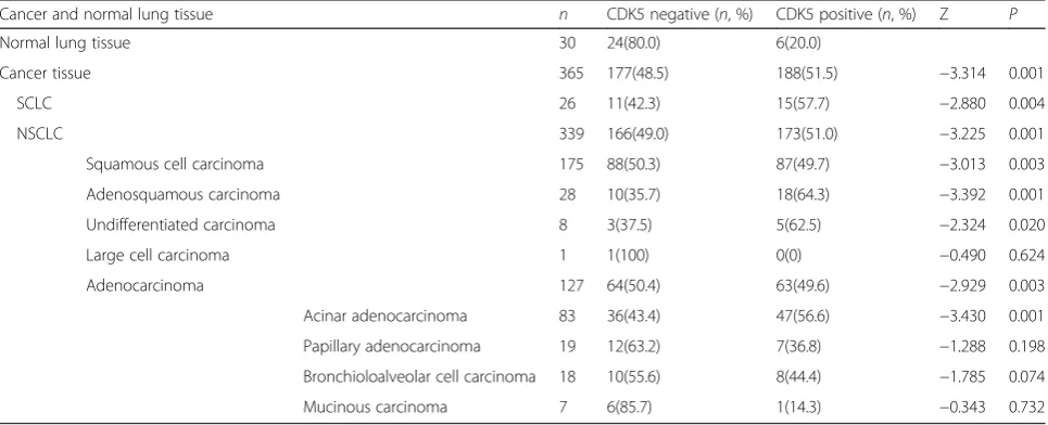
Table 2CDK5 expression in lung cancer compared with normal lung tissue
Cancer and normal lung tissue n CDK5 negative (n, %) CDK5 positive (n, %) Z P
Normal lung tissue 30 24(80.0) 6(20.0)
Cancer tissue 365 177(48.5) 188(51.5) −3.314 0.001
SCLC 26 11(42.3) 15(57.7) −2.880 0.004
NSCLC 339 166(49.0) 173(51.0) −3.225 0.001
Squamous cell carcinoma 175 88(50.3) 87(49.7) −3.013 0.003
Adenosquamous carcinoma 28 10(35.7) 18(64.3) −3.392 0.001
Undifferentiated carcinoma 8 3(37.5) 5(62.5) −2.324 0.020
Large cell carcinoma 1 1(100) 0(0) −0.490 0.624
Adenocarcinoma 127 64(50.4) 63(49.6) −2.929 0.003
Acinar adenocarcinoma 83 36(43.4) 47(56.6) −3.430 0.001
Papillary adenocarcinoma 19 12(63.2) 7(36.8) −1.288 0.198
Bronchioloalveolar cell carcinoma 18 10(55.6) 8(44.4) −1.785 0.074
Mucinous carcinoma 7 6(85.7) 1(14.3) −0.343 0.732
Non-small cell lung cancer (NSCLC) vs small cell lung cancer (SCLC)P= 0.513 Table 1The classification of lung cancer
Cancer subtype of histology
Lung cancer
SCLC
NSCLC Squamous cell carcinomas
Adenosquamous carcinomas
Large cell carcinoma
Undifferentiated carcinomas
Adenocarcinoma Acinar adenocarcinoma
Papillary adenocarcinomas
Bronchioloalveolar cell carcinomas
Mucinous carcinomas
pathological grading III (66.4 %, 87/131), CDK5 expression was higher than that in pathological grading I (25.6 %, 10/39,P< 0.001) and there was an increasing trend for CDK5 positive rate as the pathological grading in-creased (P< 0.001, Table 3). In addition, Spearman co-efficient of correlation was performed to investigate the relationship between the expression of CDK5 and clinicopathological parameters. It was showed that there were close correlations between CDK5 expres-sion and TNM stage (r= 0.155,P= 0.003), lymph node metastasis (r= 0.279,P< 0.001), and pathological grad-ing (r= 0.310, P< 0.001). A marginal correlation be-tween CDK5 expression and distal metastasis has been
found (r= 0.102, P= 0.053). Besides, no significant correlation between CDK5 expression and other clini-copathological factors was discovered, such as gender, age, and tumor diameter.
Cyclin-dependent kinases (CDK5) expression in non-small cell lung cancer (NSCLC)
When lung cancer patients were separated into two sub-groups of NSCLC and SCLC, we discovered that there was higher positive rate in NSCLC compared with normal lung tissues (P= 0.001, Table 4). Higher CDK5-positive expression was also found in the subgroups of NSCLCs including adenocarcinoma (P= 0.003), squamous cell car-cinoma (P= 0.003), adenosquamous carcinoma (P= 0.001), and undifferentiated carcinoma (P= 0.02), than that in the normal lung tissue. After adenocarcinoma was further split into four different types, remarkably higher expression of CDK5 was found in acinar adenocarcinoma as compared to normal lung tissues (P= 0.001, Table 4). When the rela-tionship between CDK 5 expression and other parameters was concerned in the patients with NSCLCs, higher CDK5 expression positive rate appeared in the advanced stages (III and IV) (66 %, 35/53) compared with the early stages (I and II) (48.3 %, 138/286,P= 0.018, Table 4) and the similar result was found in lymph node metastasis (76.5 %, 88/115) as compared to non-lymph node metastasis (37.9 %, 85/ 224, P< 0.001, Table 4). In pathological grading III, the positive expression of CDK5 was 66.2 % (86/130) in the cases of NSCLC higher than that in pathological grading I (25.6 %, 10/39)and II (42.4 %, 39/92, bothP< 0.001, Table 4). Borderline difference of CDK5 expression has been found between distal metastasis (75 %, 12/16) and non-distal me-tastasis (49.8 %, 161/323, P= 0.05, Table 4). Moreover, Spearman analysis showed that the positive CDK5 expres-sion in NSCLC was correlated with TNM stages (r= 0.129, P= 0.017), lymph node metastasis (r= 0.365, P< 0.001), and pathological grading (r= 0.307,P< 0.001). A marginal correlation between CDK5 expression and distal metastasis was also noticed (r= 0.107,P= 0.05).
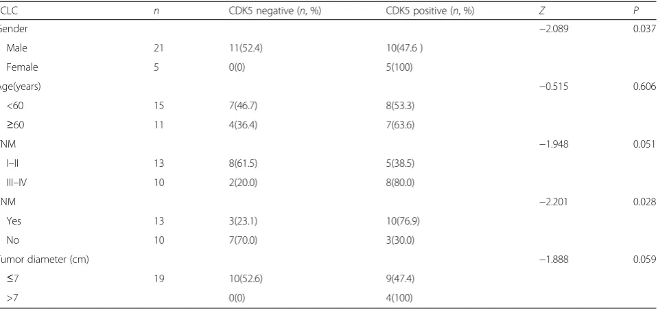
Cyclin-dependent kinases (CDK5) expression in small cell lung cancer (SCLC)
There were 26 patients of SCLC, and the positive rate of CDK5 expression was 57.7 % (15/26), significant higher compared to normal lung tissues (20 %, 6/30,P= 0.004). In the patients with SCLC, higher expression of CDK5 was found in female (100 %, 5/5) and lymph node metastasis (76.9 %, 10/13) compared with that in male (47.6 %, 10/21, P= 0.037) and without lymph node metastasis (30 %, 3/10, P= 0.028, Table 5), respectively. Spearman coefficient of correlation showed that the positive CDK5 expression in SCLC was correlated with gender (r= 0.418, P= 0.034), TNM stages (r= 0.415,P= 0.049), and lymph node metas-tasis (r= 0.469,P= 0.024, Table 5).
Fig. 1Immunohistochemical staining of CDK5 in lung tissue. Negative expression of CDK5 was found in normal lung cancer tissue (a×100,
Discussion
Cyclin-dependent kinase 5 (CDK5) is vital in neural cell migration and differentiation and is activated by p35 or p39 [24], and CDK5 is considered to be essential in neuronal cells [25, 26]. Nevertheless, as a unique mem-ber of cyclin-dependent kinases, the function of CDK5 beyond the nervous system has been demonstrated. CDK5 also regulates cell proliferation by alterant expres-sion and its downstream signaling pathways, especially in cancer cells. Up to date, there has been a growing number of evidence that CDK5 has an important effect on cancer progression [27]. The expression of CDK5 was aberrant in several cancers, and CDK5 regulated the proliferation of cancer cell in prostate cancer, medullary thyroid carcinoma, and gastric cancer [14, 15, 17]. In breast cancer, CDK5 was essential for the motility of cancer cell [11]. Moreover, CDK5 can be used to predict the prognosis of multiple myeloma [18]. In a word, the role of CDK5 in cancer is attracting increasing attention. To date, the expression of CDK5 was investigated in sev-eral cancers [13, 15, 17]. Higher expression of CDK5 was observed in hepatocellular carcinoma, ampullary adenocarcinoma, breast cancer, and medullary thyroid carcinoma, and Zachary et al. confirmed the expression
of CDK5 was upregulated in colorectal, head/neck, breast, lung, ovarian, lymphoma, prostatic, sarcoma, myeloma, and bladder cancers via the Oncomine micro-array online data mining software [11, 12, 18, 28]. How-ever, downregulated expression of CDK5 was observed in gastric cancer. Thus, CDK5 might be heterogeneously expressed in different cancers.
In the present study, immunohistochemistry on lung tissue microarrays was performed to explore the expres-sion of CDK5 in lung cancer and normal lung tissues. There was prominently higher expression of CDK5 in lung cancer, independent of various pathological sub-types, than in normal lung tissue. In the study of Liu et al., CDK5 was upregulated in cancer tissue as compared to benign pulmonary disease with a sample size of 95 non-small cell lung cancers (NSCLCs) [23]. Our study, with a bigger sample size, approximately four times, confirmed that increased expression of CDK5 could be detected in lung cancer tissue compared with normal lung tissue and further supported that CDK5 was con-sidered as an oncogene in lung cancer. No significant difference of CDK5 expression was found between NSCLC and SCLC in this current study. Although NSCLC and SCLC are commonly regarded as different Table 3CDK5 expression associated with the various clinicopathological parameters in lung cancer
Lung cancer n CDK5 negative (n, %) CDK5 positive (n, %) Z P
Gender −0.884 0.377
Male 275 137(49.8) 138(50.2)
Female 90 40(44.4) 50(55.6)
Age(years) −0.418 0.676
<60 196 96(49.0) 100(51.0)
≥60 169 81(47.9) 88(52.1)
Pathological grading 25.060a <0.001
I 39 29(74.4) 10(25.6)
II 92 53(57.6) 39(42.4)
III 131 44(33.6) 87(66.4)
TNM −2.944 0.003
I–II 299 156(52.2) 143(47.8)
III–IV 63 20(31.7) 43(68.3)
LNM −7.080 <0.001
Yes 128 30(23.4) 98(76.6)
No 234 146 (62.4) 88(37.6)
Tumor diameter (cm) −1.653 0.098
≤7 314 158(50.3) 156(49.7)
>7 48 18(37.5) 30(62.5)
Distal metastasis −1.931 0.054
Absent 346 172(49.7) 174(50.3)
Present 16 4(25.0) 12(75.0)
a
diseases owing to their distinct biology and genomic ab-normalities, the role and function of CDK5 may be con-sistent, as CDK5 level was both upregulated in NSCLC and SCLC tissues than the non-cancerous lung. How-ever, the exact role of CDK5 in SCLC needs further in-vestigation, since only a limited sample size (n= 26) was included in the current study. Taken together, CDK5 might be a potential biomarker of lung cancer despite its histology types.
The regulative mechanism of CDK5 in several cancers was investigated. CDK5 regulates DNA damage response via phosphorylating Ataxia telangiectasia mutated (ATM) kinase and thereby affecting its downstream sig-nal pathways which was crucial to progression of hepa-tocellular carcinoma [12]. In ampullary adenocarcinoma, over expression of nestin/CDK5 was involved in several oncogenic pathways (the activation of NOTCH, TGF-β1, or PDGFR pathways) that facilitated invasiveness of Table 4The correlation of CDK5 with diverse clinical clinicopathological factors in NSCLC
NSCLC n CDK5 negative (n, %) CDK5 positive (n, %) Z P
Gender −0.406 0.685
Male 254 126(49.6) 128(50.4)
Female 85 40(47.1) 45(52.9)
Age(years) −0.080 0.936
<60 181 89(49.2) 92(50.8)
≥60 158 77(48.7) 81(51.3)
Pathological grading 24.58a <0.001
I 39 29(74.4) 10(25.6)
II 92 53(57.6) 39(42.4)
III 130 44(33.8) 86(66.2)
TNM −2.376 0.018
I–II 286 148(51.7) 138(48.3)
III–IV 53 18(34.0) 35(66.0)
LNM −6.717 <0.001
Yes 115 27(23.5)) 88(76.5)
No 224 139(62.1) 85(37.9)
Tumor diameter (cm) −1.145 0.252
≤7 295 148(50.2) 147(49.8)
>7 44 18(40.9) 26(59.1)
Distal metastasis −1.962 0.05
Absent 323 162(50.2) 161(49.8)
Present 16 4(25.0) 12(75.0)
Histology 3.646a 0.456
Adenocarcinoma 127 64(50.4) 63(49.6)
Squamous cell carcinoma 175 88(50.3) 87(49.7)
Adenosquamous carcinoma 28 10(35.7) 18(64.3)
Undifferentiated carcinoma 8 3(37.5) 5(62.5)
Large cell carcinoma 1 1(100) 0(0)
Adenocarcinoma classification 6.508a 0.089
Acinar adenocarcinoma 83 36(43.4) 47(56.6)
Papillary adenocarcinoma 19 12(63.2) 7(36.8)
Broncholoalveolar cell carcinoma 18 10(55.6) 8(44.4)
Mucinous carcinoma 7 6(85.7) 1(14.3)
Pathological grading I vs. IIZ=−1.805,P= 0.071, I vs. IIIZ=−4.466,P< 0.001, II vs. IIIZ=−3.508,P< 0.001. Acinar adenocarcinoma vs. mucinousZ=−2.144,
P= 0.032. There were no differences of expression of CDK5 in other subgroups a
cancer [28]. In breast cancer, CDK5 takes part in epithelial-mesenchymal transition induced by
TGF-β1which is vital for tumor metastasis [11]. In medullary thyroid carcinoma, CDK5 is essential to tumorigenesis and progression by retinoblastoma protein (Rb) and in-hibition of Rb reduced proliferation of medullary thyroid carcinoma [15]. The mechanisms of tumorigenesis and progression in lung cancer might be similar to the mechanisms aforementioned in other cancers in consid-eration of a consistent trend of CDK5 expression. How-ever, this hypothesis needs to be verified with in vitro and in vivo studies.
Though expression of CDK5 was detected in lung can-cer tissue and regulative mechanism of CDK5 was inves-tigated in other cancers, the mechanism and exact role of CDK5 in the carcinogenesis and development of lung cancer remain unclear. A study of Korean population shows that CDK5 promoter polymorphisms contribute to the genetic susceptibility to lung cancer [20]. As one of the downstream components of the EGFR-family-signaling pathway, the gene of CDK5 was amplified in lung cancer and it might be the common mechanism of oncogene activation in carcinogenesis [16]. Tripathi et al. demonstrated that the CDK5 phosphorylates four ser-ines located N-terminal to the Rho-GTPase activating protein(Rho-GAP) domain in DLC1(deleted in lung can-cer 1), a tumor suppressor protein, and thereby activates DLC1 [22]. Through the reconstruction of an integrated genome-scale co-expression network, Bidkhori et al. ex-hibited that CDK5 played a vital role in cell cycle pro-gression in lung adenocarcinoma [19]. The studies above of CDK5 in lung cancer suggested CDK5 may play an oncogenic role in lung cancer.
In addition to the expression of CDK5 in cancer tis-sues, the relationship between CDK5 and pathological parameters has been paid more and more attention to. There were a few researches of the relationship be-tween CDK5 and clinical factors in the patients with cancers. In breast cancer, upregulated expression of CDK5 was related to higher grading (grading III) [11]. In multiple myeloma, downregulated expression of cdk5 predicted favorable overall survival after bortezo-mib treatment [18]. Liu et al. demonstrated that higher expression of CDK5 was correlated with low/ undifferentiated, high pathological stage, lymph node metastasis, shorter median survival, and lower 5-year overall survival in the patients with NSCLC [23]. Simi-larly, with a larger sample size, the consistent trend in this current study was confirmed that higher positive rate of CDK5 expression was greatly correlated with unfavorable clinicopathological parameters, including advanced TNM stage, lymphatic metastasis, and high pathological grading, which commonly indicate poorer prognosis. Thus, CDK5 might be used for the predic-tion to prognosis of lung cancer.
Further, Demelash et al. [21] demonstrated that CDK5 played a vital role in the regulation of lung cancer cell migration and invasion through Wound closure and Boyden chamber assay and certified that achaete-scute homologue-1 (ASH1), a basic tran-scription factor which was expressed in lung cancer cells with neuroendocrine features [29], could stimu-late migration of lung cancer cells through CDK5/ p35 pathway. The mechanism may support that CDK5 was closed related to lymphatic metastasis in lung cancer.
Table 5The correlation of CDK5 expression with various clinical pathological factors in SCLC
SCLC n CDK5 negative (n, %) CDK5 positive (n, %) Z P
Gender −2.089 0.037
Male 21 11(52.4) 10(47.6 )
Female 5 0(0) 5(100)
Age(years) −0.515 0.606
<60 15 7(46.7) 8(53.3)
≥60 11 4(36.4) 7(63.6)
TNM −1.948 0.051
I–II 13 8(61.5) 5(38.5)
III–IV 10 2(20.0) 8(80.0)
LNM −2.201 0.028
Yes 13 3(23.1) 10(76.9)
No 10 7(70.0) 3(30.0)
Tumor diameter (cm) −1.888 0.059
≤7 19 10(52.6) 9(47.4)
Conclusions
In summary, in this current study, the expression of CDK5 was investigated by lung tissue microarrays and immunohistochemistry. We demonstrated that CDK5 was highly expressed in lung cancer, including non-small cell lung cancer and small cell lung cancer, compared to normal lung tissue. Higher positive rate of CDK5 was associated with several clinicopathological parameters, which are representative of the progression and deterior-ation of lung cancer. These results suggest that the CDK5 expression associated with several unfavorable clinicopathological factors linked with poorer prognosis. Nevertheless, further plans are needed to explore the po-tential function of CDK5 in vitro and in vivo in the car-cinogenesis of and progression in lung cancer.
Competing interests
The authors declare that they have no competing interests.
Authors’contributions
KLW and ZYL designed the present study. YWD, LHY, and GC were responsible for immunohistochemistry and analysis of data. ZHY and XC conducted the experiment and wrote the paper. NH, CXB, and TQG contributed to statistical analysis. All authors read and approved the final manuscript.
Acknowledgements
The study was supported by the Fund of Guangxi Zhuang Autonomous Region University Student Innovative Plan (no. WLXSZX1555), China, the Fund of National Natural Science Foundation of China (NSFC 81360327), and the fund of Guangxi Provincial Health Bureau Scientific Research Project (Z2013201, Z2014055). The funders had no role in the study design, data collection and analysis, decision to publish, or preparation of the manuscript.
Received: 30 July 2015 Accepted: 26 January 2016
References
1. Barrow TM, Michels KB. Epigenetic epidemiology of cancer. Biochem Biophys Res Commun. 2014;455(1–2):70–83. doi:10.1016/j.bbrc.2014.08.002. 2. Siegel RL, Miller KD, Jemal A. Cancer statistics, 2015. CA Cancer J Clin.
2015;65(1):5–29. doi:10.3322/caac.21254.
3. Chen W, Zheng R, Zhang S, Zhao P, Zeng H, Zou X, et al. Annual report on status of cancer in China, 2010. Chin J Cancer Res. 2014;26(1):48–58. doi:10.3978/j.issn.1000-9604.2014.01.08.
4. Brothers JF, Hijazi K, Mascaux C, El-Zein RA, Spitz MR, Spira A. Bridging the clinical gaps: genetic, epigenetic and transcriptomic biomarkers for the early detection of lung cancer in the post-national lung screening trial era. BMC Med. 2013;11:168. doi:10.1186/1741-7015-11-168.
5. Jafri SH, Shi R, Mills G. Advance lung cancer inflammation index (ALI) at diagnosis is a prognostic marker in patients with metastatic non-small cell lung cancer (NSCLC): a retrospective review. BMC Cancer. 2013;13:158. doi:10.1186/1471-2407-13-158.
6. Zhang C, Huang C, Wang J, Wang X, Li K. Maintenance or consolidation therapy for non-small-cell lung cancer: a meta-analysis involving 5841 subjects. Clin Lung Cancer. 2015. doi:10.1016/j.cllc.2015.01.002 7. DeSantis CE, Lin CC, Mariotto AB, Siegel RL, Stein KD, Kramer JL, et al.
Cancer treatment and survivorship statistics, 2014. CA Cancer J Clin. 2014;64(4):252–71. doi:10.3322/caac.21235.
8. Barr Kumarakulasinghe N, Zanwijk NV, Soo RA. Molecular targeted therapy in the treatment of advanced stage non-small cell lung cancer (NSCLC). Respirology. 2015. doi:10.1111/resp.12490
9. Parums DV. Current status of targeted therapy in non-small cell lung cancer. Drugs Today. 2014;50(7):503–25. doi:10.1358/dot.2014.50.7.2185913. 10. Gerard C, Tyson JJ, Coudreuse D, Novak B. Cell cycle control by a minimal
cdk network. PLoS Comput Biol. 2015;11(2):e1004056. doi:10.1371/journal. pcbi.1004056.
11. Liang Q, Li L, Zhang J, Lei Y, Wang L, Liu DX, et al. CDK5 is essential for TGF-beta1-induced epithelial-mesenchymal transition and breast cancer progression. Scientific Reports. 2013;3:2932. doi:10.1038/srep02932. 12. Ehrlich SM, Liebl J, Ardelt MA, Lehr T, De Toni EN, Mayr D et al. Targeting
cyclin dependent kinase 5 in hepatocellular carcinoma—a novel therapeutic approach. J Hepatol. 2015. doi:10.1016/j.jhep.2015.01.031
13. Xie W, Wang H, He Y, Li D, Gong L, Zhang Y. CDK5 and its activator P35 in normal pituitary and in pituitary adenomas: relationship to VEGF expression. Int J Biol Sci. 2014;10(2):192–9. doi:10.7150/ijbs.7770.
14. Lindqvist J, Imanishi SY, Torvaldson E, Malinen M, Remes M, Orn F et al. Cyclin-dependent kinase 5 acts as a critical determinant of AKT-dependent proliferation and regulates differential gene expression by the androgen receptor in prostate cancer cells. Molecular biology of the cell. 2015. doi:10.1091/mbc.E14-12-1634
15. Pozo K, Castro-Rivera E, Tan C, Plattner F, Schwach G, Siegl V, et al. The role of Cdk5 in neuroendocrine thyroid cancer. Cancer Cell. 2013;24(4):499–511. doi:10.1016/j.ccr.2013.08.027.
16. Lockwood WW, Chari R, Coe BP, Girard L, Macaulay C, Lam S, et al. DNA amplification is a ubiquitous mechanism of oncogene activation in lung and other cancers. Oncogene. 2008;27(33):4615–24. doi:10.1038/onc.2008.98. 17. Cao L, Zhou J, Zhang J, Wu S, Yang X, Zhao X et al. Cyclin dependent
kinase 5 decreases in gastric cancer and its nuclear accumulation suppresses gastric tumorigenesis. Clin Cancer Res. 2015. doi:10.1158/1078-0432.CCR-14-1950
18. Levacque Z, Rosales JL, Lee KY. Level of cdk5 expression predicts the survival of relapsed multiple myeloma patients. Cell Cycle. 2012;11(21):4093– 5. doi:10.4161/cc.21886.
19. Bidkhori G, Narimani Z, Hosseini Ashtiani S, Moeini A, Nowzari-Dalini A, Masoudi-Nejad A. Reconstruction of an integrated genome-scale co-expression network reveals key modules involved in lung adenocarcinoma. PLoS One. 2013;8(7):e67552. doi:10.1371/journal.pone. 0067552.
20. Choi HS, Lee Y, Park KH, Sung JS, Lee JE, Shin ES, et al. Single-nucleotide polymorphisms in the promoter of the CDK5 gene and lung cancer risk in a Korean population. J Hum Genet. 2009;54(5):298–303. doi:10.1038/jhg.2009. 29.
21. Demelash A, Rudrabhatla P, Pant HC, Wang X, Amin ND, McWhite CD, et al. Achaete-scute homologue-1 (ASH1) stimulates migration of lung cancer cells through Cdk5/p35 pathway. Mol Biol Cell. 2012;23(15):2856–66. doi:10.1091/mbc.E10-12-1010.
22. Tripathi BK, Qian X, Mertins P, Wang D, Papageorge AG, Carr SA, et al. CDK5 is a major regulator of the tumor suppressor DLC1. J Cell Biol. 2014;207(5): 627–42. doi:10.1083/jcb.201405105.
23. Liu JL, Wang XY, Huang BX, Zhu F, Zhang RG, Wu G. Expression of CDK5/ p35 in resected patients with non-small cell lung cancer: relation to prognosis. Med Oncol. 2011;28(3):673–8. doi:10.1007/s12032-010-9510-7. 24. Shah K, Lahiri DK. Cdk5 activity in the brain—multiple paths of regulation.
J Cell Sci. 2014;127(Pt 11):2391–400. doi:10.1242/jcs.147553.
25. Nishimura YV, Shikanai M, Hoshino M, Ohshima T, Nabeshima Y, Mizutani K, et al. Cdk5 and its substrates, Dcx and p27kip1, regulate cytoplasmic dilation formation and nuclear elongation in migrating neurons. Development. 2014;141(18):3540–50. doi:10.1242/dev.111294.
26. Xu S, Li X, Gong Z, Wang W, Li Y, Nair BC, et al. Proteomic analysis of the human cyclin-dependent kinase family reveals a novel CDK5 complex involved in cell growth and migration. Mol Cell Proteomics. 2014;13(11): 2986–3000. doi:10.1074/mcp.M113.036699.
27. Kimura T, Ishiguro K, Hisanaga S. Physiological and pathological phosphorylation of tau by Cdk5. Front Mol Neurosci. 2014;7:65. doi:10.3389/fnmol.2014.00065.
28. Shan YS, Chen YL, Lai MD, Hsu HP. Nestin predicts a favorable prognosis in early ampullary adenocarcinoma and functions as a promoter of metastasis in advanced cancer. Oncol Rep. 2015;33(1):40–8. doi:10.3892/or.2014.3588. 29. Miki M, Ball DW, Linnoila RI. Insights into the achaete-scute homolog-1