_____________________________________________________________________________________________________
*Corresponding author: E-mail: emavelkova@yahoo.com;
27(10): 1-10, 2018; Article no.JAMMR.43917
ISSN: 2456-8899
(Past name: British Journal of Medicine and Medical Research, Past ISSN: 2231-0614, NLM ID: 101570965)
Noninvasive Antenatal Diagnosis of Fetal RhD
Status
Emilija Velkova
1*and Dijana Plaseska-Karanfilska
21
Institute for Transfusion Medicine of the Republic of Macedonia, Republic of Macedonia. 2
Macedonian Academy of Sciences and Art, Skopje, Republic of Macedonia.
Authors’ contributions
This work was carried out in collaboration between both authors. Author EV designed the study, performed the statistical analysis, wrote the protocol and wrote the first draft of the manuscript.
Author DPK managed the analyses of the study. Both authors read and approved the final manuscript.
Article Information
DOI:10.9734/JAMMR/2018/43917
Editor(s):
(1) Dr. Chan-Min Liu, School of Life Science, Xuzhou Normal University, Xuzhou City, China.
Reviewers:
(1) Shigeki Matsubara, Jichi Medical University, Japan. (2) Gangaram Akangire, University of Missouri, USA. (3)Ravneet Kaur, Government Medical College and Hospital, India. Complete Peer review History:http://www.sciencedomain.org/review-history/26846
Received 19 July 2018 Accepted 16 October 2018 Published 25 October 2018
ABSTRACT
Introduction: Fetal cell-free nucleic acids within the blood stream of a pregnant woman come from fetal genetic material which can be acquired by simple venipuncture that reduces any risk to a minimum. Fetal cell-free DNA can be detected in the mother's blood stream in the 5th gestation week at the earliest. That enables fetal genotyping at the earliest possible stage of pregnancy which is best done in the 12th gestation week.
Aim: To determine fetal RhD status at RhD negative pregnant women where the father is a heterozygote, Dd.
Materials and Methods: The research includes 1540 RhD negative pregnant women, out of which at 30 of them the RhD fetal status had been detected by a PCR technique from the mother’s plasma. The RhD fetal status was confirmed after delivery by serologic analysis at 27 newborn babies.
All research patients were submitted to serologic immunohematology testing: blood group typing of red blood cell antigens, screening of irregular anti-red blood cell antibodies. Fetal RhD status was determined by the plasma of RhD negative pregnant women using the real-time PCR technology in
the period from the 12th gestation week until the 31 gestation week. The biological fathers of all 30 fetuses were phenotyped as heterozygote to the RhD antigen. The results showed that 30% of the fetuses are RhD negative, and 70% are RhD positive.
Conclusion: The noninvasive fetal RhD genotyping is not only one precious tool in the management of RhD alloimmunised pregnancies, but it also allows antenatal anti-D immunoglobulin prophylaxis exclusiveness for only non-immunized RhD pregnant women carrying RhD positive fetus. Taking into consideration that 30% of the RhD negative pregnant women that carry a RhD negative fetus receive antenatal RhIG prophylaxis with no absolute need for it. At RhD alloimmunised pregnant women the noninvasive genotyping of the fetal blood group enables an easy and safe method in determination of a fetal risk from a hemolytic disease, and at the same time evading a vast laboratory and clinical monitoring of RhD antigen-negative fetal cases.
Keywords: Hemolytic Disease of the Newborn (HDFN); antibody; red blood cell antigen; alloimunisation; sensibilisation; anti-D Ig prophylaxis (RhIG); Rh blood group system; fetomaternal haemorrhage (FMH); fetal DNK.
1. INTRODUCTION
Cell-free fetal nucleic acids running through the blood stream of a pregnant woman demonstrate a source of fetal genetic material which can be acquired by simple venipuncture. Contrary to invasive procedures, where fetal genetic material is acquired from fetal cells directly from the
uterus through chorionic villous sampling
between the 11th and 14th gestation week, or through amniocentesis after the 15th week, this acquisition of the mother’s blood sample does not involve a procedure that can cause fetal loss [1,2].
It is well known that fetal cell-free DNA is released by trophoblastic cells that are subjected to apoptosis [3]. As a result of this apoptotic penetration fetal cell-free DNA in the mother’s blood stream is usually comprised of fragments smaller than 150 (bp) by size, demonstrating its presence as mononucleosomal DNA [4]. Firstly, fetal cell-free DNA can be detected in the mother’s blood stream in the 5th gestation week at the earliest [5,6], or 18 days after the embryo transfer[7].
Fetal cell-free DNA present within the frames of the mother’s blood stream can be found in the rich background of the mother’s cell-free DNA. The mother’s cell-free DNA is mainly of hematopoetic origin, but transplanted models displayed also other tissues as a source of cell-free DNA [8].
The middle fraction concentration of fetal cell-free DNA in maternal plasma is approximately 9,7%, 9,0% and 20,4% during the first, second and third trimester, measured by digital PCR [8].
The concentration of fetal cell-free DNA in the mother’s serum is lower than the one in the mother’s plasma because of lysis of the mother’s nucleic red blood cells during the process of coagulation that leads to increasing of the percentage of mother's DNA in the blood sample [9].
After delivery, the fetal cell-free DNA is purified by the mother's circulation within a few hours, with a semi-life of 16 minutes. This fast breakthrough shows that during pregnancy fetal cell-free DNA is constantly released in large
amounts within the mother's circulation.
Calculations suggest that there is a release rate of 2,24 × 10⁴ copies in a minute. Unlike fetal cells that can survive within the mother’s circulation for many years [9] fetal cell-free DNA has not been discovered in the blood stream of non-pregnant women [10], which makes it specific compared to the state occurring in current pregnancies.
The non-invasive foetal RhD genotyping is a precious tool in the management of RhD
allosensitised pregnancies, especially for
mothers who carry a RhD negative fetus, avoiding numerous unnecessary analyses during pregnancy.
On the other hand, antenatal anti-D
immunoglobulin prophylaxis is exclusively
intended for non-immunized RhD pregnant women carrying RhD positive fetus.
2. MATERIALS AND METHODS
Velkova and Plaseska-Karanfilska; JAMMR, 27(10): 1-10, 2018; Article no.JAMMR.43917
technique from the mother’s plasma. The RhD fetal status was confirmed after delivery by serologic immunohematology analysis at 27 newborn babies. There was also phenotyping of Rh D,C,c,E,e and K antigens including 200 samples from the fetal biological father. The
phenotypisation was performed with two
serologic mikcroaglutination techniques in
immunohematology, simultaneously: mikrogel (Biorad) and magnetic pearls (OrthoDiagnostic). The tests were done including the serum, plasma and red blood cells of vein blood, without anticoagulants and/or EDTA, not older than 24 hours.
The tests for newborns were done including vein and umbilical cord blood, without anticoagulants and/or EDTA, not older than 24 hours.
All research patients: pregnant women, biological fathers of the fetuses and newborns, were
submitted to serologic immunohematology
testing:
o Blood group typing of red blood cell antigens,
o Screening of irregular anti-red blood cell antibodies (indirect antihuman globulin test, enzyme test and direct antihuman globulin test)
The serology tests were done by
microaglutination technique - DiaMed ID card, using two monoclonal anti-D reagents out of which one of them recognises DVI phenotype that is immunogenic for RhD negative mother.
Acquired results were analysed and elaborated by different statistical methods: all statistical series according to variables of interest are tabulated and graphically displayed; the analysis of the structure of the attributable statistical series is made with the coefficients of relations, proportions and rates.
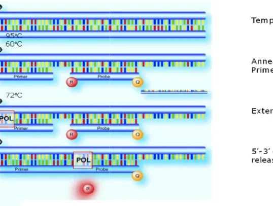
Real-time PCR technique monitors the
amplification of a targeted DNA molecule during the PCR in real time. It measured the amount of nucleic acids through the detection of a fluorescent signal which is proportional to the concentration of a double chain DNA in the PCR tube.The fluorescent signals are produced by fluorophores that are added to the reaction mixture. SYBR Green real-time PCR, SYBR Green introduces a colour that forms a complex with the double chain DNA which efficiently fluorescents. TaqMan analysis probes are
specific fluorescently marked oligonucleitic tests that also have a quencher residues and they hybridise with the targeted DNA.
The probe subsequently hydrolyses from the elongated Taq polimerasis which results in the division of the fluorophore from its quencher. In both cases, the produced fluorescent signal can be used as a measure for the initial amount of the targeted sequence in reaction. The real-time PCR fluorescent signal is noticed in “the real time” at the end of each PCR cycle. The amount of the targeted sequence is then calculated automatically.
In quantification, by using the method of a standard curve, you can measure an unknown quantity on the basis of measures for a known quantity. First, you generate a standard curve and then the values of an unknown quantity are compared to the standard curve out of which you can extrapolate values.
It represents a noninvasive method for
determination of the fetal RhD status by analysing the mother's plasma.
The fetal RhD status of the fetus was determined by RHD TaqMan system used as described by Chiu R and Coo.in 2001, in order to detect fetal RhD status. Specific primers were used to amplify egzon 5 (RHD5) and egzon 7 (RHD7) of the RHD gen, as well as the control fragment CCR5.
The primer sequences and the fluorescent probes were used from the Finning publication [11]. Cff DNA (Cell Free Fetal DNA) was extracted due to the QIAamp Circulating Nucleic Acid kit according the producers protocol.
RHD5 primers and the probe are amplified only by the RHD gene, while RHD7 primers and the probe are amplified not only by the RHD gene, but by the RHD pseudogene as well.
2.1 Reaction Mixture
Each plate for real-time PCR amplification included RHD5, RHD7 and CCR5 amplification on patients, RHD negative control and controlling genome DNA (Promega) for the generation of standard curves. The reaction evolved in a volume of 25 L, out of which 20 L reaction
mixture and 5 L сff DNA/negative control
Picture 1. Schematic display of a real
Table 1
Target Primer 5'-3' sequence
RHD5 Forward CGCCCTCTTCTTGTGGATG
Reverse GAACACGGCATTCTTCCTTTC
RHD7 Forward CAGCTCCATCATGGGCTACAA
Reverse AGCACCAGCAGCACAATGTAGA
CCR5 Forward TACCTGCTCAACCTGGCCAT
Reverse TTCCAAAGTCCCACTGGGC
Table 2. Used fluorogenic oligonucleotic probes
Target 5'-3' sequence
RHD5 TCTGGCCAAGTTTCAACTCTGCTCTGCT
RHD7 AGCTTGCTGGGTCTGCTTGGAGAGATC
CCR5 TTTCCTTCTTACTGTCCCCTTCTGGGCTC
2.2 Real-time Protocol and Amplification Conditions
The aparatus software is adjusted to the adequate template and type of samples according to the schedule that involved their plate application. The appropriate colour should be chousen on the reporter (FAM for
CCR5 and HEX for RHD7, while BHQ fluorescent quencher) as a bleacher
3. RESULTS
All samples are analysed, and standard curves are generated for each set of primers. The basic line is adjusted between the 3rd
cycle, and the threshold is adjusted to 1.0.
Schematic display of a real-time PCR reaction
Table 1. Used primers for amplification
3' sequence Final conc.
(nM)
Size of amplicon (bp)
CGCCCTCTTCTTGTGGATG 300 82
GAACACGGCATTCTTCCTTTC 300
CAGCTCCATCATGGGCTACAA 300 75
AGCACCAGCAGCACAATGTAGA 300
TACCTGCTCAACCTGGCCAT 300 91
TTCCAAAGTCCCACTGGGC 300
Used fluorogenic oligonucleotic probes
5' Label 3' Label
TCTGGCCAAGTTTCAACTCTGCTCTGCT FAM BHQ1
AGCTTGCTGGGTCTGCTTGGAGAGATC HEX BHQ1
TTTCCTTCTTACTGTCCCCTTCTGGGCTC FAM BHQ1
Protocol and Amplification
The aparatus software is adjusted to the adequate template and type of samples according to the schedule that involved their plate application. The appropriate colour should for RHD5 and while BHQ1 (non-as a bleacher.
All samples are analysed, and standard curves are generated for each set of primers. The basic and the 15th cycle, and the threshold is adjusted to 1.0.
A negative result is indicated by a Ct value (no amplification), while a positive result is when the Ct value <42 (there is amplification).
The angle of standard curves should be between -3.2 and -4.5.
Table number 3 is used for interpretation of results.
There was a determination of fetal RhD genotyping by the mother's plasma for 30 RhD negative pregnant women related to various gestation week of pregnancy, i.e. from 12 gestation week up till 31 gestation
using the method of Polymerase C (PCR).
Size of amplicon
Final conc. (nM)
166 166 166
A negative result is indicated by a Ct value ≥42 (no amplification), while a positive result is when
(there is amplification).
The angle of standard curves should be between
Table number 3 is used for interpretation of
Velkova and Plaseska-Karanfilska; JAMMR, 27(10): 1-10, 2018; Article no.JAMMR.43917
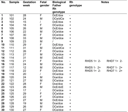
There was also RhD phenotyping from the biological father in reference to 200 samples, at which the mother was previously typed as RhD negative.All 30 pregnant women included in the research are related to a biological father phenotyped as RhD heterozygote.
Acquired results demonstrated that
9 fetuses are RhD negative and 21
RhD positive. The test was repeated
at the negative ones in the period of early pregnancy one more time after the 20th gestation week.
Table 3. Interpretation of results
Number of replicates RHD5 <Ct42
Number of replicates RHD7 <Ct42
RHD genotype
0 0 RHD negative
0 1 RHD negative
1 0 RHD negative
1 1 RHD negative if Cts>38;
2-4 2-4 Inconclusive
0,1 5,6 RHD pseudogene
5,6 5,6 RHD positive if 5-6 Cts <42 (standard
deviation should be no more than 1.5)
Table 4. RHD genotyping fetal DNA by the mother’s plasma
No. Sample Gestation week
Fetal gender
Biological Father Rh genotype
Rh genotype
Notes
1 101 28 F DcE/dce -
2 102 24 М DCe/dCe +
3 103 15 / DcE/dce +
4 104 18 F DCe/dce +
5 105 26 М DcE/dce +
6 106 22 М DCe/dce +
7 107 30 F DCe/dce +
8 108 33 М DCe/dce +
9 109 23 М / +
10 110 29 F DcE/dce +
11 111 31 М DCe/dCe +
12 112 26 F DCe/dce +
13 113 27 М DCe/dce +
14 114 31 М DcE/dcE +
15 115 21 F Dce/dce - RHD5 1+ 2- RHD7 1+ 2-
16 116 24 М DCe/dce +
17 117 12 М DCe/dce + RHD5 1- 2+ RHD7 1- 2+
18 118 22 F DcE/dce + RHD5 1+ 2- RHD7 1- 2+
19 119 20 / DCe/dce +
20 120 24 М Dсe/dce +
21 121 27 М DCe/dce +
22 122 28 F Dсe/dce +
23 123 26 М DcE/dcE +
24 124 17 / DCe/dce +
25 125 29 F DCe/dce +
26 126 22 F DCe/dce -
27 127 27 М DcE/dce +
28 128 19 М Dсe/dce +
29 129 26 М DCe/dce +
Picture 2. Amplification plot of a real-time PCR related to RHD gene from cff DNA of a positive RHD fetus
Picture 3. Amplification plot of a real-time PCR related to RHD gene from cff DNA of a negative RHD fetus
4. DISCUSSION
Developed countries have introduced routine
antenatal anti-D prophylaxis since 1960,
combined with RhIG administration in high risk situations during pregnancy and delivery, such as miscarriage, terminate pregnancy, invasive antenatal diagnosis procedures, external version, Caesarean section, etc [12,13,14]. It was noted that there is a significant decrease in the rate of anti –D immunization and anti –D perinatal mortality [15].
Further decrease had been noticed in several
studies that researched routine antenatal
application of RhIG in order to prevent immunisation of undiscovered FMH during the
last trimester of pregnancy [14,15]. But even if postnatal and antenatal prophylaxis is
combined, 0.1-0.3% of the women are at risk, and they still create RhD antibodies [16,17]. If the prevention risk factors that contribute to the rest of the immunisation can be identified, it will be possible to get even a greater decrease of HDFN.
Velkova and Plaseska-Karanfilska; JAMMR, 27(10): 1-10, 2018; Article no.JAMMR.43917
man who is also RhD negative. There is no need of anti-D prophylaxis for them.
Undeniably, we should avoid application of antenatal prophylaxis for those women. There are opinions that it might require additional analysis that is not necessary, but it is a fact that they should be saved from possible side effects during application of anti-D, as well as avoid the trauma of unnecessary application and risks.
Nevertheless, it is necessary for the RhD negative woman to identify the biological father, and he should determine his Rh phenotype and possible genotype.
It has been discovered that 56% of Rh positive white race individuals are heterozygote to the RhD antigen. Thus, when the biological father is heterozygote RhD positive, there is 50% chance for the fetus to be Rh negative.
If the father is antigen negative, there is no chance for the fetus to be antigen positive, and therefore there is no risk of HBFN that can be caused by the mother’s alloantibodies.
Nowadays it is possible to determine the antigen fetal status by a DNA test through the mother’s plasma if the father is heterozygote antigen positive, in case of allosensitization of the mother by D,C,c,E and K antibodies.
Traditionally, genotyping of the fetal blood group is done by amniocentesis.
This invasive procedure involves a risk of miscarriage, can potentially cause maternal sensitization and increases the risk of FMH that can produce a larger amount of antibodies [18,19].
The discovery of cell-free fetal DNA in the plasma of pregnant women (since the end of the 20th century) demonstrated one noninvasive and safe method for determination of fetal blood group genotype with almost 100% accuracy [20,21].
At the moment, the noninvasive fetal RhD genotyping at D-allosensitized pregnant women is a routine clinical practice in several European states.
Moreover, following the grand research of fetal RhD genotyping since 2010, Denmark and Holland introduced a national program for fetal
RhD screening related to the determination of the
fetal D antigen in order to apply
immunoprophylaxis at not immunised D-negative pregnant women [22]. There was also a development of noninvasive genotyping analysis in determination of fetal blood group for the rest of clinically relevant red blood cell antigens.
In our research, we have used vein maternal blood taken in EDTA. Genotyping was done in
the maternal plasma because fraction
concentration of cell-free fetal DNA within the maternal serum is lower than the one in the maternal plasma (because of lyses of maternal nucleic red blood cells during coagulation) which produces percentage increase of maternal DNA in the blood sample [23,24,25].
The research included 1540 RhD negative
pregnant women with serologic
immunohematology testing. In order to determine fetal RhD status it is necessary to primarily determine the RhD zygote type of the biological father. Unfortunately, the biological father is sometimes difficult to determine if based on the women’s report/declaration, besides only ½ of them can be a heterozygote. Only 30 pregnant women volunteered to participate in the research out of the total number of fetuses with RhD heterozygote biological father from the results acquired by testing samples of 200 fathers.
The total amount of 30 RhD negative pregnant women was tested by Real time PCR between the 12 and 31 gestation week. All pregnant women including the ones at which we have detected Allosensitization to the D antigen involved phenotyping of the Rh blood group system of the biological father.
The research related to the determination of the fetal RhD status included 30 case at which the biological father was a heterozygote, while the extraction of fetal DNA in the maternal serum was confirmed.
Regarding the fetal RhD screening, samples of the postnatal umbilical cord were sent to our lab in order to determine RhD serology of the newborn. Serology of the umbilical cord was done at 90% of the cases. Fetal RhD typing was positive at 20 out of 20 samples by plasma from women that had delivered a D-positive baby, accordingly the result is 100% sensitivity. Out of the total amount of 7 samples of plasma by women that had delivered a D-negative baby, fetal RhD typing was negative at 6 samples.
In reference to science literature, false negative results appear because of lack of enough DNA in the system of detection, because of failure in extraction, low fetal DNA concentration in the plasma sample, or unsuccessful PCR or mixed sample. False negative results represent the main concern in fetal RhD screening analysis since anti-D immunoprophylaxis will be unjustly excluded from the treatment of D-negative pregnant women that can result in risk of immunisation and potential morbidity and mortality by HDFN in subsequent pregnancies. According to statistics, this percentage is minimal and it amounts to 0.06% [26].
Numerous studies succeeded in fetal RhD genotyping at RhD negative mothers with almost 100% accuracy. The standard methodology is available and there are established international exterior schemes of quality.
In the last several years, a couple of research studies for RhD typing that use automated DNA extraction and accurate robotic handling were successfully presented, achieving a sensitivity of 99.7% to 99.9% [8,25,26].
Recently in many developed countries, routine antenatal anti-D prophylaxis where anti-D immunoglobulin is applied between 28 – 34 gestation week, became a standard care for
D-negative pregnant women. This practice
combined to postnatal prophylaxis brought about further reduction of maternal immunisation for more than 50% [27,28,29] Never the less, we have to pay attention to the unnecessary use of antenatal prophylaxis in women who carry RhD negative baby.
5. CONCLUSION
Collected data demonstrate that the noninvasive fetal RhD genotyping is not only a precious tool in the management of RhD allosensitized pregnancies, but also allows antenatal RhIG
prophylaxis exclusively for those not immunised RhD negative pregnant women that carry RhD positive fetus.
The analysis of results that we have acquired in our study reveal the fact that 30% of RhD negative pregnant women receive antenatal RhIG prophylaxis, and have no need of it. Besides, these pregnant women can be saved
from further unnecessary testing during
pregnancy since there is no risk of HBFN.
Pregnant women that were allosensitized in their previous pregnancies and/or transfusions, and have a confirmation of fetal RhD negative status, there is no risk of HBFN, and no further testing is necessary.
However, if the fetus inherits the implicated antigen, it implies the need of timely and careful follow up of fetal anaemia through a series of titers from maternal antibodies and other activities, fetal measures by Doppler ultrasound at peak systolic velocity in the middle cerebral artery, and finally intrauterine sample of fetal blood with possibility of intrauterine transfusion on time.
CONSENT AND ETHICAL APPROVAL
As per university standard guideline participant consent and ethical approval has been collected and preserved by the authors.
COMPETING INTERESTS
Authors have declared that no competing interests exist.
REFERENCES
1. Grootkerk-Tax MGHM, Soussan AA, de
Haas M, Maaskant–van Wijk PA, van der Schoot CE. Evaluation of prenatal RHD typing strategies on cell-free fetal DNA
from maternal plasma. Transfusion. 2006;
46:2142–8.
2. Avent ND. RHD genotyping from maternal
plasma: Guidelines and technical
challenges. Methods Mol Biol. 2008; 444:185–201.
3. van Wijk IJ, de Hoon AC, Jurhawan R,
Tjoa ML, Griffioen S, Mulders MA, et al. Detection of apoptotic fetal cells in plasma of pregnant women. Clin Chem. 2000; 46:729
4. Lapaire O, Holzgreve W, Oosterwijk JC,
Velkova and Plaseska-Karanfilska; JAMMR, 27(10): 1-10, 2018; Article no.JAMMR.43917
on trophoblasts in the maternal circulation. Placenta. 2007;28:1-5.
5. van der Schoot CE, Soussan AA,
Koelewijn J, Bonsel G, Page-Christiaens GCML, de Haas M. Non-invasive antenatal RHD typing.Transfus Clin Biol. 2006; 13:53–7.
6. Finning K, Martin P, Summers J, Massey
E, Poole G, Daniels G. Effect of high throughput RHD typing of fetal DNA in maternal plasma on use of anti-RhD immunoglobulin in RhD negative pregnant
women: Prospective feasibility study.
British Medical Journal. 2008;336:816–8. 7. Muller SP, Bartels I, Stein W, Emons G,
Gutensohn K, Kohler M et al. The determination of the fetal D status from maternal plasma for decision making on Rh prophylaxis is feasible. Transfusion. 2008;48:2292–301.
8. Birch L, English CA, O’Donoghue K,
Barigye O, Fisk NM, Keer JT. Accurate and robust quantification of circulating fetal and total DNA in maternal plasma from 5 to 41 weeks of gestation. Clin Chem. 2005;51:312–20.
9. Oosterwijk JC. Prenatal diagnosis on fetal cells from maternal blood: Approaches and perspectives. Eur J Obster Gynecol Reprod Biol. 1999;82:169-70.
10. Lo YM, Hjelm NM, Fidler C, Sargent IL, Murphy MF, Chamberlain PF et al.Prenatal diagnosis of fetal RhD status bymolecular analysis of maternal plasma. N Engl J Med. 1998;339:1734–8.
11. Finning et al. Effect of high throughput RHD typing of fetal DNA in maternal plasma on use of anti-RhD immunoglobulin
in RhD negative pregnant women:
prospective feasibility study. British
Medical Journal. 2008;336:816-818.
12. American College of Obstetricans and
Gynecologists. ACOG practice bulletin. Prevention of Rh D alloimmunization. Number 4, May 1999 (replaces educational bulletin Number 147, October 1990).
Clinical management guidelines for
obstetrician-gynecologists. American
College of Obstetrics and Gynecology. Int J Gynaecol Obstet. 1999;66(1):63-70
13. Vandenbussche FPHA, Klumper FJ.[Red
cell immunisation and pregnancy]. Dutch Society of Obstetrics and Gynaecology; 2002 [Dutch].
14. NICE. NICE issues guidance for
RhD-negative women during pregnancy.
2002/024 ed.2002
15. Kumpel BM. On the immunologic basis of
Rh immune globulin (anti-D) prophylaxis.
Transfusion Journal.
2006;46(9):1652-1656.
16. Engelfreit CP, Reesink HW, Judd WJ,
Ulander VM, Kuosmanen M, Koskinen S, et al. Current status of immunoprophylaxis with anti-D immunoglobin. Vox Sangvin. 2003;85(4):328-337.
17. Koelewijn JM, de Haas M,Vrijkotte TG,
Bonsel GJ, Van der Schoot CE. One single dose of 200 microg of antenatal RhIg halves the risk of anti-D immunization and hemolytic disease of the fetus and
newborn in the next pregnancy.
Transfusion Journal.
2008;48(8):1721-1729.
18. Avent N. Fetal genotyping. In: Hadley A.G,
Soothill P, editors. Alloimune disorders of pregnancy. Anaemia, thrombocytopenia and neutropenia in the fetus and newborn. Cambridge: Cambridge University Press. 2002;121-140.
19. Mujezinovic F, Alfirevic Z.
Procedure-related complications of amniocentesis
and chorionic villous sampling: A
systematic review [published erratum
appears in Obstet Gynecol.2008;111:779].
Obstet Gynecol. 2007;110:687–94.
20. Geifman-Holtzman O, Grotegut CA,
Gaughan JP. Diagnostic accuracy of noninvasive fetal Rh genotyping from maternal blood – a meta-analysis. Am J Obstet Gynecol. 2006;195:1163–73.
21. Daniels G, Finning K, Martin P, Massey E.
Noninvasive prenatal diagnosis of fetal blood group phenotypes: Current practice and future prospects. Prenat Diagn. 2009; 29:101–7.
22. Clausen F, Christiansen M, Steffensen R, Jorgensen S, Nielsen C, Jakobsen M et al. Report of the first nationally implemented clinical routine screening for fetal RHD in D– pregnant women to ascertain the requirement for antenatal RhD prophylaxis. Transfusion; 2011.
23. Lo YM, Tein MS, Lau TK, Haines CJ,
Leung TN, Poon PM et al. Quantitative analysis of fetal DNA in maternal plasma and serum: Implications for noninvasive prenatal diagnosis. Am J Hum Genet. 1998;62:768–75.
24. Lee TH, Montalvo L, Chrebtow V, Busch
serum than in plasma. Transfusion. 2001;41:276–82.
25. Fernando MR, Chen K, Norton S,
Krzyzanowski G, Bourne D, Hunsley B et al. A new methodology to preserve the original proportion and integrity of cell-free fetal DNA in maternal plasma during sample processing and storage. Prenat Diagn. 2010;30:418–24.
26. Chan KCA, Zhang J, Hui ABY, Wong N,
Lau TK, Leung TN et al. Size distributions of maternal and fetal DNA in maternal plasma. Clin Chem. 2004;50:88-92.
27. Engelfriet CP, Reesink HW, Judd WJ,
Ulander VM, Kuosmanen M, Koskinen S,
et al. Current status of immunoprophylaxis with anti-D immunoglobin. Vox Sang. 2003;85:328–37.
28. Koelewijn JM, de Haas M, Vrijkotte TGM, Bonsel GJ, van der Schoot CE. One single dose of 200 microg of antenatal RhIG halves the risk of anti-D immunization and hemolytic disease of the fetus and
newborn in the next pregnancy.
Transfusion. 2008;48:1721–9.
29. Pilgrim H, Lloyd-Jones M, Rees A. Routine
antenatal anti-D prophylaxis for RhD-negative women: a systematic review and
economic evaluation. Health Technol
Assess. 2009;13:iii, ix–iii,103.
_________________________________________________________________________________ © 2018 Velkova and Plaseska-Karanfilska; This is an Open Access article distributed under the terms of the Creative Commons Attribution License (http://creativecommons.org/licenses/by/4.0), which permits unrestricted use, distribution, and reproduction in any medium, provided the original work is properly cited.
Peer-review history: