_____________________________________________________________________________________________________ SCIENCEDOMAIN international
www.sciencedomain.org
Diagnostic Algorithm for the Risk of Dermatosis
Occurrence in Athletes
V. A. Zaborova
1, V. G. Arzumanian
2, K. G. Gurevich
3*, M. V. Ivkina
3, A. G. Globa
4and E. E. Achkasov
11Chair of Physical Therapy and Sports Medicine, Sechenov First Moscow State Medical University,
Moscow, Russia. 2
Laboratory of Fungal and Bacterial Physiology, Mechnikov Research Institute of Vaccines and Serums, Russian Academy of Medical Sciences (Institutions of the Russian Academy of Medical Sciences), Moscow, Russia. 3UNESCO Chair Healthy Lifestyle Is the Key to Successful Development, Moscow State University of
Medicine and Stomatology,Russia. 4Institute of Agricultural Biotechnology, Russian Academy of Agricultural Sciences,Russia.
Authors’ contributions
This work was carried out in collaboration between all authors. Author VAZ designed the study and managed the literature searches, author VGA performed the statistical analysis and wrote the protocol, author KGG managed the analyses of the study, author MVI prepared biophysical test systems, author AGG prepared microbiological test systems, author EEA wrote the first draft of the manuscript. All authors read and approved the final manuscript.
Article Information
DOI: 10.9734/BJMMR/2015/16636 Editor(s): (1)Rui Yu, Environmental Sciences and Engineering, Gillings School of Global Public Health, The University of North Carolina at Chapel Hill, USA.
Reviewers: (1)Anonymous, The Netherlands.
(2)Anonymous, Nigeria. (3)Jaspinder Kaur, ECHS Polyclinic, India. Complete Peer review History:http://www.sciencedomain.org/review-history.php?iid=1115&id=12&aid=8982
Received 9th February 2015 Accepted 7th April 2015 Published 27th April 2015
ABSTRACT
Summary: The investigation had the aim to evaluate the effect of professional sports on the functional condition of the skin in order to develop a diagnostic algorithm for the risk of skin diseases occurrence in athletes. We examined 182 people: 126 athletes and 56 non athletes within the period of 2013 to 2014. Athletes were divided into 3 groups comparable in the sex and age composition, with the average age of 23.4±0.6 years old. The 1st group comprised 77 athletes of
water sports (swimmers, water polo players and pentathlets), the 2nd group comprised 49 not aquatics athletes. We used microbiological tests: Method of contact inoculation, washing off method, a testing system in the real time PCR format and biophysical tests: Corneometry and sebumetry.
Results: The species composition of the staphylococcal microflora in the 1st and 2nd groups (χ2=22.7, p<0.001), аs well as that in the 1st and 3rd groups (χ2=12.8, p<0.05) was positively different. In the 1st group S. aureus prevailed, in the 2nd group the S. epidermidis, and in the 3rd group - both. The highest dissemination of the Malassezia species yeast was revealed in the 1st group (61.5%) and in the 2nd group (61.8%). The frequency for revealing P. acnes, granulosum and avidum both in athletes and in the control group was 100%. The indices of corneometry and sebumetry on the athletes skin, independently of the water factor effect, were positively lower than in the 3rd group (t=1.7, p<0.05).
Keywords: Athletes in aquatic and non-aquatic sports; functional condition of the skin.
1. INTRODUCTION
Professional activity in the sport of high achievements is characterized by a high probability for the athletes to be subjected to the effect of harmful and dangerous factors of the operating environment and of the operating process, mainly of a higher heaviness and intensity of their labor [1]. There are some transient states that weaken a human organism and cause the origination of diseases. Among these are: Physical and emotional overstrain, incomplete recovery after loads, stress, unfavorable and sharply modifying environment conditions, that are inherent constituents of the professional sport [2]. Various factors of chemical and physical origin are able to have an effect on the onset and on the course of professional skin diseases. A mechanical injury of the integuments promotes the penetration of irritants, including allergens, from the environment. Often athletes contact a whole set of substances that comprise chemical compounds with an irritating, sensitizing and toxic effect, able to reinforce the effects of each other [3]. Individual reactivity of the integument depends on the condition of the barrier function, which is determined by the composition of the epidermis lipids, concentration of chlorine ions, acidity, as well as by the genetic ability of cells to produce antimicrobial peptides [4].
Healthy skin is a good barrier against various agents, but under unfavorable conditions, it looses its protective function. The water-lipid mantle protects the skin against penetration of exogenous substances and pathogenic microorganisms as well as protects the same against losses. In the case of disturbance of the skin barrier function, not only the penetrability from outside is increased but that from inside as
well, that is why the determination of the water transcutaneous loss level and of its content in the skin represents an important characteristic. Disturbance of structure or function of any component of the epidermal barrier, such as reduced content of lipids, moistening substances, and decrease of water level in the horny layer to less than 10% results in the origination of xeroderma. In a healthy human, the barrier function is recovered at 60% only after 12 hours, the complete recovery taking 72 hours [5]. In professional athletes, the modifications of biophysical parameters can be more stable, hence the recovery to normal values is highly improbable. At the same time, it is known that after the end of the sport career, the health of the most of professional athletes gets worse abruptly [6]. A group of risks for disturbances of the skin barrier properties is constituted by water discipline athletes, that spend several hours in swimming pools every day. They undergo a regular contact of the skin with disinfectants, which favor dehydration and defatting of the upper epidermis layers, decreasing by the same the protective function and resulting in xeroderma [7].
microorganisms as well as the study of the biophysical parameters will enable to reveal professional peculiarities of the skin in athletes, which will help to prevent any transformation of transient states to a disease and to preserve the athletes’ health for achieving higher results. The aim of this study was to assess the effect of dealing with professional sports on the functional condition of the skin to develop a diagnostic algorithm for the risk of dermatosis in athletes.
2. METHODS OF THE STUDY
2.1 Participants of the Study
To achieve the goal indicated, we examined 182 people. Between them, the 1st group comprised 77 athletes whose professional activity was related to trainings in water, the 2nd group comprised 49 athletes the training conditions of which were not related to water, and the 3rd group comprised 56 individuals not engaged in sports. The groups were comparable in the members’ sex and age composition, with the average age of 23.4±0.6 years old. The members of the 1st and 2nd groups had a comparable sport experience duration that was 12.2±0.2 years on the average.
Considering the sport specialization, the conditions and the training regime, the 1st group was divided into 3 subgroups. The subgroup 1A comprised 22 swimmers, sport swimming being an individual sport where the only trigger factor is water, the subgroup 1B comprised 23 water polo players representing a team sport where, besides the effect of the water factor, a permanent contact between the athletes is present and the 1C subgroup comprised 32 pentathlon athletes for whom swimming is only one of the pentathlon disciplines. The water sports group athletes had everyday trainings in pools presenting the same swimming pool characteristics.
The athletes were examined at several sport facilities: In the track and field athletics arena of the RGUFKSMiT State University, in the pools of the Open Joint Stock Company “Luzhniki” and of the Sports School GBOU DODSN SDUSShOR “Severnyi”, in the laboratory “Informational Technologies in Sports” of the Moscow MFTI Institute within the period of 2013 to 2014.
2.2 Methods of a Contact Inoculation on Touch Plates
The biological material was picked up from the skin in the middle of the chest. To determine
staphylococcal microflora, we used touch plates representing plastic sterile containers containing selective media. The agar layer of a touch plate was applied to the skin for 20 seconds and then the containers were placed into a thermostat to calculate grown colonies after a few hours. The susceptibility to the antibacterial agents was determined by the disk-diffusion procedure according to the Kirby-Bauer method with the help of standardized disks containing antibiotics.
2.3 Washing off Methods
To simultaneously study staphylococcal and yeast microflora, the washing off method was used. Samples were taken from a skin area of 9 cm2 in the chest middle while rubbing this area with a sterile cotton tampon wetted in an alkali-phosphate buffer. The washing off method we used enabled us to determine the quantitative and qualitative composition of various microorganisms inhabiting the skin, except for propionic bacteria.
2.4 PCR Methods
2.5 Biophysical Methods
The moisture level of the superficial skin layers and the lipid content were measured with a “Skin-o-mat” (Cosmomed Corp., Germany). Hydration was determined by the corneometry method, and the volume of skin fat on the skin surface was determined by the sebumetry method. The parameters were measured on 4 areas: front, chest, back, hand. The study was carried out at room temperature, at the state of physical rest, at air humidity of 40-60%.
2.6 Statistical Analysis
The results obtained were processed statistically by a program built into Microsoft Excel. The program evaluated the following parameters: Arithmetic means, mean deviations, and correlation coefficients.
3. RESULTS AND DISCUSSION
3.1. Microbiological Indices
3.1.1 Staphylococci
A comparative analysis of the skin microbiocenosis for the 1st, 2nd and 3rd groups revealed that the species composition of the staphylococcal microflora of the 1st and 2nd groups (χ2=22.7, p<0.001), аs well as that of the
1st and 3rd groups (χ2=12.8, p<0.05) is positively different. In athletes the activity of which was related to water, the conventionally pathogenic Staphylococcus aureus prevailed; in athletes dealing with non-aqueous disciplines, the epidermal Staphylococcus prevailed, and in individuals not engaged in sports, both Staphylococcus aureus and the Staphylococcus epidermidis were detected (Table 1).
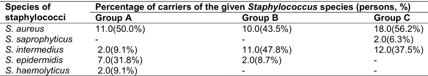
A frequent infection with Staphylococcus aureus in athletes the activity of which is related to water enables to classify with a group with a high risk of dermatological diseases [10,11]. It was established that S. aureus prevailed in swimmers, and in water polo players and in pentathlon athletes S. aureus and S. intermedius prevailed. The results of the test χ2 demonstrated that the species composition of the staphylococcal microflora on the swimmers’ skin differs from that of the water polo players and of the pentathlon athletes (χ2=22.5, p<0.001) (Table 2).
In water polo players and in pentathlon athletes the infection with S. aureus was superior to the norm, while the swimmers did not show positive differences compared to established limits. It seems to be related to the peculiarities of the sports: In particular, swimmers have no direct contact with other athletes during trainings and competitions, while water polo is a team sport
Table 1. Occurrence of Staphylococcus species detected on the skin of subjects in the groups
studied
Species of staphylococci
Percentage of carriers of the given Staphylococcus species (persons, %)
Group I Group II Group III
S. aureus 38.0(49.3%) 16.0(32.6%) 18.0(32.1%)
S. saprophyticus 2.0(2.6%) 4.0(8.2%) 3.0(5.4%)
S. intermedius 26.0(33.8%) 4.0(8.2%) 12.0(21.4%)
S. epidermidis 9.0(11.7%) 25.0(51.0%) 12.0(21.4%)
S. haemolyticus 2.0(2.6%) - 11.0(19.7%)
The Ist group comprised 77 athletes of water sports; the IInd group comprised 49 not aquatics athletes; the IIIrd group comprised 56 non athletes
Table 2. Occurrence of Staphylococcus species detected on the skin of the athletes engaged
in aquatic sports
Species of staphylococci
Percentage of carriers of the given Staphylococcus species (persons, %)
Group A Group B Group C
S. aureus 11.0(50.0%) 10.0(43.5%) 18.0(56.2%)
S. saprophyticus - - 2.0(6.3%)
S. intermedius 2.0(9.1%) 11.0(47.8%) 12.0(37.5%)
S. epidermidis 7.0(31.8%) 2.0(8.7%) -
S. haemolyticus 2.0(9.1%) - -
where the contact between the athletes is inevitable. According to the Student’s criterion, the level of infection with S. aureus was the highest in pentathlon athletes and was positively different from that of the swimmers (t=1.8, p<0.05), which can also be related to the fact that besides swimming, the athletes are trained as well with other disciplines of the modern pentathlon.
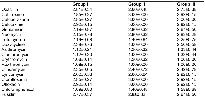
The study of the S. aureus susceptibility to methicillin (oxacillin) showed that methicillin resistant strains of S. aureus (MRSA) were revealed only in athletes the activity of which is related to water, which does not correspond to the data of other authors that revealed the MRSA strains as the most often in representatives of disciplines not related to water [12,13]. However, we revealed MRSA-strains as well in the pentathlon athletes for whom swimming is only a part of pentathlon while other disciplines making part of the modern pentathlon (racing, fencing, shooting and running) have their effect on the skin microbiocenosis. It is consistent with literature data about the MRSA strains spreading in athletes dealing with fencing [14]. On the basis of the data obtained, one can select medicines to treat dermatosis associated with Staphylococcus aureus: The 1st group athletes have to avoid macrolids or chloramphenicol, the 2nd group athletes have to avoid as well tetracycline-based drugs, while the pentathlon athletes have to exclude oxacillin due to a high widespread of the MRSA strains (Table 3).
3.1.2 Malassezia yeast
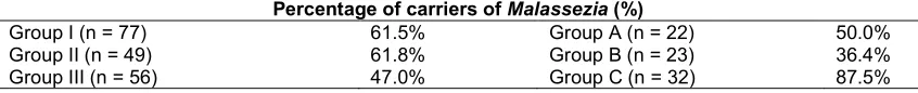
It was revealed that the highest infection with Malassezia was revealed in the groups of athletes both for water sports (61.5%) and for non-aquatic sports (61.8%). Between the 1st group athletes, the highest occurrence frequency of the Malassezia species yeast was revealed in pentathlon athletes (87.5%), it was of 50% between the swimmers, and the least infection with Malassezia was observed in water polo athletes (36.4%). In the 2nd group, the highest degree of the Malassezia occurrence was observed in soccer players, these indices having a significant statistical difference with those of the control group (p ≤ 0.05). In the 3rd group, the infection with Malassezia was significantly lower (47.0%) (Table 4).
According to our observations, the clinical picture of various malasseziosis in athletes has some differences. Their manifestations are most often multiple, symmetrical, they are located on the neck, chest, back, abdomen and in the axillary cavities. Chronic eruptions are represented as maculae: On the chest and abdomen of a brownish color, 0.3 to 1 cm diameter, with rounded shapes that alternate on the back with whitish color maculae of a higher size and of whimsical configurations. The absence of seasonal fluctuations and a scarce desquamation on the maculae surface are to be classified with the peculiarities of the heterochromatic lichen formation.
Table 3. Sensitivity of St. aureus to antibiotics and antiseptics in the Groups
Group I Group II Group III
Oxacillin 2.81±0.34 2.60±0.48 2.75±0.38
Cefuroxime 2.85±0.27 3.00±0.00 2.92±0.15
Cefoperazone 2.85±0.27 3.00±0.00 3.00±0.00
Cefotaxime 2.92±0.15 3.00±0.00 2.92±0.15
Gentamicin 2.19±0.87 2.80±0.32 2.67±0.50
Neomycin 2.15±0.78 2.80±0.32 2.83±0.28
Tetracycline 2.19±0.68 1.40±0.64 2.25±0.75
Doxycycline 2.38±0.76 1.00±0.00 2.50±0.58
Azithromycin 1.12±0.21 1.20±0.32 1.33±0.44
Clarithromycin 1.12±0.20 1.00±0.00 1.33±0.44
Erythromycin 1.08±0.14 1.20±0.32 1.00±0.00
Roxithromycin 1.08±0.15 1.00±0.00 1.00±0.00
Clindamycin 2.35±0.65 2.40±0.72 2.42±0.78
Lyncomycin 2.62±0.56 2.60±0.64 2.92±0.15
Ciprofloxacin 2.85±0.27 3.00±0.00 2.92±0.15
Ofloxacin 2.92±0.14 3.00±0.00 2.92±0.15
Chloramphenicol 1.69±0.80 1.40±0.48 1.58±0.68
Revealing micellar forms of the Malassezia yeast has a prognostic importance to assess the welfare of a considered ecosystem [15].
3.1.3 Determination of propionic bacterial species
The determination of the species diversity for propionic bacteria on the skin of athletes by the quantitative PCR method showed that the frequency of revealing the three species of propionic bacteria: Propionibacterium acnes, granulosum and avidum in a considered skin area both in athletes and in the control group was of 100%. It is not conform to literature data obtained with the use of traditional methods where this index for P. acnes varied from 46 to 100%, for P. granulosum, from 0 to 85%, and for P. avidum, from 0 to 52% [16]. Besides, the infection (on the average) represented: In the group of athletes: P. acnes of 1.6*105 cells/сm2, P. granulosum of 9.4*102 cells/сm2, P. avidum of 5.8*104 cells/сm2, in the control group, P. acnes of 3.8*105 cells/сm2, P. granulosum of 9.4*102 cells/сm2, P. avidum of 7.7*104 cells/сm2. As it appears, in the control group said index was 2 times higher than that of the group of athletes only in the case of P. acnes (Table 5).
No significant correlative relationship was observed between the abundance of propionic bacteria and the biophysical indices in all the groups. So, our proposed method for studying the subpopulation composition of propionic bacteria provides for additional possibilities in the assessment of the skin microbiota condition. The main difference from existing methods is that the bacteria genome is determined regardless of their viability.
3.2 Biophysical Indices
While determining the moisture of superficial skin layers, decreased indices were observed in most of the examined people. The corneometry
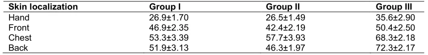
method gave the data as follows: The hand skin was very dry in the 1st and 2nd groups and dry in those of the 3rd group. The skin of the front in the 1st and 3rd groups was found dry, and in those of the 2nd group as very dry. In the 2nd and 3rd groups, the measuring of the chest skin hydration demonstrated a normal skin, while in athletes the activity of which is related to water, it was dry. The skin of the back in all the athletes was found dry, and it was normal in individuals not engaged in sports. In the 2nd group, the skin hydration was assessed as follows: The hand and front skin was dry in swimmers, while it was very dry in water polo players and in pentathlon athletes. The chest skin in pentathlon athletes and in water polo players was found dry, in the swimmers it was normal, while the skin of the back was very dry in pentathlon athletes and normal in swimmers and in water polo players. Trustworthy differences were found between the swimmers and the water polo players (p<0.05), as well as between the swimmers and the pentathlon athletes (p<0.001). Thus, between all the athletes, the pentathlon athletes demonstrated the most significant modifications of the corneometry indices: The skin of the chest was dry, the skin of the hand, front and back was very dry. The corneometry indices modifications in the swimmers were less manifested: The skin of the hand and of the front was dry, the skin of the chest and the back being normal (Table 6).
The determination of the lipid content on the skin surface showed that in all the examined people, the skin was dry. The lipid content indices in athletes of non-aqueous sports were lower than in athletes training in swimming pools (t=1.8, p<0.05). The most significant decrease of indices was revealed in pentathlon athletes (t=1.7, p<0.05). The corneometry and sebumetry indices of the athletes’ skin, independently of the water factor effect, were positively lower than those of individuals not engaged in sports (t=1.7, p<0.05) (Table 7).
Table 4. Occurrence of Malassezia detected on the skin of subjects in the groups studied
Percentage of carriers of Malassezia (%)
Group I (n = 77) 61.5% Group A (n = 22) 50.0%
Group II (n = 49) 61.8% Group B (n = 23) 36.4%
Group III (n = 56) 47.0% Group C (n = 32) 87.5%
Table 5. Determination of propionic bacterial species
Group III Group I, II
P. acnes 3,8*105 cells/сm2 1,6*105 cells/сm2
P. granulosum 9,4*102 cells/сm2 9,4*102 cells/сm2
Table 6. Determination of the moisture of superficial skin layers (standard units)
Skin localization Group I Group II Group III
Hand 26.9±1.70 26.5±1.49 35.6±2.90
Front 46.9±2.35 42.4±2.19 50.4±2.50
Chest 53.3±3.39 57.7±3.93 68.3±2.18
Back 51.9±3.13 46.3±1.97 72.3±2.17
Considering the fact that the washing off method used to determine the infection with staphylococcal and micellar microflora was carried out on a skin area in the middle of the chest, we used the corneometry and sebumetry indices of the same area in order to reveal correlative relations. A direct mean power correlation was revealed between the corneometry indices and the skin infection with S. intermedius (r= 0.7) in the 2nd group patients, and an inverse mean power correlation was revealed between the corneometry indices and the skin infection in the 3rd group patients with S. aureus (r= -0.7). An inverse mean-power correlation was revealed in water polo players between the age and the corneometry indices (r= -0.7), a direct mean-power correlation was revealed in water polo players between the corneometry and sebumetry indices (r = 0.76), a direct mean-power correlation was revealed in pentathlon athletes between the skin infection with S. aureus and the sebumetry indices (r= 0.62), a direct mean-power correlation was revealed in swimmers between the infection with
S. epidermidis and the corneometry indices (r= 0.69).
Contrary to the generally accepted opinion about a high correlation between the volume of the Malassezia yeast and the lipid content, we obtained the data as follows: Correlation factor (r) between the occurrence and the sebumetry, assessed after comparison of all the examined groups was equal to 0.447, the same index between the skin infection degree and sebumetry was of 0.425. At the same time, correlation is observed between the degree of skin infection and the skin hydration r = 0.694. However, no relationship between the occurrence of Malassezia and the hydration was observed, r = 0.135. A direct mean-power correlation was observed between the occurrence and the infection degree r = 0.582, as well as a high correlation between the age and the occurrence r = 0.773. Decrease of biophysical indices enables to arrive at a conclusion of the insufficiency of the epidermal barrier, while the prevailing content of Staphylococcus aureus in athletes of water
sports, at the conclusion of a disturbance in the colonization resistance of the skin. The consequence of the disturbances revealed resides in the development of the skin dryness and in an eventual infection. In turn, recovery of the skin barrier function is accompanied by a lower degree of infection with Staphylococcus aureus and the appearance of saprophyte strains [17].
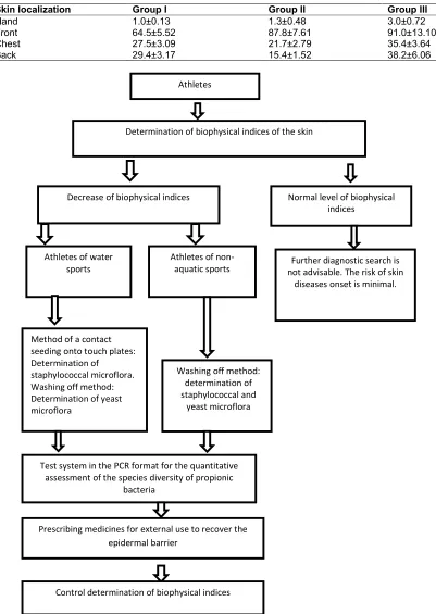
First stage of the pre-nosological diagnosis of the athlete skin is the determination of just biophysical indices. When following the methods we have carried out, it is advisable to start the assessment of the functional condition of the skin just with the measuring of the skin hydration by the method of corneometry and with that of lipid content on the skin surface by the method of sebumetry. We have developed a diagnostic algorithm that can be recommended for carrying out a profound medical examination of athletes.
As shown in the diagram, at a normal level of skin biophysical indices in an athlete, regardless of the specialization, further diagnostic search is not advisable. When revealing some decrease of biophysical indices, it is recommended, at a next stage, to determine the skin microbiocenosis condition according to the methods as follows:
For the athletes whose activity is not related to water: determination of the species composition and the degree of skin infection with staphylococcal and yeast microflora by the washing off method.
For the athletes of water sports: The method of a contact inoculation on touch plates to determine the species composition and the degree of skin infection with the staphylococcal microflora, and determining the frequency of occurrence and the degree of infection with yeast microflora by the washing off method.
Table 7. Determination of the lipid content on the skin surface (mkg/sm2)
Skin localization Group I Group II Group III
Hand 1.0±0.13 1.3±0.48 3.0±0.72
Front 64.5±5.52 87.8±7.61 91.0±13.10
Chest 27.5±3.09 21.7±2.79 35.4±3.64
Back 29.4±3.17 15.4±1.52 38.2±6.06
Diagram of the diagnostic algorithm for the risk of dermatosis onset in athletes Athletes
Determination of biophysical indices of the skin
Decrease of biophysical indices
Athletes of water sports
Athletes of non-aquatic sports
Method of a contact seeding onto touch plates: Determination of
staphylococcal microflora. Washing off method: Determination of yeast microflora
Washing off method: determination of staphylococcal and
yeast microflora
Prescribing medicines for external use to recover the epidermal barrier
Control determination of biophysical indices
Normal level of biophysical indices
Further diagnostic search is not advisable. The risk of skin
diseases onset is minimal.
Test system in the PCR format for the quantitative assessment of the species diversity of propionic
The algorithm provided enables to reveal the groups of risks for onset of dermatologic diseases.
4. CONCLUSION
Thus, professional sport activities have a significant effect on the functional condition of the skin that changes depending on the conditions and the regime of training. It is to note that between all the athletes we have examined, the pentathlon athletes have the most manifested decrease of biophysical indices as well as a high degree of skin infection with the conditionally pathogenic Staphylococcus aureus, which enables to classify them in a group with a high risk of the dermatosis development. Using the data obtained, it is possible to give practical recommendations to doctors, coaches and their athletes. According to this diagnostic algorithm, it is easy to solve the problem of prescribing medicines for external use that recover the epidermal barrier. In this case, it is advisable to control the biophysical indices not earlier than 3 weeks after starting the treatment with medicines for external use. When revealing skin diseases related to S. aureus it is obligatory to determine the susceptibility to antimicrobial medicines, in particular the MRSA strains. When selecting antibiotics for the 1st group athletes, it is necessary to avoid macrolides and chloramphenicol, and for the 2nd group athletes, the use of tetracycline medicines is to be avoided as well, due to the existence of resistant strains. It is of current interest to further study, at our opinion, the functional skin condition in athletes of various sports to allow an early diagnosis and to prevent the development of professional dermatosis.
ETHICAL APPROVAL
Ethical expert’s opinion about this research was approved by the decision No 31 of the local Ethics Committee of the FGU VNIIFK Institute on the 17.08.2010. All the athletes gave written informed consent before the study.
ACKNOWLEDGEMENTS
Thanks to the leadership of the to the swimming pools of the Open Joint Stock Company “Luzhniki” and of the Sport School GBOU DODSN SDUSShOR “Severnyi”, and special thanks to the laboratory “Informational Technologies in Sports” of the Moscow MFTI Institute.
COMPETING INTERESTS
Authors have declared that no competing interests exist.
REFERENCES
1. Suvorov VG, Achkasov EE, Kurshev VV, Lazareva IA, Sultanova OA, Krasavina TV. Legal and organizational principles of medical rehabilitation of patients with occupational diseases. Sports medicine: Research and Practice. 2014;1:74-79. (in Russian).
2. Polievskyi SA, Ivanov AA, Grigorieva OV, Sivtsev IN. About the diagnosis and monitoring of the physical health and of the sport condition for athletes students. Theory and Practice of the Physical Culture. 2005;3:24-26. (in Russian). 3. Chikin VV. Peculiarities of the dermatosis
development in people having contacts with substances presenting an irritating and allergenic effect. Medicine of Labor and Industrial Ecology. 2001;11(12-16) (in Russian).
4. Zaborova VA, Arzumanyan VG, Gurevich KG, Terekhova MV. Effect of the physical culture and sport exercises on the integument condition in people dealing with water sports. Russian Journal of Cutaneous and Venereal Diseases. Moscow. 2012;1:56-58. (in Russian). 5. Ernandes EI, Margolina AA, Petrukhina
AO. Lipid barrier of the skin and cosmetics. Third edition, completed. Moscow: Clavel Company. 2005;400. (in Russian).
6. Ivko OA. Effect of the sport traumatism on the life quality of sport veterans at middle and old ages: Ph.D. in biol. Dissert. St. Petersb. 2007;107. (in Russian).
7. Goriachkina LA, Kashkin KP. Clinical allergology and immunology. Moscow: Miclosh. 2009;273-287. (in Russian). 8. Shenderov BA. Medical microbial ecology
and functional diet. Social, economic and clinical consequences of the unbalance in the microbial ecology of the man and the animals. Moscow: “Grant”. 1998;2:412. (in Russian).
10. Barna Z, Kádár M. The risk of contracting infectious diseases in public swimming pools. A review. Ann Ist Super Sanita. 2012;48(4):374-386.
11. Bonini M, Bodina A, Bonali D, Bascucci B, Pellino P, Castaldi S. Investigation and comparison of behaviours of adults and children in swimming pools. Ann Ig. 2011;23(4):319-328.
12. Bowers AL, Huffman GR, Sennett BJ. Methicillin-resistant Staphylococcus aureus infections in collegiate football players. Medicine & Science in Sports & Exercise. 2008;40(8):1362-7.
13. Buss BF, Mueller SW, Theis M, Keyser A, Safranek TJ. Population-based estimates of methicillin-resistant Staphylococcus aureus (MRSA) Infections among High School Athletes—Nebraska, 2006–2008. The Journal of School Nursing. 2009;25(4): 282-291.
14. Centers for Disease Control and Prevention (CDC) Methicillin-resistant
Staphylococcus aureus infections among competitive sports participants: Colorado, Indiana, Pennsylvania and Los Angeles County, 2000–2003. MMWR Morbidity and Mortality Weekly Report. 2003;52(33): 793-795.
15. Zaborova VA, Arzumanyan VG, Gurevich KG. Malasseziosis in athletes. Russian Journal of Cutaneous and Venereal Diseases. Moscow. 2013;6:55-59. (in Russian).
16. Cogen AL, Nizet V, Gallo RL. Skin microbiota: A source of disease or defence? Br. J. Dermatol. 2008;158(3): 442–455.
17. Gysleva OR. Morphological characteristic of microbial communities and peculiarities of the epidermal barrier in patients with the atopic dermatitis: Ph.D. in boil. Dissert. GOU VPO “Military Medical Academy. St. Petersburg. 2011;127. (in Russian).
© 2015 Zaborova et al.; This is an Open Access article distributed under the terms of the Creative Commons Attribution License (http://creativecommons.org/licenses/by/4.0), which permits unrestricted use, distribution, and reproduction in any medium, provided the original work is properly cited.
Peer-review history:
The peer review history for this paper can be accessed here: