Clinical Ophthalmology
Iatrogenic retinal breaks during 20-gauge
vitrectomy for proliferative diabetic retinopathy
Yumi Kamura1 Yukihiro Sato2 Yuzou Deguchi3 Fumihiko Yagi3
1Department of Ophthalmology,
Division of Visual Sciences, Nihon University School of Medicine, Tokyo, Japan; 2Department of Diabetes
Center, Jichi Medical University, Tochigi, Japan; 3Department of
Ophthalmology, Toho University Sakura Medical Center, Chiba, Japan
Correspondence: Yumi Kamura Department of Ophthalmology, Division of Visual Sciences, Nihon University School of Medicine, 30-1 Ohyaguchi kamimachi, Itabashi-ku, Tokyo, 173-8610, Japan
Tel +81 3 3972 8111 ext 2531 Fax +81 3 5995 3495
Email kamura.yumi@nihon-u.ac.jp
Background: We classified iatrogenic retinal break formation during 20-gauge pars plana vitrectomy for proliferative diabetic retinopathy into three types according to the mechanism of development, and evaluated the association of each type with postoperative complications. This is the largest series of such patients published to date.
Methods: This was a retrospective comparative study of 760 eyes from 609 cases who underwent primary 20-gauge vitrectomy for proliferative diabetic retinopathy and were followed-up for at least 6 months after surgery. Postoperatively, the eyes were classified as having vitreous hemor-rhage only (group 1), fibrovascular membrane without traction retinal detachment (group 2), or fibrovascular membrane with traction retinal detachment (group 3).
Results: The overall incidence of iatrogenic retinal breaks was 29%. Fibrovascular membrane dissection was associated with retinal break formation in 50 of the eyes in group 3, an incidence which was significantly higher than that in group 2 (P , 0.001). Posterior vitreous detachment creation and peripheral vitreous shaving were associated with retinal break formation in 8% of eyes overall, and oral dialysis occurred in 2%. Postoperatively, vitreous hemorrhage requiring washout, neovascular glaucoma, recurrent retinal detachments, and fibrovascular proliferation at the sclerotomy sites occurred in 4%, 4%, 3%, and 1%, respectively, of all eyes. Outcomes of eyes with these postoperative complications, other than vitreous hemorrhage, were poor. Multiple regression analysis revealed retinal break formation during fibrovascular membrane dissection to be significantly related to postoperative vitreous hemorrhage (P = 0.019), recurrent retinal detachments (P , 0.001), and neovascular glaucoma (P = 0.048). Oral dialysis was also significantly related to postoperative vitreous hemorrhage (P = 0.001).
Conclusion: Iatrogenic retinal break formation during fibrovascular membrane dissection was more likely to be the cause of poor outcomes than peripheral retinal breaks or oral dialysis.
Keywords: iatrogenic retinal breaks, 20-gauge vitrectomy, proliferative diabetic retinopathy, postoperative complications
Introduction
Vitreous surgery for proliferative diabetic retinopathy has been reported to be closely associated with severe intraoperative and postoperative complications.1 Iatrogenic breaks develop via different mechanisms, such as oral dialysis and during proliferative
fibrovascular membrane dissection.2 To our knowledge, there has been only one
previ-ous study in which iatrogenic retinal breaks were classified according to mechanisms of development.2 In that study, breaks were divided into two groups, ie, posterior breaks and peripheral breaks, to allow incidence comparisons between 20-gauge and 23-gauge vitrectomy for proliferative diabetic retinopathy. In our study, iatrogenic breaks were classified into three types, ie, breaks occurring during fibrovascular membrane dissection,
Dove
press
O r I g I N A L r E S E A r C h
open access to scientific and medical research
Open Access Full Text Article
Clinical Ophthalmology downloaded from https://www.dovepress.com/ by 118.70.13.36 on 21-Aug-2020
For personal use only.
Number of times this article has been viewed
This article was published in the following Dove Press journal: Clinical Ophthalmology
those occurring during the induction of posterior vitreous detachment or peripheral vitreous dissection, and oral dialysis related to the sclerotomy sites. Their incidences and associa-tions with major postoperative complicaassocia-tions were evaluated in the largest published series to date of patients undergoing 20-gauge vitrectomy for proliferative diabetic retinopathy. These evaluations may provide basic data allowing compari-son between 20-gauge vitrectomy and micro-incision vitreous surgery for proliferative diabetic retinopathy.
Materials and methods
The subjects consisted of 609 consecutive patients (760 eyes) who underwent their first operation for proliferative diabetic retinopathy between August 2001 and September 2003 at Nihon University Itabashi Hospital and between October 2003 and March 2008 at Sakura Medical Center Toho Uni-versity Hospital. All were followed up for at least 6 months after their surgery. There were 385 males and 224 females
of mean age 57.3 ± 11.4 (range 24–84) years.
Vitreous surgery was performed using a 20-gauge three-port vitrectomy system. When there was no posterior vitreous detachment, it was induced. Proliferative membrane dissec-tion using vertical or horizontal scissors and intraocular pho-tocoagulation were performed, and intraocular tamponade was
added when necessary. In phakic patients aged $ 50 years,
cataract surgery was simultaneously performed. The opera-tions were performed by 15 surgeons.
Patients with neovascular glaucoma before surgery, those who underwent surgery to treat macular edema or to remove submacular hard exudates, and those with pre-existing retinal breaks including combined traction and rhegmatogenous retinal detachment were excluded. No patients were treated with bevacizumab (antivascular endothelial growth factor) either preoperatively or postoperatively.
The subjects were classified postoperatively according to their disease type into three groups, ie, group 1 with vitreous hemorrhage only, group 2 with fibrovascular membranes requiring treatment without traction retinal detachment, and group 3 with fibrovascular membrane and traction retinal detachments.
Iatrogenic breaks were classified into breaks occurring during fibrovascular membrane dissection (breaks during membrane dissection), breaks occurring during the induc-tion of posterior vitreous detachment or peripheral vitreous dissection (peripheral breaks), and oral dialysis related to the sclerotomy sites.
The postoperative complications evaluated were vitreous rebleeding requiring washout on reoperation (rebleeding),
neovascularization at the sclerotomy site, retinal detachment due to reproliferation with or without retinal breaks, and neovascular glaucoma. This retrospective study was per-formed using medical records. Visual acuities were measured using a standard Japanese decimal visual acuity chart.
Statistical analysis
The χ2 test, mxn χ2 test, Fisher’s exact probability test, logistic regression analysis, and multiple comparisons with the Bonferroni method were used to perform the statistical
analysis. P , 0.05 was regarded as being statistically
sig-nificant. StatView 5.0 (SAS Institute Inc, Cary, NC, USA) was the statistical analysis software package used.
Results
Patients
Group 1 consisted of 239 patients (259 eyes), group 2 con-sisted of 286 patients (318 eyes), and group 3 of 161 patients
(183 eyes). The mean ages of the patient were 60.0 ± 10.9,
56.2 ± 11.0, and 53.6 ± 12.0 years, respectively.
Incidence of iatrogenic breaks
All cases
Iatrogenic breaks occurred in 28.5% of all 760 eyes. Breaks during membrane dissection developed in 31.5% of eyes in groups 2 and 3 after membrane dissection. Peripheral breaks occurred in 7.5% and oral dialysis in 2.1% of all eyes.
According to group
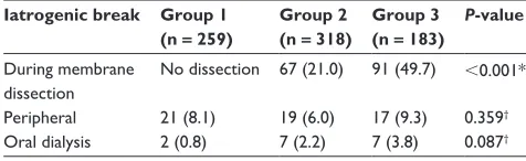
Iatrogenic breaks occurred in 8.9%, 29.2%, and 62.8% of groups 1, 2, and 3, respectively (see Table 1). Breaks dur-ing membrane dissection were significantly more common in group 3 than in group 2 (P , 0.001, χ2 test). Incidences of peripheral breaks and oral dialysis did not differ
signifi-cantly among the three groups (P = 0.359 and P = 0.087,
respectively, mxn χ2 test).
Table 1 Incidence of each type of iatrogenic break
Iatrogenic break Group 1 (n = 259)
Group 2 (n = 318)
Group 3 (n = 183)
P-value
During membrane dissection
No dissection 67 (21.0) 91 (49.7) ,0.001*
Peripheral 21 (8.1) 19 (6.0) 17 (9.3) 0.359†
Oral dialysis 2 (0.8) 7 (2.2) 7 (3.8) 0.087† Notes: Group 1, vitreous hemorrhage only; group 2, fibrovascular membranes requiring treatment without traction retinal detachment; group 3, fibrovascular membrane with traction retinal detachments. Eyes (%). Some eyes showed multiple types of breaks. †mxn χ2 test; *χ2 test.
Dovepress
Kamura et al
Clinical Ophthalmology downloaded from https://www.dovepress.com/ by 118.70.13.36 on 21-Aug-2020
Postoperative complications
All cases
Evaluation of postoperative complications in all 760 eyes showed rebleeding in 3.6%, neovascularization at the sclero-tomy site in 1.3%, retinal detachment due to reproliferation in 2.8%, and neovascular glaucoma in 3.9%.
According to group
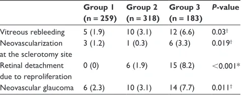
The incidence of rebleeding and that of neovascular
glau-coma were higher in group 3 than in group 1 (P = 0.013 and
P = 0.008, respectively, Bonferroni method; Table 2). Further-more, the incidence of neovascularization at the sclerotomy site and that of retinal detachment due to reproliferation was higher in group 3 than in group 2 (P = 0.007 and P = 0.001, respectively, Bonferroni method, χ2 test).
Associations between visual outcomes after rebleeding and neovascularization
at sclerotomy site
Vitreous rebleeding was observed in 27 eyes, ie, 10 eyes with and 17 eyes without neovascularization at the sclerotomy site. Final visual acuity was ,0.1 in all 10 eyes showing neovascularization at the sclerotomy site, and in 41.2% of the 17 eyes without this complication. The final visual acuity was poorer in the presence of neovascularization at the sclerotomy site (P = 0.002, Fisher’s exact probability test). Neovascular glaucoma developed in 80.0% of the 10 eyes showing neovascularization at the sclerotomy site and 35.3% of the 17 eyes without this complication, being significantly more
frequent in the presence of neovascularization (P = 0.031,
Fisher’s exact probability test).
Associations between visual outcomes after retinal detachment due to reproliferation and neovascular glaucoma
Retinal detachment due to reproliferation occurred in 21 eyes. Reoperation for retinal detachment due to reproliferation was performed 1–3 (mean 1.4) times, and retinal reattachment
was achieved in only 38% of these eyes. Final visual acuity
was ,0.1 in 25% of the eight eyes showing retinal
reattach-ment, but was ,0.1 in all 13 eyes without retinal
reattach-ment, with poorer visual acuity in the latter (P , 0.001, Fisher’s exact probability test). None of the eight eyes show-ing retinal reattachment developed neovascular glaucoma, but this complication did occur in 38.5% of the 13 eyes without retinal reattachment. However, the incidences did not differ significantly between the two groups (P = 0.063, Fisher’s exact probability test).
Associations between iatrogenic breaks
and postoperative complications
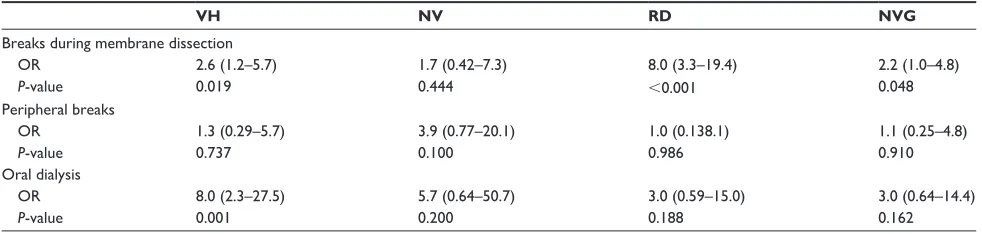
Associations between iatrogenic breaks and postoperative complications were evaluated by logistic regression analysis. Breaks during membrane dissection were significantly asso-ciated with vitreous rebleeding (P = 0.019), retinal detach-ment due to reproliferation (P , 0.001), and neovascular
glaucoma (P = 0.048; Table 3). Peripheral breaks were not
associated with postoperative complications, while oral dialysis was associated with rebleeding (P = 0.001).
Discussion
Vitreous surgery for proliferative diabetic retinopathy is associated with severe intraoperative and postoperative
com-plications, which might cause worsening of visual outcomes.1
Iatrogenic retinal breaks and intraocular bleeding are major intraoperative complications.1 However, iatrogenic breaks include breaks occurring by different mechanisms, such as breaks at the posterior pole during fibrovascular membrane dissection, those in the peripheral retina during posterior vitreous detachment induction or thorough dissection of the peripheral vitreous, and oral dialysis associated with instru-ment insertion and removal, and their influences on outcomes would presumably differ. To our knowledge, there has been only one previous study in which iatrogenic retinal breaks
were classified according to the mechanisms of development.2
However, the relationships between iatrogenic breaks and postoperative complications were not evaluated.
Herein, we clarified the associations between iatrogenic breaks and major postoperative complications in the larg-est series of patients undergoing 20-gauge vitrectomy for proliferative diabetic retinopathy published to date. These evaluations may provide basic data allowing comparison between 20-gauge vitrectomy and microincision vitreous surgery for proliferative diabetic retinopathy.
Among postoperative complications, vitreous rebleed-ing consistently develops at a predictable rate after vitreous
Table 2 Postoperative complications
Group 1 (n = 259)
Group 2 (n = 318)
Group 3 (n = 183)
P-value
Vitreous rebleeding 5 (1.9) 10 (3.1) 12 (6.6) 0.03†
Neovascularization at the sclerotomy site
3 (1.2) 1 (0.3) 6 (3.3) 0.019†
retinal detachment due to reproliferation
0 (0) 6 (1.9) 15 (8.2) ,0.001*
Neovascular glaucoma 6 (2.3) 10 (3.1) 14 (7.7) 0.011† Notes: Eyes (%). Some eyes showed multiple types of breaks. †mxn χ2 test; *χ2 test.
Dovepress Iatrogenic retinal breaks during 20-gauge vitrectomy
Clinical Ophthalmology downloaded from https://www.dovepress.com/ by 118.70.13.36 on 21-Aug-2020
surgery for proliferative diabetic retinopathy despite its minimal influences on visual outcomes.1 In contrast, neovas-cularization at the sclerotomy site, retinal detachment due to reproliferation, and neovascular glaucoma are considered to carry poor prognoses.1,3–5
In this study, certain iatrogenic breaks occurred in about 30% of all cases. Studies in more than 100 eyes have shown the incidence of iatrogenic breaks in 20-gauge surgery to
range from 33% to 41%.3,6,7 Breaks developed in about 30%
of our cases during fibrovascular membrane dissection. The incidence was significantly higher in those with traction retinal detachment. This difference might be attributable to traction retinal detachment inducing atrophy and fragility of the detached retina, thereby allowing the development of iatrogenic retinal breaks.
The incidence of peripheral breaks developing during induction of posterior vitreous detachment or peripheral vitreous dissection, and that of oral dialysis related to the sclerotomy sites, did not differ significantly among the three groups. The incidence of peripheral breaks during the induc-tion of posterior vitreous detachment or peripheral vitreous dissection is reportedly 6%,2 while those with oral dialysis
have been shown to be 8%2 and 6%.8 Concerning
postopera-tive complications, the incidence of each complication was highest in group 3, and the incidences of retinal detachment due to reproliferation and neovascular glaucoma were about 8% each.
The incidence rates of each of these complications were similar to those obtained by Oda et al,3 who reported the highest incidence of postoperative complications in the traction retinal detachment group and that incidences of reproliferation and neovascular glaucoma each exceeded 10%. Therefore, cases with fibrovascular membranes should be closely followed up. If progressive membrane contrac-tion likely to cause traccontrac-tion retinal detachment is detected, surgical intervention should be considered. Evaluating the
association between iatrogenic breaks and postoperative complications may provide useful information for the management of patients who have already developed this detachment. Oda et al3 evaluated the association between iatrogenic breaks and postoperative complications, and the final visual acuity was ,0.1 in 30% of cases developing iatrogenic retinal breaks, and the odds ratio as a risk factor for final visual acuity , 0.1 was 1.9. Yorston et al7 reported iatrogenic breaks to be a risk factor for worsening of post-operative visual acuity. However, in their study, the types of iatrogenic breaks were not evaluated. In the present study, retinal breaks during fibrovascular membrane dissection were significantly associated with postoperative rebleeding, retinal detachment due to reproliferation, and neovascular glaucoma. Furthermore, there was a significant association between oral dialysis and rebleeding. Therefore, if the incidences of iatrogenic breaks during membrane dissection and oral dialysis can be lowered, the incidences of these postoperative complications can likely be reduced as well.
Despite providing basic data on iatrogenic retinal breaks during 20-gauge vitrectomy for proliferative diabetic retin-opathy in the largest published patient series to date, this study is limited by being retrospective and non-randomized. A prospective, randomized clinical trial is needed for com-parison between 20-gauge vitrectomy and micro-incision vitreous surgery for proliferative diabetic retinopathy.
Disclosure
The authors report no conflicts of interest in this work.
References
1. Eliott D, Lee MS, Abrams GW. Proliferative diabetic retinopathy: principles and techniques of surgical treatment. In: Ryan SJ, editor. Retina, 4th ed. St Louis, MO: Mosby; 2006.
2. Issa SA, Connor A, Habib M, Steel DHW. Comparison of retinal breaks observed during 23 gauge transconjunctival vitrectomy versus con-ventional 20 gauge surgery for proliferative diabetic retinopathy. Clin Ophthalmol. 2011;20:109–114.
Table 3 Iatrogenic breaks and postoperative complications
VH NV RD NVG
Breaks during membrane dissection
Or 2.6 (1.2–5.7) 1.7 (0.42–7.3) 8.0 (3.3–19.4) 2.2 (1.0–4.8)
P-value 0.019 0.444 ,0.001 0.048
Peripheral breaks
Or 1.3 (0.29–5.7) 3.9 (0.77–20.1) 1.0 (0.138.1) 1.1 (0.25–4.8)
P-value 0.737 0.100 0.986 0.910
Oral dialysis
Or 8.0 (2.3–27.5) 5.7 (0.64–50.7) 3.0 (0.59–15.0) 3.0 (0.64–14.4)
P-value 0.001 0.200 0.188 0.162
Abbreviations: Vh, vitreous rebleeding; NV, neovascularization at sclerotomy site; rD, retinal detachment due to reproliferation; NVg, neovascular glaucoma; Or, odds ratio (95% confidence interval).
Dovepress
Kamura et al
Clinical Ophthalmology downloaded from https://www.dovepress.com/ by 118.70.13.36 on 21-Aug-2020
Clinical Ophthalmology
Publish your work in this journal
Submit your manuscript here: http://www.dovepress.com/clinical-ophthalmology-journal Clinical Ophthalmology is an international, peer-reviewed journal covering all subspecialties within ophthalmology. Key topics include: Optometry; Visual science; Pharmacology and drug therapy in eye diseases; Basic Sciences; Primary and Secondary eye care; Patient Safety and Quality of Care Improvements. This journal is indexed on
PubMed Central and CAS, and is the official journal of The Society of Clinical Ophthalmology (SCO). The manuscript management system is completely online and includes a very quick and fair peer-review system, which is all easy to use. Visit http://www.dovepress.com/ testimonials.php to read real quotes from published authors. 3. Oda H, Konno K, Mitsui K, Kawamata E, Hiraoka T, Miki D. Recent
outcome of vitreous surgery for diabetic retinopathy. Nippon Ganka Gakkai Zasshi. 2005;109:603–612. Japanese.
4. Charles S, Calzada J, Wood B. Diabetic retinopathy. In: Charles S, editor. Vitreous Microsurgery, 5th ed. Philadelphia, PA: Wolters Kluwer/ Lippincott Williams & Wilkins; 2011.
5. Sawa H, Ikeda T, Matsumoto Y, Niiya A, Kinoshita S. Neovascularization from scleral wound as cause of vitreous rebleeding after vitrectomy for proliferative diabetic retinopathy. Jpn J Ophthalmol. 2000;44: 154–160.
6. Higuchi A, Yamada H, Kawai E, et al. Vitrectomy for proliferative diabetic retinopathy. Nippon Ganka Gakkai Zasshi. 2005;109:134–141. Japanese.
7. Yorston D, Wickham L, Benson S, Bunce C, Sheard R, Charteris D. Predictive clinical features and outcomes of vitrectomy for proliferative diabetic retinopathy. Br J Ophthalmol. 2008;92:365–368.
8. Oyakawa RT, Schachat AP, Michels RG, Rice TA. Complications of vitreous surgery for diabetic retinopathy. I. Intraoperative complications. Ophthalmology. 1983;90:517–521.
Dovepress
Dove
press
Iatrogenic retinal breaks during 20-gauge vitrectomy
Clinical Ophthalmology downloaded from https://www.dovepress.com/ by 118.70.13.36 on 21-Aug-2020